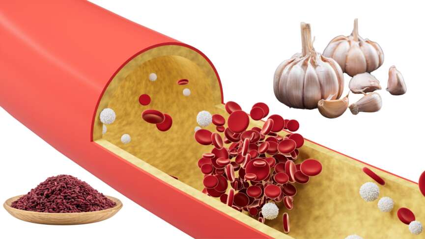
Explore the untold secrets of Red Yeast Rice and Garlic for effectively reducing cholesterol levels in this captivating video. Uncover the remarkable benefits of these natural remedies, supported by scientific evidence. Don’t miss out – click to watch now!
Unveiling the Causes of Hyperthyroidism. Explore the underlying factors that trigger hyperthyroidism, including autoimmune disorders like Graves’ disease, thyroid nodules, excessive iodine intake, and more. Understand how these causes impact your thyroid function and gain insights into preventive measures for maintaining thyroid health.
Uncover the Secrets: Watch our Informative Video on the 6 Signs of High Cholesterol! Are you concerned about your cholesterol levels? We’ve got you covered with our engaging video that reveals the top 6 signs indicating high cholesterol. In this captivating presentation, we delve into the symptoms you should pay attention to. From persistent chest pain to frequent fatigue, we highlight the key indicators that warrant your immediate attention. Take charge of your health by tuning into our video today and gain the knowledge you need to make informed decisions. Don’t hesitate, watch now and empower yourself with the tools to safeguard your well-being! – E Healthy Info
Are you curious about sleep apnea and its different types? Look no further! This comprehensive 1-minute video provides valuable insights into sleep apnea, covering its definition, types, and specific effects on your health. Discover the characteristics of obstructive sleep apnea, where breathing is repeatedly blocked during sleep, leading to disrupted rest and potential health complications. Explore central sleep apnea, a condition characterized by a lack of effort in breathing due to a communication breakdown between the brain and respiratory muscles. Uncover the symptoms of sleep apnea and gain knowledge about available treatment options. Empower yourself with the understanding necessary to identify, manage, and seek appropriate care for sleep apnea. Start your journey toward restful sleep and better health today.
Heart attacks are a leading cause of death worldwide, and they can happen to anyone, regardless of age or gender. Recognizing the warning signs of a heart attack is crucial for timely treatment and better outcomes. This informative video will discuss the top 5 warning symptoms of a heart attack that you should never ignore. These symptoms can be subtle and life-threatening, from chest pain and discomfort to shortness of breath and nausea. Don’t wait until it’s too late – watch this video and learn how to identify the signs of a heart attack and take steps to protect your heart health.
A stroke occurs when blood flow to a part of the brain is disrupted, either by a blockage (ischemic stroke) or bleeding (hemorrhagic stroke). This deprives brain cells of oxygen and nutrients, causing them to die. As a result, the functions controlled by that part of the brain are affected, such as movement, speech, and cognition. The symptoms of a stroke can vary depending on the area of the brain affected but may include weakness or numbness on one side of the body, difficulty speaking, vision problems, and loss of balance or coordination.
Atopic dermatitis, or eczema, is a chronic skin condition that causes dry, itchy, and inflamed skin.
People with atopic dermatitis often have a weakened skin barrier, making their skin more sensitive to irritants and allergens.
Atopic dermatitis is common in children but can also affect adults. It is not contagious.
Atopic dermatitis is linked to a combination of genetics, an overactive immune response, and environmental triggers.
Moisturizing your skin daily helps lock in moisture, soothe dryness, and keep your atopic dermatitis under control.
What is Atopic Dermatitis?
Atopic dermatitis, also known as atopic eczema, is a condition that causes the skin to become dry, itchy, and inflamed. Atopic dermatitis is the most common chronic inflammatory skin disease. It usually begins in infancy or childhood but can develop at any age. [1]
Types of Atopic Dermatitis
There are many different types of atopic dermatitis. The most common ones are:
1. Classic Atopic Dermatitis
This is the most common form, often starting in childhood. It causes dry, itchy, and inflamed skin, usually on the face, elbows, and knees. [2]
2. Contact Dermatitis
This occurs when your skin reacts to allergens or irritants like soaps, fragrances, or metals. It causes redness, itching, and sometimes blisters. [3]
3. Seborrheic Dermatitis
This type affects oily areas like the scalp, face, and chest, causing redness, scaling, and dandruff-like flakes. It is linked to yeast overgrowth and genetics. [4]
4. Nummular Dermatitis
This type appears as round, coin-shaped patches of irritated skin, often after skin injuries like cuts or insect bites. It is more common in adults. [5]
Causes of Atopic Dermatitis
Atopic dermatitis happens due to a mix of genetics, immune system issues, and environmental factors.
1. Genetics
If your family has a history of eczema, asthma, or allergies, you will likely develop atopic dermatitis. If one parent has atopic dermatitis, your risk of developing it is 50%, increasing to 80% if both parents are affected. Similarly, a gene mutation can compromise your skin barrier, leading to dryness and increased sensitivity to irritation. [6]
2. Immune System Dysfunction
Your immune system may overreact to harmless substances, leading to inflammation, redness, and itching. [7]
3. Loss of Skin Barrier
A weak skin barrier allows moisture to escape and irritants, allergens, and bacteria to enter, making your skin dry and easily inflamed. [6]
4. Allergens and Irritants
Pollen, dust mites, pet dander, soaps, and fragrances can trigger flare-ups by irritating your skin. [8]
5. Climate and Weather
Cold, dry air can worsen eczema by drying out your skin, while hot weather and sweating can cause itching and irritation. [6]
6. Stress
Emotional stress can trigger or worsen eczema by increasing inflammation in your body. [9]
Triggers for Atopic Dermatitis
People with atopic dermatitis may report periods with less marked symptoms followed by periods when symptoms are more severe, known as flare-ups. There may be various triggers that may lead to such flare-ups, such as strong soaps, detergents, skin care products, perfumes, makeup products, fabrics like wool or linen, pollen, mold, dust mites, animal dander, tobacco smoke, stress, anger, anxiety, low humidity, dry skin, sweating, or skin infections. [10]
Symptoms of Atopic Dermatitis
The most common symptom of atopic dermatitis is itching. Other common symptoms include:
Red, dry, and scaly patches of skin.
Rashes that may ooze or weep clear fluid.
Bleeding from rashes upon scratching/itching.
Thickening and hardening of skin.
Skin creases on the palms of the hand (lichenification).
Progression of Atopic Dermatitis
Atopic dermatitis usually starts with dry, itchy skin that becomes inflamed due to repeated scratching. In the early stages, the skin appears red and irritated and may have small bumps that ooze fluid. As the condition progresses, persistent scratching and rubbing cause the skin to thicken and become rough, a process called lichenification. This thickened skin develops deep lines, a leathery texture, and darker or lighter patches than the surrounding skin. Lichenified lesions are more resistant to treatment and can cause chronic itching and discomfort, often worsening in flare-ups. [11]
Diagnosis of Atopic Dermatitis
Examination of the lesion is the initial and the most important way to diagnose the conditions. You might need repeated examinations by your doctor to recognize the type of atopic dermatitis. Other tests that might be helpful to diagnose eczema are:
1. Dermoscopy
A dermoscopy is a tool your doctor may use to examine your skin closely. This handheld device magnifies the skin’s surface and helps the doctor identify patterns and small details that may not be visible to the naked eye. Dermoscopy can help distinguish atopic dermatitis from other skin conditions, ensuring an accurate diagnosis. [12]
2. Allergy Tests
Since atopic dermatitis can be triggered by allergens, your doctor might suggest allergy testing. Skin prick tests or blood tests can help identify environmental or food-related triggers that might worsen your symptoms, like pollen, dust, or certain foods. These tests can assist in managing flare-ups by avoiding triggers. [13]
3. Patch Testing
Patch testing is a special allergy test that may be done if your doctor suspects that a contact allergen could trigger your symptoms. This test involves applying small amounts of different substances to your skin to see if any cause irritation or allergic reactions. It helps identify potential triggers that might be worsening your eczema. [14]
4. Skin Biopsy
In some rare cases, a skin biopsy may be performed when the diagnosis is unclear. A small skin sample is removed and examined under a microscope to rule out other skin diseases with similar symptoms, such as psoriasis or contact dermatitis. [15]
Treatment of Atopic Dermatitis
Various treatment options are available, and the choice is made based on the type, stage, and severity of the condition.
1. Moisturizers
One of the most important aspects of treating atopic dermatitis is regularly using moisturizers. These help lock moisture into your skin, which is key because dry skin can trigger or worsen flare-ups. Moisturizers come in different forms, such as creams, ointments, or lotions. Ointments are thicker and work better for severe dryness, while creams are lighter and can be used more frequently. It’s best to apply moisturizers right after bathing when your skin is still damp, as this helps to trap moisture in. Emollient-rich moisturizers can help create a protective barrier that reduces water loss and soothes irritated skin. [16]
2. Topical Steroids
Topical corticosteroids are often prescribed to reduce inflammation and itchiness. These medications are available in different strengths, from mild to potent, depending on the severity of the condition. You have to use them sparingly and only during flare-ups to avoid side effects like skin thinning. A milder steroid may be preferred for sensitive areas, like the face or groin. [17]
3. Topical Calcineurin Inhibitors
If your condition is severe or you need to use steroids for a very long period, topical calcineurin inhibitors, such as tacrolimus or pimecrolimus, can be your alternative. These medications help to control inflammation by inhibiting the immune system’s inflammatory response without the risks associated with steroids. They’re typically used in sensitive areas like the face or eyelids. [17]
4. Systemic Medications
For more severe cases of atopic dermatitis, doctors might prescribe oral medications, such as oral corticosteroids or immunosuppressive drugs like cyclosporine. These suppress the immune system’s overactive response and reduce inflammation. Systemic medications, however, are typically used as a last resort due to potential side effects. [18]
5. Antihistamines
Since it is one of the most common symptoms of auto dermatitis, antihistamines are often recommended. They work by blocking histamine, a substance released by your immune system during an allergic reaction, which can cause itching. These can be helpful, especially at night, as they may also have a sedative effect to help with sleep when itching is severe. [17]
6. Phototherapy
Phototherapy (or light therapy) involves exposing the skin to controlled amounts of ultraviolet (UV) light under medical supervision. This treatment can help reduce inflammation and improve symptoms, particularly in severe cases of atopic dermatitis that don’t respond to topical treatments. It’s commonly used in cases where the condition is widespread and persistent. [18]
7. Antibiotics
Antibiotics are not needed for dermatitis, but with the cracks in the skin because of atopic dermatitis, there is a very high chance of infection. Atopic dermatitis is commonly associated with infections caused by Staphylococcus aureus. Managing these infections can help prevent the condition from worsening. [19]
8. Lifestyle Changes
Making lifestyle adjustments is crucial in managing atopic dermatitis. This includes avoiding known triggers like harsh soaps, certain fabrics (such as wool), and hot water. Using gentle, fragrance-free skin care products and wearing soft, breathable clothes can also help minimize irritation. Additionally, managing stress through relaxation techniques like meditation or yoga can prevent flare-ups since stress is a known trigger. [20]
Conditions Associated with Atopic Dermatitis
People with atopic dermatitis may be more likely to develop other health conditions.
Asthma and allergic rhinitis (hay fever) are common, often alongside eczema.
Skin infections occur more frequently due to a weakened skin barrier, which allows bacteria, viruses, and fungi to enter.
Food allergies are also more common, especially in children with severe eczema.
Sleep disturbances can result from persistent itching, leading to fatigue and affecting daily life. Some people may also experience mental health challenges like anxiety and depression due to the chronic nature of the disease. [21]
Questions To Ask Your Doctor
How do I find out about my triggers for eczema?
Can I use over-the-counter skin care products for atopic dermatitis?
How long should I use the medication for atopic dermatitis?
Clebak KT, Helm L, Uppal P, Davis CR, Helm MF. Atopic Dermatitis. Medical Clinics of North America [Internet]. 2024 May 28 [cited 2025 Feb 25];108(4):641–53. Available from: https://pubmed.ncbi.nlm.nih.gov/38816108
Chu DK, Schneider L, Asiniwasis RN, Boguniewicz M, De Benedetto A, Ellison K, et al. Atopic dermatitis (eczema) guidelines: 2023 American Academy of Allergy, Asthma and Immunology/American College of Allergy, Asthma and Immunology Joint Task Force on Practice Parameters GRADE– and Institute of Medicine–based recommendations. Annals of Allergy, Asthma & Immunology [Internet]. 2023 Dec 18;132(3). Available from: https://pubmed.ncbi.nlm.nih.gov/38108679
Jackson JM, Alexis A, Zirwas M, Taylor S. Unmet needs for patients with seborrheic dermatitis. Journal of the American Academy of Dermatology [Internet]. 2022 Dec 17 [cited 2023 Apr 16];0(0). Available from: https://pubmed.ncbi.nlm.nih.gov/36538948
Böhner A, Manja Jargosch, Müller NS, Garzorz-Stark N, Pilz C, Lauffer F, et al. The neglected twin: Nummular eczema is a variant of atopic dermatitis with codominant TH2/TH17 immune response. Journal of Allergy and Clinical Immunology. 2023 Aug 1;152(2):408–19.Available from: https://pubmed.ncbi.nlm.nih.gov/37119871
Chu DK, Schneider L, Asiniwasis RN, Boguniewicz M, De Benedetto A, Ellison K, et al. Atopic dermatitis (eczema) guidelines: 2023 American Academy of Allergy, Asthma and Immunology/American College of Allergy, Asthma and Immunology Joint Task Force on Practice Parameters GRADE– and Institute of Medicine–based recommendations. Annals of Allergy, Asthma & Immunology [Internet]. 2023 Dec 18;132(3).Available from: https://pubmed.ncbi.nlm.nih.gov/38108679
Savva M, Papadopoulos NG, Stamatis Gregoriou, Spyridoula Katsarou, Papapostolou N, Makris M, et al. Recent Advancements in the Atopic Dermatitis Mechanism. Frontiers in bioscience. 2024 Feb 22;29(2):84–4.Available from: https://pubmed.ncbi.nlm.nih.gov/38420827
Zhang J, Li G, Guo Q, Yang Y, Yang J, Feng X, et al. Allergens in Atopic Dermatitis. Clinical Reviews in Allergy & Immunology [Internet]. 2025 Feb 10 [cited 2025 Feb 25];68(1).Available from: https://pubmed.ncbi.nlm.nih.gov/39924626
Waitek M, Weisshaar E. Psychological aspects in occupational dermatology. Dermatologie (Heidelberg, Germany) [Internet]. 2025 Feb;76(2):57–63. Available from: https://pubmed.ncbi.nlm.nih.gov/39841259/
Amin MM, Elkady HM. Prevalence of common sensitizing aeroallergens in patients with atopic dermatitis. Cytokine. 2023 Feb;162:156087.Available from: https://pubmed.ncbi.nlm.nih.gov/36470066
Torres T, Ferreira EO, Gonçalo M, Mendes-Bastos P, Selores M, Filipe P. Update on Atopic Dermatitis. Acta Médica Portuguesa [Internet]. 2019 Sep 2;32(9):606. Available from: https://pubmed.ncbi.nlm.nih.gov/31493365/
Errichetti E, Stinco G. Dermoscopy in General Dermatology: A Practical Overview. Dermatology and Therapy. 2016 Sep 9;6(4):471–507. Available from: https://pubmed.ncbi.nlm.nih.gov/27613297/
Frati F, Incorvaia C, Cavaliere C, Di Cara G, Marcucci F, Esposito S, et al. The skin prick test. Journal of Biological Regulators and Homeostatic Agents [Internet]. 2018 Jan 1;32(1 Suppl. 1):19–24. Available from: https://pubmed.ncbi.nlm.nih.gov/29552869/
Wu PA. The Importance of Education When Patch Testing. Dermatologic Clinics [Internet]. 2020 Jul 1 [cited 2021 Mar 16];38(3):351–60. Available from: https://pubmed.ncbi.nlm.nih.gov/32475513/
Greenwood JD, Merry SP, Boswell CL. Skin Biopsy Techniques. Primary Care [Internet]. 2022 Mar 1;49(1):1–22. Available from: https://pubmed.ncbi.nlm.nih.gov/35125151/
Katibi OS, Cork MJ, Flohr C, Danby SG. Moisturizer therapy in prevention of atopic dermatitis and food allergy: To use or disuse? Annals of Allergy, Asthma & Immunology. 2022 May;128(5):512–25. Available from: https://pubmed.ncbi.nlm.nih.gov/35235817/
Frazier W, Bhardwaj N. Atopic Dermatitis: Diagnosis and Treatment. American Family Physician [Internet]. 2020 May 15;101(10):590–8. Available from: https://pubmed.ncbi.nlm.nih.gov/32412211/
Davis DM, Drucker AM, Alikhan A, Bercovitch L, Cohen D, Darr JM, et al. Guidelines of care for the management of atopic dermatitis in adults with phototherapy and systemic therapies. Journal of The American Academy of Dermatology. Available from: https://pubmed.ncbi.nlm.nih.gov/37943240/
Wang V, Boguniewicz J, Boguniewicz M, Ong P. The infectious complications of atopic dermatitis. Annals of Allergy, Asthma & Immunology [Internet]. 2021 Jan 1;126(1):3–12. Available from: https://pubmed.ncbi.nlm.nih.gov/32771354/
Chu DK, Koplin JJ, Ahmed T, Islam N, Chang CL, Lowe AJ. How to prevent Atopic Dermatitis (Eczema) in 2024: theory and evidence. The Journal of Allergy and Clinical Immunology: In Practice [Internet]. 2024 May 2;12(7). Available from: https://pubmed.ncbi.nlm.nih.gov/38703820/
Davis DMR, Drucker AM, Alikhan A, Bercovitch L, Cohen DE, Darr JM, et al. AAD Guidelines: awareness of comorbidities associated with atopic dermatitis in adults. Journal of the American Academy of Dermatology. Available from: https://pubmed.ncbi.nlm.nih.gov/35085682/
Duchenne muscular dystrophy is a condition that weakens muscles over time. It typically presents in early childhood and commonly affects boys.
Duchenne muscular dystrophy occurs because of a problem with a gene that helps make a protein needed to keep muscles strong and healthy. Without this protein, muscles start to break down, causing trouble with walking, running, or even doing everyday things.
As the condition progresses, the muscles in the heart and lungs can also weaken, making breathing harder.
The diagnosis of Duchenne muscular dystrophy is made with the help of genetic testing and muscle biopsy.
The condition has no cure, and treatment can only help manage symptoms.
What is Duchenne Muscular Dystrophy?
Duchenne muscular dystrophy (DMD) is a genetic disorder marked by progressive muscle degeneration and weakness. It can be inherited, meaning it gets transferred from parents to the child, and is known to be one of the most severe forms of inherited muscular dystrophy. [1] It affects approximately 1 in 3500 male births worldwide. [2]
Causes of Duchenne Muscular Dystrophy
DMD is an X-linked recessive disorder caused by alteration in the dystrophin gene, which encodes the protein dystrophin. [3] The gene makes a protein called dystrophin, important for keeping muscle cells strong. [4] The mutation of this gene leads to various muscle disorders, and DMD is one of the most common pathologies. [5] Without dystrophin protein, muscles become more easily damaged, leading to weakness and wasting. Over time, muscle tissue is replaced by fat and scar tissue. [6]
Symptoms of Duchenne Muscular Dystrophy
The first signs and symptoms of DMD may appear as early as 4 months; most signs develop around 2 to 3 years. [7]
1. Proximal Muscle Weakness
In the case of DMD, muscle weakness selectively affects the limb muscles closer to the trunk. This is known as proximal muscle weakness. [8] The muscles of the hip, pelvic areas, and thighs are affected first, followed by the involvement of the shoulder muscles. Children often exhibit difficulty with activities such as running, jumping, and climbing stairs. [9,10]
DMD often involves the proximal muscles, resulting in difficulty standing up due to weakness in the hip and thigh muscles. They use their hands to “walk” up their body to compensate. They push off from the floor to their knees and then use their hands to push themselves upright. [11]
2. Calf Hypertrophy
In most patients, the calves appear enlarged. This is due to pseudohypertrophy, which replaces muscular tissue with fat and connective tissue. Hypertrophy of the tongue and muscles of the forearm can also be seen, although it is less common. [12]
3. Delayed Motor Milestones
Children suffering from DMD have delayed milestones such as sitting, standing, and walking. For instance, a delay in the age of first independent walking is common in DMD. [13]
4. Cognitive Delays
Cognitive delays and intellectual impairment are seen in most people with DMD. Children with DMD experience delays in speaking and other cognitive functions. [14]
5. Skeletal Deformities
DMD can present with low bone mineral density and a higher risk of bone fractures. Moreover, corticosteroid therapy, an authorized DMD treatment, may have a variety of consequences on the health of the bones in DMD patients. [15]
6. DMD-Associated Cardiomyopathy
Features of cardiomyopathy usually develop in the early teens. Dilated cardiomyopathy may present with different symptoms, such as persistent tachycardia, signs of heart failure, and arrhythmia. [16]
Diagnosis of Duchenne Muscular Dystrophy
The diagnosis of DMD usually begins with your doctor noticing signs like muscle weakness or delayed milestones, such as trouble walking or climbing stairs. If your doctor suspects DMD, they may start by asking about your family history since this condition is often inherited. They might also perform a physical exam to check for specific signs, like calf muscles that look large but feel weak.
Your doctor may recommend a blood test to measure your creatine kinase (CK) levels to confirm the diagnosis. CK is a protein that leaks into your bloodstream when muscles are damaged, and high levels can be an early clue for DMD. Serum CK is elevated even before the development of clinical features. Levels of serum CK peak by the age of two and are often 10 to 20 times the normal limit. Other muscle enzymes, such as aldolase, may also be elevated. If your CK levels are elevated, your doctor may suggest genetic testing. This test looks for mutations in the dystrophin gene, which is the most common cause of DMD. It’s a simple test done by taking a blood or saliva sample. [17]
In some cases, your doctor might also recommend a muscle biopsy, where a small sample of muscle tissue is taken to look for the presence of dystrophin protein. This is less common now, as genetic testing is usually enough to confirm DMD. If needed, imaging tests like MRI can also help your doctor assess the extent of muscle damage. [1]
Treatment of Duchenne Muscular Dystrophy
Currently, there is no cure for DMD, but it can be managed symptomatically with medications and surgery in some cases.
1. Glucocorticoid Therapy
Glucocorticoids reduce the rate of muscle damage. Prednisone and deflazacort have been shown to prolong ambulation and delay the onset of secondary complications. Prednisone is used in patients over four years of age whose muscle function is declining. Deflazacort is another alternative with a better side effect profile. The major side effects of glucocorticoids include weight gain, bone mineralization, and behavioral changes. [18, 19]
2. Cardioprotective Agents
Angiotensin-converting enzyme (ACE) inhibitors, angiotensin receptor blockers (ARBs), and beta-blockers are used to manage cardiomyopathy. These agents prevent the onset of heart failure. Overt cases of heart failure are managed with digoxin and diuretics. [20]
3. Physiotherapy
Physiotherapy is the first choice for weakness. Physiotherapy options are based on the patient’s requirements. In advanced cases with contractures, surgery is suggested. Surgery is also used to correct scoliosis and improve pulmonary function.
4. Emerging Disease-Modifying Therapies Include
Emerging therapies for DMD offer new hope by targeting the root cause of the condition, mutations in the dystrophin gene. Gene therapy is a promising area where scientists aim to deliver healthy copies of the dystrophin gene to muscle cells, helping them produce the protein needed to strengthen muscles. Another exciting approach is exon skipping, a technique using specialized medicines to skip over faulty parts of the gene, allowing the body to create a functional, though shorter, version of dystrophin. Other treatments focus on reducing inflammation and protecting muscle tissue from further damage, such as medications targeting specific proteins or pathways involved in muscle breakdown. These therapies are still being studied in clinical trials, but they represent a big step forward in improving the lives of individuals with DMD. [19]
Prognosis of Duchenne Muscular Dystrophy
The prognosis for DMD has improved over the years due to advances in medical care, but it remains a serious and life-limiting condition. DMD is a progressive disease, meaning it worsens over time as muscle weakness spreads throughout the body. Most children with DMD lose the ability to walk usually by the age of 12, the onset of scoliosis around 14 years. They may face complications such as heart and lung problems needing ventilatory support by late teens. The most common causes of death in individuals with DMD are respiratory failure and heart failure due to the weakening of the muscles involved in breathing and heart function. With proper medical management, including physical therapy, many individuals with DMD can live into their 20s or 30s, and some even longer. [21]
Can Duchenne muscular dystrophy be prevented?
DMD cannot be entirely prevented because it is caused by genetic mutations, and about a third of cases happen randomly without a family history of the condition. However, genetic counseling and testing can help identify if you or your partner are carriers of the gene before planning a family. This can help assess the risk of passing the condition on to your child. For families with a history of DMD, prenatal testing or preimplantation genetic diagnosis during in-vitro fertilization can detect the mutation early. [21]
Questions To Ask Your Doctor
How often should my child have check-ups to monitor their muscle, heart, and lung health?
When to seek urgent medical care in Duchenne muscular dystrophy?
When should we start planning for mobility aids, like wheelchairs or braces, and which options are best?
Are there any clinical trials that might benefit my child?
How do we plan for my child’s education and daily life to adapt to their physical needs?
Is my other child at risk?
References
Venugopal V, Pavlakis S. Duchenne Muscular Dystrophy [Internet]. PubMed. Treasure Island (FL): StatPearls Publishing; 2022. Available from: https://pubmed.ncbi.nlm.nih.gov/29493971/
Bez Batti Angulski A, Hosny N, Cohen H, Martin AA, Hahn D, Bauer J, et al. Duchenne muscular dystrophy: disease mechanism and therapeutic strategies. Front Physiol 2023;14. https://doi.org/10.3389/FPHYS.2023.1183101.
Tsuda T. Clinical Manifestations and Overall Management Strategies for Duchenne Muscular Dystrophy. Methods Mol Biol 2018;1687:19–28. https://doi.org/10.1007/978-1-4939-7374-3_2.
Shrestha S, Munakomi S. Gower Sign [Internet]. PubMed. Treasure Island (FL): StatPearls Publishing; 2020 [cited 2020 Nov 25]. Available from: https://pubmed.ncbi.nlm.nih.gov/31082017/
Wonkam-Tingang E, Nguefack S, Esterhuizen AI, Chelo D, Wonkam A. DMD-related muscular dystrophy in Cameroon: Clinical and genetic profiles. Mol Genet Genomic Med 2020;8. https://doi.org/10.1002/MGG3.1362.
Norcia G, Lucibello S, Coratti G, Onesimo R, Pede E, Ferrantini G, et al. Early Gross Motor Milestones in Duchenne Muscular Dystrophy. J Neuromuscul Dis 2021;8:453–6. https://doi.org/10.3233/JND-210640.
Cyrulnik SE, Fee RJ, De Vivo DC, Goldstein E, Hinton VJ. Delayed Developmental Language Milestones in Children with Duchenne’s Muscular Dystrophy. J Pediatr 2007;150:474. https://doi.org/10.1016/J.JPEDS.2006.12.045.
Morgenroth VH, Hache LP, Clemens PR. Insights into bone health in Duchenne muscular dystrophy. Bonekey Rep 2012;1. https://doi.org/10.1038/BONEKEY.2012.5.
Mavrogeni SI, Markousis-Mavrogenis G, Papavasiliou A, Papadopoulos G, Kolovou G. Cardiac Involvement in Duchenne Muscular Dystrophy and Related Dystrophinopathies. Methods Mol Biol 2018;1687:31–42. https://doi.org/10.1007/978-1-4939-7374-3_3.
Ke Q, Zhao ZY, Mendell JR, Baker M, Wiley V, Kwon JM, et al. Progress in treatment and newborn screening for Duchenne muscular dystrophy and spinal muscular atrophy. World J Pediatr 2019;15:219–25.https://doi.org/10.1007/S12519-019-00242-6.
Shimizu-Motohashi Y, Komaki H, Motohashi N, Takeda S, Yokota T, Aoki Y. Restoring Dystrophin Expression in Duchenne Muscular Dystrophy: Current Status of Therapeutic Approaches. J Pers Med 2019;9. https://doi.org/10.3390/JPM9010001.
D’Ambrosio ES, Mendell JR. Evolving Therapeutic Options for the Treatment of Duchenne Muscular Dystrophy. Neurotherapeutics 2023;20:1669–81. https://pubmed.ncbi.nlm.nih.gov/37673849/
Weber FJ, Latshang TD, Blum MR, Kohler M, Wertli MM. Prognostic factors, disease course, and treatment efficacy in Duchenne muscular dystrophy: A systematic review and meta-analysis. Muscle Nerve 2022;66:462–70. https://doi.org/10.1002/MUS.27682.
Guillain-Barré syndrome is a rare but serious condition in which the immune system attacks nerves. It often occurs after infections like Campylobacter jejuni, which are usually linked to food poisoning.
It can cause paralysis of the limbs and muscles of respiration and can cause life-threatening complications requiring ventilator support.
Up to 20% of patients may experience persistent neurological deficits, and a subset of these individuals may be severely disabled.
Diagnosis can be made clinically, and study of the spinal fluid that circulates in the brain and spinal cord.
Guillain-Barré syndrome has no cure, but treatments like plasma exchange and intravenous immunoglobulin therapy can help reduce symptoms and speed up recovery.
What is Guillain-Barré syndrome (GBS)?
Guillain-Barré syndrome (GBS) is a neurological condition where the immune system mistakenly attacks the peripheral nervous system, [1] which consists of nerves connecting the brain and spinal cord to the rest of the body. [2] GBS often follows a respiratory or gastrointestinal infection, commonly triggered by a bacteria named Campylobacter jejuni, though other organisms are also involved.[3]
Nerves have a central core called the axon, surrounded by a protective myelin sheath that speeds up nerve signals. In GBS, the immune system attacks and damages the myelin and sometimes the axons, causing rapid nerve damage. This disrupts communication between the brain and body over hours, days, or weeks, causing pain, numbness, and muscle weakness. [4]
GBS can affect anyone, but it is slightly more common in adults and men. However, depending on other risk factors, it can also occur in children. [5]
Types of GBS
GBS has several types, each with distinct features. The most common type, Acute Inflammatory Demyelinating Polyneuropathy, starts with sudden leg weakness that can spread to your arms and face. [6] It is more common in East Asia. [7] Other types can affect movement, sensation, or coordination. [8] Some may target the muscles around your eyes, while others might cause weakness in your throat, neck, or upper arms. Each type varies in how it impacts your body. [9, 10, 11]
Causes and Risk Factors of GBS
The exact causes of Guillain-Barré syndrome are unclear, but it likely involves an autoimmune reaction triggered by various factors:
1. Bacterial or Viral Infection
Guillain-Barré Syndrome often follows respiratory illnesses or stomach infections. Common triggers include the flu, Epstein-Barr virus, or Campylobacter jejuni bacteria, which is found in undercooked poultry. [12] These infections can confuse your immune system, leading it to mistakenly attack your nerves. [13, 14]
2. Surgery and Trauma
Sometimes, surgery or trauma can trigger GBS, especially when it affects your nerves. This may happen because the body’s immune system becomes highly active during recovery and mistakenly targets nerve tissue. [15]
3. Vaccinations
In rare cases, certain vaccines, such as the flu vaccine, rabies, meningococcal disease, and COVID-19 vaccine, may be linked to Guillain-Barré Syndrome. While this connection is very uncommon, it’s thought that vaccines might trigger an overactive immune response in some individuals. [16, 17, 18]
Symptoms of GBS
Guillain-Barré syndrome (GBS) symptoms usually begin 1-3 weeks after an infection. They get worse over 2-4 weeks, then stay the same for a while before gradually improving. [19] Common symptoms include:
1. Weakness
You may experience sudden weakness that starts in your legs and moves up to your arms and face. In severe cases, this can lead to complete muscle paralysis. You might also feel tingling or a pins-and-needles sensation, usually starting in your toes and fingers and spreading upward. [20] You might find it hard to smile or shut your eyes tightly, especially if both sides of your face are affected.
2. Pain
You might feel pain in your lower back and legs, with burning, tingling, or shock-like sensations in your arms and legs. This pain often starts early, sometimes before muscle weakness, and can spread to your upper body as the condition worsens. Some people find the pain very distressing or even excruciating, and it may be severe enough to need strong painkillers like opioids. [21]
3. Difficulty Swallowing and Speaking
You may have trouble swallowing food and drinks, which can cause choking or a feeling that something is stuck in your throat. Your speech might be slurred or unclear, making it hard for others to understand you. [22]
4. Disrupted Muscle Coordination
GBS commonly causes coordination problems and a wobbly feeling. It can also affect automatic body functions, like blood pressure and heart rhythm, which can be serious. Some people may experience weak breathing muscles and might need a ventilator.
Diagnosis of GBS
Diagnosing GBS can be tricky because its symptoms are similar to other conditions.
1. Blood Tests
Blood work can rule out other conditions that mimic GBS, such as infections or metabolic disorders, but it doesn’t directly diagnose the syndrome.
2. Nerve Conduction Velocity Test (NCV)
It checks how well nerves send signals. In GBS, damaged nerves send signals more slowly because the myelin sheath is damaged. [23]
3. Lumbar Puncture
In this test, a small amount of fluid is taken from your spinal cord. The fluid is called cerebrospinal fluid. This fluid is then tested to see if the results are consistent with GBS. If you have GBS, the fluid usually shows higher protein levels without an increase in white blood cells. This is a key diagnostic clue. [24]
4. Immunological Test
Anti-ganglioside antibodies in GBS help identify specific antibodies that attack gangliosides. Gangliosides are molecules found in the nervous system, and their role is connected to the onset of GBS. [25]
5. Imaging
Sometimes, a nerve ultrasound or an MRI scan of the spinal cord or brain can help determine if something else is causing muscle weakness. [26]
Treatment of GBS
Currently, there is no cure for GBS, but treatments can lessen its severity and speed up recovery. There are two main treatments to stop nerve damage caused by the immune system:
1. Plasma Exchange
Plasma exchange is a useful treatment for GBS, especially for those with moderate to severe cases. This treatment involves removing plasma (the liquid part of your blood) and replacing it with a substitute like albumin or fresh frozen plasma. It helps by removing harmful antibodies attacking your nerves, reducing inflammation, and potentially speeding recovery. [27]
2. Intravenous Immunoglobulin Therapy (IVIg)
IVIG is a treatment where healthy antibodies are given through a vein to neutralize the harmful ones attacking your nerves. It is often as effective as plasma exchange and easier to administer. [28]
Recovery from GBS
While most people with GBS recover well, some may have lasting effects. Studies show that many patients are symptom-free within a few years, but up to 20-40% may still experience weakness, pain, or fatigue. [29] Although the risk of death is low, outcomes can be influenced by factors like age, early severe symptoms, and the need for a ventilator. Long-term quality of life might be affected, especially for those who need intensive care. Children usually recover with minor issues, but severe cases can impact their daily life and development. [30]
Aftercare in GBS
Close monitoring and support are essential in GBS, especially in the early stages. Breathing support is often a priority because GBS can weaken the muscles you use to breathe. In severe cases, a ventilator might temporarily help you breathe until your muscles regain strength. Heart rate and blood pressure may also need to be monitored closely, as GBS can affect your nervous system, leading to sudden changes. If swallowing becomes difficult, a feeding tube might be used to ensure you get the nutrition your body needs during recovery. [31]
As you stabilize, care focuses on helping you regain strength and independence. Physical therapy plays a big role in rebuilding your muscles and improving mobility. Eating and drinking are also important as you recover, and you might need to work with a speech therapist if swallowing remains a challenge. Emotional support is just as crucial. GBS can be overwhelming, and talking to a counselor or joining a support group can help you cope with the physical and emotional effects. [31]
Complications of GBS
GBS can lead to various complications in different parts of the body:
1. Respiratory Complications
Around 20-30% of patients may need a ventilator to help them breathe because their breathing muscles are weak. Trouble swallowing can also cause pneumonia from food or liquid entering the lungs. [32]
2. Cardiovascular Complications
Nervous system issues can change blood pressure and heart rhythm, which can be life-threatening. [33]
3. Neurological Complications
Some patients may have long-lasting muscle weakness that affects their ability to move. Sensory problems like tingling or pain can also continue and may need ongoing care. [32]
4. Long-Term Sequelae
Many patients may still have trouble walking independently after six months. Ongoing fatigue is common and can affect daily activities. The stress of long-term disability and pain can also cause depression and lower quality of life. [34]
Questions To Ask Your Doctor?
What lifestyle changes can I make to lower the risk of getting GBS?
Are there alternative ways to manage my GBS pain without using pain medications without risking addiction?
Can GBS trouble me again?
Will I need long-term care or follow-up appointments?
Are there support groups or communities where I can connect with others who have GBS?
Waxenbaum JA, Varacallo M, Reddy V. Anatomy, Autonomic Nervous System [Internet]. Nih.gov. StatPearls Publishing; 2023. Available from: https://www.ncbi.nlm.nih.gov/books/NBK539845/
Khedr EM, Shehab MM, Mohamed MZ, Mohamed KO. Early electrophysiological study variants and their relationship with clinical presentation and outcomes of patients with Guillain-Barré syndrome. Scientific Reports [Internet]. 2023 Aug 26 [cited 2023 Nov 17];13(1):14000. Available from: https://www.nature.com/articles/s41598-023-41072-x
Hiew FL, Ramlan R, Viswanathan S, Puvanarajah S. Guillain-Barré Syndrome, variants & forms fruste: Reclassification with new criteria. Clinical Neurology and Neurosurgery. 2017 Jul;158:114–8. Available from: https://pubmed.ncbi.nlm.nih.gov/28514704/
Wakerley BR, Uncini A, Yuki N, GBS Classification Group, GBS Classification Group. Guillain-Barré and Miller Fisher syndromes–new diagnostic classification. Nature Reviews Neurology [Internet]. 2014 Sep 1 [cited 2020 Feb 25];10(9):537–44. Available from: https://www.ncbi.nlm.nih.gov/pubmed/25072194
Iqbal R, Asad MJ, Mahmood RT, Shah MB, Nawaz Z, Siddiqi S, et al. Antecedent infections, recent developments and future directions in Guillain-Barré syndrome. Journal of the Pakistan Medical Association. 2022 Nov 5; Available from: https://pubmed.ncbi.nlm.nih.gov/36842019/
Nachamkin I, Allos BM, Ho T. Campylobacter Species and Guillain-Barré Syndrome. Clinical Microbiology Reviews. 1998 Jul 1;11(3):555–67. Available from: https://pubmed.ncbi.nlm.nih.gov/9665983/
Sivadon‐Tardy V, Orlikowski D, Porcher R, Sharshar T, Durand M, Enouf V, et al. Guillain‐Barré Syndrome and Influenza Virus Infection. Clinical Infectious Diseases. 2009 Jan;48(1):48–56. Available from: https://pubmed.ncbi.nlm.nih.gov/19025491/
Finsterer J. Triggers of Guillain–Barré Syndrome: Campylobacter jejuni Predominates. International Journal of Molecular Sciences. 2022 Nov 17;23(22):14222. Available from: https://pubmed.ncbi.nlm.nih.gov/36430700/
Abuawwad MT, Mohammad J.J. Taha, Taha AJ, Yasmeena Abdelall Kozaa, Obaida Falah, Abuawwad IT, et al. Guillain-Barré Syndrome After COVID-19 Vaccination: A Systematic Review and Analysis of Case Reports. Clinical neurology and neurosurgery. 2024 Feb 1;108183–3. Available from: https://pubmed.ncbi.nlm.nih.gov/38401232/
Shui IM, Rett M, Weintraub E, Marcy M, Amato AA, Sheikh S, et al. Guillain-Barré Syndrome Incidence in a Large United States Cohort (2000–2009). Neuroepidemiology. 2012 Jan 1;39(2):109–15. Available from: https://pubmed.ncbi.nlm.nih.gov/22846726/
Taly A, Rao S, Karkare K, Sinha S. Temporal profile of pain and other sensory manifestations in Guillain-Barre′ syndrome during ten days of hospitalization. Neurology India. 2011;59(5):712. Available from: https://pubmed.ncbi.nlm.nih.gov/22019656/
Faloona J, Walsh‐Kelly CM. Upper airway dysfunction—an unusual presentation of Guillain-Barré syndrome. Annals of Emergency Medicine. 1992 Apr 1;21(4):437–9. Available from: https://pubmed.ncbi.nlm.nih.gov/1554187/
Antonino Uncini, Yuki N. Electrophysiologic and immunopathologic correlates in Guillain–Barré syndrome subtypes. Expert Review of Neurotherapeutics. 2009 Jun 1;9(6):869–84. Available from: https://pubmed.ncbi.nlm.nih.gov/19496690/
Expósito J, Carrera L, Natera D, Nolasco G, Nascimiento A, Ortez C. [Guillain-Barré syndrome and other autoimmune neurophaties: current therapy]. Medicina [Internet]. 2022 Aug 30 [cited 2024 Jul 17];82 Suppl 3:82–8. Available from: https://pubmed.ncbi.nlm.nih.gov/36054864
Zhu W, Li K, Cui T, Yan Y. Detection of anti-ganglioside antibodies in Guillain-Barré syndrome. Annals of Translational Medicine [Internet]. 2021 Jan [cited 2021 Nov 9];0(0). Available from: https://atm.amegroups.com/article/viewFile/67827/pdf
Grimm A, Décard BF, Schramm A, Anne-Katrin Pröbstel, Rasenack M, Hubertus Axer, et al. Ultrasound and electrophysiologic findings in patients with Guillain–Barré syndrome at disease onset and over a period of six months. Clinical Neurophysiology. 2016 Feb 1;127(2):1657–63. Available from: https://pubmed.ncbi.nlm.nih.gov/26220732
Bellanti R, Rinaldi S. Guillain-Barré syndrome: A comprehensive review. European Journal of Neurology [Internet]. 2024 May 30;31(8):e16365. Available from: https://pubmed.ncbi.nlm.nih.gov/38813755/
Hughes RAC, Wijdicks EFM, Barohn R, Benson E, Cornblath DR, Hahn AF, et al. Practice parameter: Immunotherapy for Guillain-Barre syndrome: Report of the Quality Standards Subcommittee of the American Academy of Neurology. Neurology [Internet]. 2003 Sep 22 [cited 2019 Nov 17];61(6):736–40. Available from: https://n.neurology.org/content/61/6/736.full
Jens Witsch, Galldiks N, Bender A, Kollmar R, Bösel J, Carsten Hobohm, et al. Long-term outcome in patients with Guillain–Barré syndrome requiring mechanical ventilation. Journal of Neurology. 2013 Jan 9;260(5):1367–74. Available from: https://link.springer.com/article/10.1007/s00415-012-6806-x
Devi AK, Randhawa MS, Bansal A, Angurana SK, Malhi P, Nallasamy K, et al. Long-Term Neurological, Behavioral, Functional, Quality of Life, and School Performance Outcomes in Children with Guillain Barre Syndrome Admitted to PICU. Pediatric Neurology. 2022 Nov; Available from: https://dx.doi.org/10.2139/ssrn.4133371
Liu S, Dong C, Ubogu EE. Immunotherapy of Guillain-Barré syndrome. Human Vaccines & Immunotherapeutics. 2018 Jun 28;14(11):1–12. Available from: https://pubmed.ncbi.nlm.nih.gov/29953326
Gupta S, Verma R, Sethi R, Garg RK, Malhotra HS, Sharma PK, et al. Cardiovascular complications and its relationship with functional outcomes in Guillain–Barré syndrome. QJM: An International Journal of Medicine. 2019 Aug 29;113(2).Available from: https://pubmed.ncbi.nlm.nih.gov/31504947
Bastien Estublier, Hélène Colineaux, Arnaud C, Pascal P, Éloïse Baudou, Chaix Y, et al. Long‐term outcomes of paediatric Guillain–Barré syndrome. Developmental Medicine & Child Neurology. 2023 Jul 27;66(2):176–86. Available from: https://pubmed.ncbi.nlm.nih.gov/37501281/
Pulmonary hypertension is a condition in which the blood pressure in the lungs’ arteries becomes higher than normal. This can cause the heart to work harder to pump blood through the lungs.
Various conditions, such as heart disease, lung disease, or blood clots in the lungs, can cause pulmonary hypertension.
Measuring pressure in your arteries with right heart catheterization is the gold standard for diagnosing pulmonary hypertension.
There’s no cure for pulmonary hypertension, but medications, oxygen therapy, and lifestyle changes can help manage symptoms and improve quality of life.
Pregnancy in women with pulmonary hypertension is associated with high maternal and fetal risks, and careful management by a multidisciplinary team is essential to mitigate these risks.
What is Pulmonary Hypertension?
Pulmonary hypertension occurs when the blood pressure in the pulmonary arteries is higher than it should be. [1] These arteries carry blood from your heart to the lungs to get oxygen. When they are too narrow, the heart has to work harder to push blood through them. Over time, this extra work can weaken the heart. [2,3]
Pulmonary hypertension can be caused by other diseases, such as Chronic Obstructive Pulmonary Disease (COPD), heart disease, or conditions like blood clots in the lungs. Oftentimes, the cause of pulmonary hypertension is unknown and is called idiopathic pulmonary hypertension. [4]
Types of Pulmonary Hypertension
Pulmonary hypertension is grouped into five main types based on its underlying cause.
Group 1, simply called pulmonary arterial hypertension (PAH), occurs when the blood vessels in the lungs become narrow or stiff, often due to genetic conditions, autoimmune diseases, or unknown causes.
Group 2 is linked to left heart disease, like heart valve problems or heart failure, where pressure builds up in the lungs because the heart can’t pump blood efficiently.
Group 3 is caused by lung conditions such as COPD or sleep apnea, where the lungs struggle to handle oxygen properly.
Group 4 occurs when blood clots block the arteries in your lungs, known as chronic thromboembolic pulmonary hypertension (CTEPH).
Group 5 includes cases where the exact cause is unclear but may be linked to rare conditions like blood disorders or metabolic diseases. [5, 6]
Risk Factors and Causes of Pulmonary Hypertension
Pulmonary hypertension can develop due to various underlying conditions, including:
1. Family History
If someone in your family has pulmonary hypertension, you might be at a higher risk due to genetic factors. Genes like BMPR2 are linked to an increased risk of PAH and can run in families. [7]
2. Heart Disease
Conditions that affect the left side of your heart, like heart valve problems or heart failure, can increase pressure in the blood vessels of your lungs over time, leading to pulmonary hypertension. Structural heart defects present from birth can also increase blood flow and pressure in your lungs, causing pulmonary hypertension. These defects include left to right shunt defects or left heart obstructive lesions such as ventricular septal defect (VSD), atrial septal defect (ASD), and persistent ductus arteriosus (PDA). [8,9]
3. Lung Disease
Chronic lung problems such as COPD, interstitial lung disease, or sleep apnea can damage your lungs and make it harder for blood to flow through, raising the risk. [10]
4. Blood Clots in the Lungs:
A history of blood clots blocking the arteries in your lungs can lead to chronic high blood pressure in the lung’s vessels, especially if left untreated. [11]
5. Autoimmune Disease
Conditions like scleroderma, lupus, or rheumatoid arthritis can cause inflammation and damage to your lungs and blood vessels, increasing the chances of pulmonary hypertension. [12]
6. Liver Disease
Severe liver conditions, like cirrhosis, can affect blood flow in your body and lead to increased pressure in your lung arteries, a condition known as portopulmonary hypertension. [13]
7. Drug and Toxin Exposure
Using illegal drugs like methamphetamines or certain chemotherapy drugs has been linked to the development of pulmonary hypertension. [14]
8. Living at High Altitudes
If you live at a high elevation for an extended time, the lower oxygen levels can strain your lungs and increase the risk of developing pulmonary hypertension. [15, 16,17]
Symptoms of Pulmonary Hypertension
Pulmonary hypertension can present with no symptoms, particularly in its early stages. This asymptomatic presentation can lead to delayed diagnosis. For instance, individuals with heritable pulmonary arterial hypertension may report no symptoms, and the condition might be suspected incidentally due to abnormal findings on physical examination on a routine doctor’s visit. The nonspecific symptoms of pulmonary hypertension, such as exertional dyspnea and fatigue, contribute to the diagnostic challenge, and some patients may remain asymptomatic until the disease progresses. Some common symptoms of pulmonary hypertension are as follows:
1. Shortness of Breath
You might notice that you get out of breath easily, especially when climbing stairs or walking short distances. Over time, this can happen even while you’re resting. [18]
2. Fatigue
Feeling drained is common because your heart and lungs work harder to pump blood. About 57% of patients with pulmonary hypertension experience significant fatigue, which is the second most debilitating symptom. [19]
3. Chest Pain or Tightness:
You may feel pressure or discomfort in your chest, especially during physical activity. This can be similar to the angina and compression of one of the arteries supplying the heart, which is found to be the cause of such pain. [20, 21]
4. Swelling
Your ankles, legs, or even your belly might swell. This happens because your heart struggles to pump blood effectively, causing fluid buildup. [21]
5. Dizziness or Fainting
You might feel lightheaded or even faint, particularly when you’re active. This occurs because your brain isn’t getting enough blood flow. [22]
6. Heart Palpitations
You may feel like your heart is racing, fluttering, or skipping beats due to the strain on your heart. [21]
7. Dry Cough
This is one of the symptoms caused by the compression of your airway by the dilated pulmonary arteries. This cough is generally persistent and increases with time. [23]
Diagnosis of Pulmonary Hypertension
Key Diagnostic Tests for Pulmonary Hypertension are:
1. Blood Test
Pulmonary hypertension is not linked to any particular abnormalities in your blood. However, the stretch on your heart might increase brain natriuretic peptide (BNP) levels, which can also be elevated in other conditions. [24]
2. Chest X-Ray
A chest X-ray provides a simple way to examine your heart and lungs. In pulmonary hypertension, the pulmonary arteries may appear enlarged, and the right side of the heart may look bigger. It can also help identify other lung conditions, such as interstitial lung disease or scarring, which might contribute to your symptoms. [25]
3. Electrocardiogram (ECG)
An ECG records the electrical activity of your heart. It’s a quick and painless test that can detect signs of strain on the right side of your heart caused by increased pressure in the pulmonary arteries. Your doctor will look for abnormalities, such as changes in heart rhythm or evidence of an enlarged right ventricle. [26]
4. Transthoracic Doppler Echocardiography (TTE)
This test is a key step in diagnosing pulmonary hypertension. It uses sound waves to create moving images of your heart. The echocardiogram helps your doctor estimate the pressure in your pulmonary arteries and assess how well your heart is pumping. It can also reveal structural abnormalities, such as thickened heart walls or valve problems, which can occur in pulmonary hypertension. [26]
5. Computed Tomography (CT) and Magnetic Resonance Imaging (MRI)
Advanced imaging techniques like CT or MRI provide detailed pictures of your heart and lungs. A CT scan can detect blood clots in your lungs or structural abnormalities, while an MRI can provide a more detailed look at the heart’s structure and function. They also help to create a baseline assessment of the disease severity. [26, 27]
6. Ventilation-Perfusion (V/Q) Scan
A VQ scan, or ventilation-perfusion scan, is a test that helps diagnose pulmonary hypertension, especially when caused by CTEPH. It has two parts: inhale a harmless gas to check how air moves through your lungs (ventilation), and a small tracer is injected into your vein to track blood flow (perfusion). Comparing these scans helps identify blocked blood flow areas that may point to clots. [28, 29]
7. Right Heart Catheterization
This is the gold standard for diagnosing pulmonary hypertension. A thin tube (catheter) is inserted into a vein in your neck or groin and guided to your heart and pulmonary arteries. This procedure directly measures the pressure in your pulmonary arteries and assesses how well your heart functions. It also tests how your heart responds to medications or other treatments. [30, 31]
8. Pulmonary Function Tests (PFTs)
This test measures how well the lungs work and helps identify any lung diseases that might contribute to or occur alongside PH. [32]
9. Exercise Testing, Six-Minute Walk Test (6MWT)
This test measures how far a person can walk in six minutes. It’s a simple way to evaluate exercise tolerance and how well the lungs and heart work together. [33]
Treatment of Pulmonary Hypertension
Pulmonary hypertension (PH) is a chronic and potentially life-threatening condition that requires a comprehensive treatment approach. Treatment strategies vary based on the underlying cause and severity of PH, and they typically include medications, lifestyle modifications, and in some cases, surgical interventions.
1. Medications
Medications are the main treatment for pulmonary hypertension. These can include vasodilators, which relax and widen blood vessels, improving blood flow and reducing pressure in the lungs. [34] Drugs like prostacyclins, endothelin receptor antagonists, and phosphodiesterase-5 inhibitors may be used to help lower pulmonary artery pressure. [35] Diuretics are also prescribed to reduce fluid buildup in the body, which can relieve symptoms like swelling and difficulty breathing. Additionally, blood thinners (anticoagulants) may be recommended to prevent blood clots, especially in Chronic thromboembolic pulmonary hypertension (CTEPH) cases. [36]
2. Oxygen Therapy
For patients with low oxygen levels, oxygen therapy can help improve breathing and reduce the strain on the heart and lungs. It involves using a portable oxygen tank or concentrator to provide supplemental oxygen, particularly during physical activity or sleeping. This treatment can prevent complications related to low oxygen, such as fatigue, dizziness, and organ damage. [37]
3. Lifestyle Changes
While medications are essential, lifestyle adjustments can also support treatment. A low-sodium diet helps prevent fluid retention, which can worsen symptoms of PH. Regular but light exercise can improve cardiovascular health and help manage symptoms. Avoiding high altitudes is also important because the lower oxygen levels at high altitudes can exacerbate breathing problems and increase stress on the heart. [38]
4. Surgical Interventions
In severe cases of pulmonary hypertension that do not respond to medications, surgical treatments may be necessary. Atrial septostomy is a procedure that creates a hole between the heart’s chambers to relieve pressure in the lungs, helping improve blood flow. [39,40] For patients with CTEPH, surgery to remove blood clots or a pulmonary thromboendarterectomy may be an option. In the most severe cases, a lung transplant may be considered when other treatments are no longer effective. [41]
5. Treating Underlying Conditions
Treating the underlying condition is vital if pulmonary hypertension is secondary to another disease. For instance, managing heart diseases like left-sided heart failure or lung conditions such as COPD can significantly impact pulmonary hypertension. Additionally, treating conditions like autoimmune diseases or infections can help control pulmonary hypertension symptoms and prevent further complications. [42]
Complications of Pulmonary Hypertension
Pulmonary hypertension can lead to several serious complications if left untreated. As the pressure in the lungs’ blood vessels increases, the heart has to work harder to pump blood through the lungs, which can cause the right side of the heart to enlarge and weaken, a condition known as right heart failure. [7] Additionally, pulmonary hypertension can increase the risk of blood clots, leading to a higher chance of stroke or other clot-related issues. [4] In severe cases, it can cause organ damage due to reduced oxygen levels in the body. Left untreated, pulmonary hypertension can significantly impact the quality of life and shorten life expectancy.
Pregnancy and Pulmonary Hypertension
Pregnancy and pulmonary hypertension can be a risky combination because of the strain pregnancy places on your heart and lungs. Pulmonary hypertension causes high blood pressure in the blood vessels of your lungs, which forces your heart to work much harder to pump blood. During pregnancy, the body’s blood volume increases significantly, further stressing your heart and lungs. This can lead to serious complications such as heart failure, arrhythmias, or low oxygen levels, both for you and your baby. Pregnancy in women with pulmonary hypertension is associated with high maternal and fetal risks, and careful management by a multidisciplinary team is essential to mitigate these risks. [43]
Questions To Ask Your Doctor
How can I manage stress or anxiety related to having Pulmonary Hypertension?
How often do I need to have follow-up appointments?
What should I do if I experience a sudden change in symptoms or feel unwell?
Are there lifestyle changes I should make to help manage my symptoms?
Are there any support groups or resources for people with pulmonary hypertension?
References
Hoeper MM, Bogaard HJ, Condliffe R, Frantz R, Khanna D, Kurzyna M, et al. Definitions and Diagnosis of Pulmonary Hypertension. J Am Coll Cardiol 2013;62:D42–50. https://pubmed.ncbi.nlm.nih.gov/24355641/
Balistrieri A, Makino A, Yuan JXJ. Pathophysiology and pathogenic mechanisms of pulmonary hypertension: role of membrane receptors, ion channels, and Ca2+ signaling. Physiol Rev 2023;103:1827–97. https://pubmed.ncbi.nlm.nih.gov/36422993/
Hoeper MM, Ghofrani HA, Grünig E, Klose H, Olschewski H, Rosenkranz S. Pulmonary Hypertension. Dtsch Arztebl Int 2017;114:73. https://pubmed.ncbi.nlm.nih.gov/28241922/
Oldroyd SH, Manek G, Sankari A, Bhardwaj A. Pulmonary Hypertension [Internet]. PubMed. Treasure Island (FL): StatPearls Publishing; 2023. Available from: https://pubmed.ncbi.nlm.nih.gov/29494070/
Guha A, Amione-Guerra J, Park MH. Epidemiology of Pulmonary Hypertension in Left Heart Disease. Prog Cardiovasc Dis 2016;59:3–10. https://pubmed.ncbi.nlm.nih.gov/27402130/
Alakhfash AA, Alqwaiee A, Alakhfash GA, Alhajjaj A, Almesned AA. Pulmonary hypertension associated with congenital heart disease; clinical decision scenario. Respir Med Case Rep 2020;31. https://pubmed.ncbi.nlm.nih.gov/33294357/
Seeger W, Adir Y, Barberà JA, Champion H, Coghlan JG, Cottin V, et al. Pulmonary hypertension in chronic lung diseases. J Am Coll Cardiol 2013;62. https://pubmed.ncbi.nlm.nih.gov/24355635/
McLaughlin V V., Archer SL, Badesch DB, Barst RJ, Farber HW, Lindner JR, et al. ACCF/AHA 2009 Expert Consensus Document on Pulmonary Hypertension: A Report of the American College of Cardiology Foundation Task Force on Expert Consensus Documents and the American Heart Association Developed in Collaboration With the American College of Chest Physicians; American Thoracic Society, Inc.; and the Pulmonary Hypertension Association. J Am Coll Cardiol 2009;53:1573–619. https://pubmed.ncbi.nlm.nih.gov/19332472/
Khangoora V, Bernstein EJ, King CS, Shlobin OA. Connective tissue disease-associated pulmonary hypertension: A comprehensive review. Pulm Circ 2023;13:e12276. https://pubmed.ncbi.nlm.nih.gov/38088955/
Shoma Bommena, Fallon MB. Pulmonary Complications of Portal Hypertension. Clinics in Liver Disease [Internet]. 2024 May 1 [cited 2024 Dec 25];28(3):467–82. Available from: https://pubmed.ncbi.nlm.nih.gov/38945638
McGee M, Whitehead N, Martin J, Collins N. Drug-associated pulmonary arterial hypertension. Clin Toxicol (Phila) 2018;56:801–9. https://pubmed.ncbi.nlm.nih.gov/29508628/
Mirrakhimov AE, Strohl KP. High-altitude Pulmonary Hypertension: an Update on Disease Pathogenesis and Management. Open Cardiovasc Med J 2016;10:19. https://pubmed.ncbi.nlm.nih.gov/27014374/
Nagaoka M, Goda A, Takeuchi K, Kikuchi H, Finger M, Inami T, et al. Nocturnal Hypoxemia, But Not Sleep Apnea, Is Associated With a Poor Prognosis in Patients With Pulmonary Arterial Hypertension. Circulation Journal 2018;82:3076–81. https://pubmed.ncbi.nlm.nih.gov/30333436/
Castro-Anon O, Golpe R, Perez-De-Llano LA, Lopez González MJ, Escalona Velasquez EJ, Perez Fernández R, et al. Haemodynamic effects of non-invasive ventilation in patients with obesity-hypoventilation syndrome. Respirology 2012;17:1269–74. https://pubmed.ncbi.nlm.nih.gov/22897169/
Tartavoulle TM, Karpinski AC, Aubin A, Kluger BM, Distler O, Saketkoo LA. Multidimensional fatigue in pulmonary hypertension: prevalence, severity and predictors. ERJ Open Res 2018;4. https://pubmed.ncbi.nlm.nih.gov/29577043/
Cabral DR, Guimarães T, Almeida AG, Campos P, Martins SR, da Silva PC, et al. Chest pain in a patient with pulmonary arterial hypertension. Revista Portuguesa de Cardiologia 2022;41:73–80. https://pubmed.ncbi.nlm.nih.gov/36062685/
Barst RJ. Evaluation and Treatment for Angina in Pulmonary Arterial Hypertension. American Journal of Medicine 2004;116:427–8. https://pubmed.ncbi.nlm.nih.gov/15006594/
Agboola KM, Kowlgi GN, Csecs I, DuBrock HM, Cajigas HR, Allison TG. Bezold–Jarisch reflex mediated syncope in pulmonary arterial hypertension: An illustrative case series. Pulm Circ 2022;12. https://pmc.ncbi.nlm.nih.gov/articles/PMC9618286/
Achouh L, Montani D, Garcia G, Jaïs X, Hamid AM, Mercier O, et al. Pulmonary arterial hypertension masquerading as severe refractory asthma. European Respiratory Journal 2008;32:513–6. https://pubmed.ncbi.nlm.nih.gov/18669791/
Casserly B, Klinger JR. Brain natriuretic peptide in pulmonary arterial hypertension: biomarker and potential therapeutic agent. Drug Des Devel Ther 2009;3:269. https://pubmed.ncbi.nlm.nih.gov/20054445/
Broncano J, Bhalla S, Gutierrez FR, Vargas D, Williamson EE, Makan M, et al. Cardiac MRI in Pulmonary Hypertension: From Magnet to Bedside. Radiographics 2020;40:982–1002. https://pubmed.ncbi.nlm.nih.gov/32609599/
D’Armini AM. Diagnostic advances and opportunities in chronic thromboembolic pulmonary hypertension. Eur Respir Rev 2015;24:253–62. https://pubmed.ncbi.nlm.nih.gov/26028637/
Galiè N, Humbert M, Vachiery JL, Gibbs S, Lang I, Torbicki A, et al. 2015 ESC/ERS Guidelines for the diagnosis and treatment of pulmonary hypertension: The Joint Task Force for the Diagnosis and Treatment of Pulmonary Hypertension of the European Society of Cardiology (ESC) and the European Respiratory Society (ERS): Endorsed by: Association for European Paediatric and Congenital Cardiology (AEPC), International Society for Heart and Lung Transplantation (ISHLT). Eur Heart J 2016;37:67–119. https://pubmed.ncbi.nlm.nih.gov/26320113/
Gonzalez-Hermosillo LM, Cueto-Robledo G, Roldan-Valadez E, Graniel-Palafox LE, Garcia-Cesar M, Torres-Rojas MB, et al. Right Heart Catheterization (RHC): A Comprehensive Review of Provocation Tests and Hepatic Hemodynamics in Patients With Pulmonary Hypertension (PH). Curr Probl Cardiol 2022;47. https://pubmed.ncbi.nlm.nih.gov/35948196/
Adhyapak SM, Konda A, K T T, Shivakumar, Varghese K. Right heart catheterization in idiopathic pulmonary hypertension: An all-inclusive necessity. Curr Probl Cardiol 2024;49. https://pubmed.ncbi.nlm.nih.gov/38750992/
Xiong J, Li J, Huang Y, Yang F, Wu R. The Role of Pulmonary Function Test for Pulmonary Arterial Hypertension in Patients with Connective Tissue Disease. Dis Markers 2022;2022. https://pmc.ncbi.nlm.nih.gov/articles/PMC9536996/
Benza RL, Kanwar MK, Raina A, Scott J V., Zhao CL, Selej M, et al. Development and Validation of an Abridged Version of the REVEAL 2.0 Risk Score Calculator, REVEAL Lite 2, for Use in Patients With Pulmonary Arterial Hypertension. Chest 2021;159:337. https://pubmed.ncbi.nlm.nih.gov/32882243/
Barnes H, Brown Z, Burns A, Williams T. Phosphodiesterase 5 inhibitors for pulmonary hypertension. Cochrane Database Syst Rev 2019;2019. https://pubmed.ncbi.nlm.nih.gov/30701543/
Kenny M, Clarke MM, Pogue KT. Overview of Riociguat and Its Role in the Treatment of Pulmonary Hypertension. J Pharm Pract 2022;35:437–44. https://pubmed.ncbi.nlm.nih.gov/33084500/
Squires RH, Ng V, Romero R, Ekong U, Hardikar W, Emre S, et al. Evaluation of the pediatric patient for liver transplantation: 2014 practice guideline by the american association for the study of liver diseases, american society of transplantation and the north american society for pediatric gastroenterology, hepatolo. Hepatology. 2014 Jun 26;60(1):362–98. https://pubmed.ncbi.nlm.nih.gov/24782219
Stout KK, Daniels CJ, Aboulhosn JA, Bozkurt B, Broberg CS, Colman JM, et al. 2018 AHA/ACC Guideline for the Management of Adults with Congenital Heart Disease: a Report of the American College of Cardiology/American Heart Association Task Force on Clinical Practice Guidelines. Circulation [Internet]. 2019 Apr 2;139(14). Available from: https://www.ahajournals.org/doi/10.1161/CIR.0000000000000603
Sandoval J, Gaspar J, Pulido T, Bautista E, Martínez-Guerra ML, Zeballos M, et al. Graded balloon dilation atrial septostomy in severe primary pulmonary hypertension. A therapeutic alternative for patients nonresponsive to vasodilator treatment. J Am Coll Cardiol 1998;32:297–304. https://pubmed.ncbi.nlm.nih.gov/9708453/
Khan MS, Memon MM, Amin E, Yamani N, Khan SU, Figueredo VM, et al. Use of Balloon Atrial Septostomy in Patients With Advanced Pulmonary Arterial Hypertension: A Systematic Review and Meta-Analysis. Chest 2019;156:53. https://pubmed.ncbi.nlm.nih.gov/30910639/
Rothman AMK, Vachiery JL, Howard LS, Mikhail GW, Lang IM, Jonas M, et al. Intravascular Ultrasound Pulmonary Artery Denervation to Treat Pulmonary Arterial Hypertension (TROPHY1): Multicenter, Early Feasibility Study. JACC Cardiovasc Interv 2020;13:989–99. https://pubmed.ncbi.nlm.nih.gov/32327095/
Al-Omary MS, Sugito S, Boyle AJ, Sverdlov AL, Collins NJ. Pulmonary Hypertension Due to Left Heart Disease: Diagnosis, Pathophysiology, and Therapy. Hypertension 2020;75:1397–408. https://pubmed.ncbi.nlm.nih.gov/32336230/
Anand Maligireddy, Jabri A, Zghouzi M, Chaitanya Rojulpote, VanAken G, Chaitra Janga, et al. Maternal and Fetal Outcomes in Pulmonary Hypertension During Pregnancy: A Contemporary Nationwide Analysis. The American Journal of Cardiology [Internet]. 2024 Apr 23 [cited 2025 Jan 13];221:113–9. Available from: https://pubmed.ncbi.nlm.nih.gov/38663575