Blog

  • Myocarditis – Causes, Symptoms, and Treatments You Must Know

    Myocarditis – Causes, Symptoms, and Treatments You Must Know

    Highlights on Myocarditis

    1. Myocarditis is inflammation of the heart muscle, often caused by infections, immune responses, or toxins. It can affect the heart’s ability to pump blood efficiently.
    2. Viral infections like influenza are the most common cause of myocarditis, but they can also be triggered by bacterial infections, autoimmune diseases, toxins, and even medications.
    3. Myocarditis can cause chest pain, shortness of breath, and fatigue and can lead to complications like heart failure that might require heart transplantation.
    4. Myocarditis typically improves on its own or with treatment, often resulting in full recovery.
    5. Myocarditis can reoccur, especially if the underlying cause isn’t fully addressed, leading to long-term damage to the heart.

    What is Myocarditis?

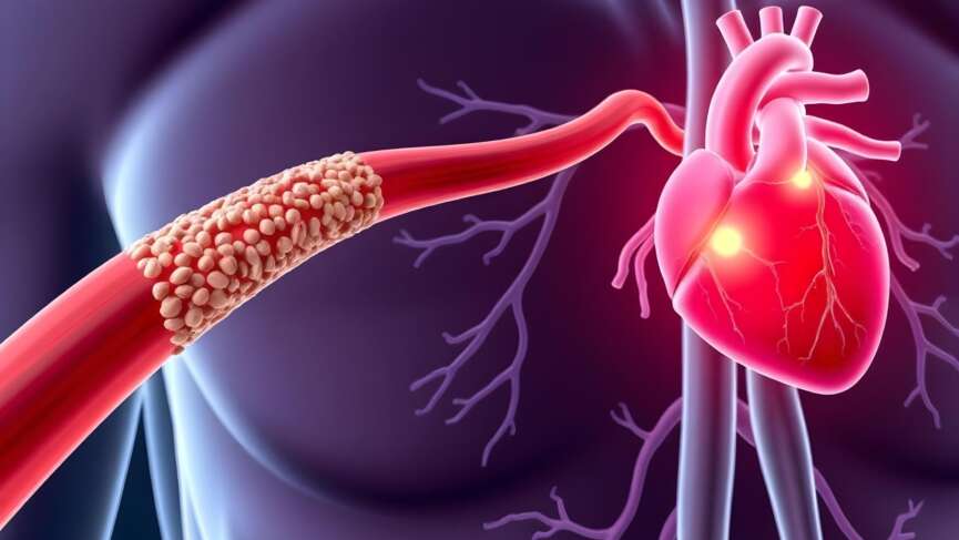
    Myocarditis is the inflammation of the middle muscular layer of your heart. Inflammation means your heart muscle has swelling and irritation when fighting an infection, injury, or something harmful. This inflammation can affect your heart’s ability to pump blood and maintain normal rhythm. The condition affects people of all ages and can range from mild cases with few symptoms to life-threatening complications. [1]

    It can happen suddenly when caused by infection, toxins, or drugs. But when the trigger is autoimmune diseases like lupus or rheumatoid arthritis, it affects the heart more slowly, causing a long-term effect like heart failure and cardiomyopathy. [1]

    Causes and Risk Factors of Myocarditis

    Commonly identified causes and risk factors of myocarditis include:

    1. Age

    Myocarditis is more common in young adults, especially males, but it can affect people of any age. Children and infants are also vulnerable, particularly during viral outbreaks. [2]

    2. Viral, Bacterial, Fungal, or Protozoan Infection

    Viral infection is the most common cause of myocarditis, directly infecting cardiac cells by releasing cytokines such as IL-1, IL-6, and TNF-α or inducing an immune-mediated response. Infections caused by various viruses, including influenza, herpes, adenovirus, coxsackievirus, parvovirus B19, and even SARS-CoV-2 (the virus responsible for COVID-19), can cause myocarditis. [1]

    3. Toxins and Radiation

    Exposure to toxins and radiation can lead to myocarditis by directly damaging the heart muscle or triggering inflammation. Substances like excessive alcohol, recreational drugs such as cocaine, heavy metals like lead or mercury, and industrial chemicals can harm heart tissues and cause inflammation. Similarly, radiation therapy, particularly when targeting the chest area for cancer treatment, can unintentionally damage the heart muscle and lead to myocarditis. [3]

    4. Autoimmune Diseases

    Autoimmune diseases, such as lupus or rheumatoid arthritis, can cause your immune system to attack healthy tissues, including the heart muscle. Myocarditis may also occur due to autoimmune reactions triggered by infections or other conditions. [4]

    5. Rheumatic Fever

    Rheumatic fever is a condition that can develop if you’ve had an untreated or poorly treated strep throat infection. It happens when your immune system, mistakenly attacks healthy tissues, including your heart. If this affects your heart muscle, it can lead to myocarditis, causing swelling and irritation in your heart. [5]

    6. Side Effects of Medicines and Vaccination

    Some medicines and vaccinations can, in rare cases, cause myocarditis as a side effect. Certain medications, such as chemotherapy drugs, antibiotics, or immune-suppressing treatments, may trigger an inflammatory response in your heart muscle. Vaccinations, including some for COVID-19, have been linked to very rare cases of myocarditis, typically occurring in younger males after their second dose. [6]

    Symptoms of Myocarditis

    The symptoms of myocarditis can vary widely, and sometimes, there might be no noticeable symptoms at all. When symptoms do occur, they may include:

    1. Chest Pain

    You may feel sharp, stabbing, or pressure-like pain in your chest, similar to what happens during a heart attack. This occurs due to inflammation of the heart muscle and can worsen with physical activity or deep breaths. [7]

    2. Shortness of Breath

    You might struggle to catch your breath, especially when lying flat or after mild activity. This happens because the heart isn’t pumping blood effectively, causing fluid buildup in your lungs. [7]

    3. Fatigue

    Feeling unusually tired or weak, even after routine tasks, is common with myocarditis. This is due to the heart’s inability to supply oxygen-rich blood to your body efficiently. [8]

    4. Irregular Heartbeat

    You might notice your heart racing, fluttering, or skipping beats. This indicates that the inflamed heart muscle interferes with your heart’s normal electrical signals. [9]

    5. Swelling

    Swelling in your legs, ankles, or feet may occur due to fluid buildup caused by the heart’s reduced ability to pump blood. Swelling may also appear around your abdomen or even in the veins in your neck. [7]

    6. Flu-like Symptoms

    If a virus triggers your myocarditis, you might first experience symptoms like fever, sore throat, headache, muscle aches, or general discomfort. These often precede heart-related symptoms.

    7. Syncope

    You could feel dizzy or faint, especially during physical activity or when standing up suddenly. This happens because reduced blood flow to your brain can result from your heart’s inability to pump effectively. [7]

    When to Seek Help if You Have Myocarditis

    Knowing when to call for help is important if you have myocarditis. You should dial 911 immediately if your chest pain feels tight or heavy, lasts more than 15 minutes, or starts spreading to your arms, back, neck, or jaw. It’s also important to seek urgent help if you feel short of breath or nauseous along with the pain. These could be signs of a serious problem that needs immediate attention, so don’t wait to get the care you need. [10]

    Diagnosis of Myocarditis

    The inflammation caused by myocarditis can cause chest pain similar to that in a heart attack, making the diagnosis difficult. The following tests might be done to diagnose myocarditis:

    1. Blood Tests

    A complete blood count may reveal an elevated white blood cell count, indicating an infection or inflammation. Tests like troponin and creatine kinase measure heart enzymes that are released when your heart muscle is damaged, helping to confirm myocarditis. C-reactive protein and erythrocyte sedimentation rate can show your body’s inflammation level. If a viral infection is suspected, blood tests can identify specific viruses or antibodies that may have triggered the condition. [7]

    2. Electrocardiography (EKG)

    EKG is a test that checks your heart’s rhythm by recording its electrical activity. It can detect certain changes that might suggest myocarditis, although these changes aren’t specific to the condition. Importantly, even if your ECG looks normal, it doesn’t rule out myocarditis completely. [10]

    3. Chest X-ray

    A chest X-ray gives a clear image of your heart and lungs, helping to check for signs like an enlarged heart or fluid buildup in your lungs, which can happen when your heart isn’t pumping well. While it can’t confirm myocarditis independently, a chest X-ray helps your doctor rule out other conditions that can mimic myocarditis. [11]

    4. Echocardiography

    An echocardiogram, or echo, is a test that uses sound waves to create detailed images of your heart. An echo can help see how well your heart is pumping and check for any areas of damage. It can also show if your heart is enlarged or if there’s fluid around it, which are signs of inflammation. This test is painless and non-invasive, and it gives information about how your heart is functioning. [12]

    5. Cardiac Magnetic Resonance Imaging (MRI)

    An MRI scan creates detailed images of the heart’s shape and structure. It’s especially useful for assessing heart function and detecting inflammation or scar tissue caused by myocarditis. [13]

    6. Positron Emission Tomography (PET) Scan

    A PET  scan involves using radioactive dyes to produce body images, showing how the heart functions. Typically, it’s done alongside a CT or MRI scan for better assessment. [11]

    7. Heart Muscle Biopsy

    If other tests are inconclusive, endomyocardial biopsy is done. During this procedure, tiny samples of heart muscle are taken. It helps identify the extent and type of inflammation linked to myocarditis. However, this procedure is uncommon. [14]

    Treatment of Myocarditis

    Myocarditis typically improves on its own or with treatment, often resulting in full recovery. If a viral infection causes myocarditis, the treatment typically focuses on managing symptoms and allowing your body to recover. This includes rest, avoiding strenuous activities to give your heart time to heal, and hydration to support overall health. Your doctor might also recommend pain relievers like acetaminophen or nonsteroidal anti-inflammatory drugs (NSAIDs) to reduce discomfort and inflammation.  [710]

    1. Corticosteroids and Immunosuppressants

    Corticosteroids (such as prednisone) or immunosuppressants (like azathioprine or methotrexate) are used in the treatment of myocarditis. These medications help reduce the immune system’s activity and limit inflammation, improving heart function. However, their use depends on the underlying cause of myocarditis, and they are often reserved for more severe cases. [7]

    2. Heart Failure Medications

    When myocarditis causes the heart to weaken, making it less effective at pumping blood, you might be given medication to assist your heart. These include angiotensinogen converting enzyme (ACE) inhibitors (e.g., enalapril), which relax blood vessels, reduce blood pressure, and improve blood flow, and beta-blockers (like metoprolol), which slow down the heart rate and reduce the heart’s workload. Diuretics (such as furosemide) may also be used to remove excess fluid from your body, especially if you have swelling in your legs, abdomen, or around the lungs. [10]

    3. Antiarrhythmic Medications

    Myocarditis can cause irregular heart rhythms, which can be life-threatening. Antiarrhythmic medications, like amiodarone or lidocaine, which help restore a normal heart rhythm, may be used to manage this. In some cases, if arrhythmias are severe or unresponsive to medication, your doctor may recommend implanting a pacemaker to regulate the heart’s rhythm and ensure it beats at a consistent pace. [9]

    4. Antibiotics (for bacterial myocarditis)

    If a bacterial infection causes your myocarditis, your doctor will prescribe antibiotics to treat the infection. The type of antibiotic depends on the specific bacteria causing the condition. For example, penicillin may be used for infections caused by certain bacteria, while more broad-spectrum antibiotics may be needed for others. Early antibiotic treatment is important to prevent further heart damage. [15]

    5. Ventricular Assist Device (VAD)

    VADs are special devices that help weakened hearts pump blood throughout the body. It is usually performed as a bridge to a heart transplant. [9]

    6. Heart Transplant (in severe cases)

    In extremely severe cases of myocarditis, where the heart is no longer able to function effectively despite other treatments, a heart transplant may be necessary. This is typically considered only when the heart failure is life-threatening, and all other options have been exhausted. A heart transplant is a complex procedure with risks, but it can offer a chance at life for patients with advanced heart failure due to myocarditis. [16]

    7. Extracorporeal Membrane Oxygenation (ECMO)

    ECMO, a specialized heart-lung support device, can significantly improve survival rates for patients with severe myocarditis, especially when conventional treatments aren’t effective. [17]

    Complications of Myocarditis

    If left untreated, myocarditis can cause can lead to serious consequences over time. The inflammation in your heart can weaken its ability to pump blood, leading to heart failure, where your heart struggles to meet your body’s needs.

    You might also develop life-threatening arrhythmias. In severe cases, myocarditis can cause cardiogenic shock, where your heart can’t pump enough blood to keep your organs working. Scarring from the inflammation may also lead to chronic heart damage, permanently weakening your heart. That’s why it is important to seek medical care early and follow your treatment plan to avoid these long-term complications. [18]

    ‌Recurrence of Myocarditis

    Myocarditis can sometimes come back, even after you’ve recovered from the initial episode. This is called recurrence and can happen if the underlying cause, like a viral infection or autoimmune condition, isn’t fully resolved or flares up again. Recurrence may bring similar symptoms, such as chest pain, fatigue, or shortness of breath, and can further stress your heart. If you experience any new or returning symptoms, it’s important to inform your doctor immediately. Early treatment of recurrent myocarditis can help protect your heart and prevent long-term damage. [18]

    Prevention of Myocarditis

    Preventing myocarditis starts with taking steps to protect your heart from infections and other triggers. Keeping up with your vaccinations, such as the flu shot and COVID-19 vaccine, can reduce the risk of viral infections that might lead to myocarditis. Practicing good hygiene, like washing your hands regularly and avoiding close contact with sick individuals, helps prevent the spread of germs. If you’re taking medications, use them only as prescribed to avoid harmful side effects, and stay away from substances like alcohol and recreational drugs that can damage your heart. Following your doctor’s treatment plan for autoimmune conditions can lower the chances of inflammation affecting your heart. [19]

    Questions To Ask Your Doctor

    1. How long will it take for my heart to recover?
    2. Should my family members be screened for any genetic or infectious causes?
    3. What warning signs should I watch for that might suggest my condition is worsening?
    4. Is it safe for me to engage in physical exercise or sports activities with myocarditis?
    5. Do I need to avoid certain activities or foods during my recovery?

    References

    1. Kühl, U., Pauschinger, M., Seeberg, B., Lassner, D., Noutsias, M., Poller, W., & Schultheiss, H. P. (2005). Viral persistence in the myocardium is associated with progressive cardiac dysfunction. Circulation, 112(13), 1965–1970. Available from: https://doi.org/10.1161/CIRCULATIONAHA.105.548156
    2. Keller K, Sagoschen I, Konstantinides S, Gori T, Münzel T, Hobohm L. Incidence and risk factors of myocarditis in hospitalized patients with COVID‐19. Journal of Medical Virology. 2023 Mar;95(3). Available from: https://pubmed.ncbi.nlm.nih.gov/36892180
    3. Nguyen LS, Cooper LT, Kerneis M, Funck-Brentano C, Silvain J, Brechot N, et al. Systematic analysis of drug-associated myocarditis reported in the World Health Organization pharmacovigilance database. Nature Communications. 2022 Jan 10;13(1). Available from:‌ https://pubmed.ncbi.nlm.nih.gov/35013204
    4. Nielsen JC, Lin YJ, de Oliveira Figueiredo MJ, Sepehri Shamloo A, Alfie A, Boveda S, et al. European Heart Rhythm Association (EHRA)/Heart Rhythm Society (HRS)/Asia Pacific Heart Rhythm Society (APHRS)/Latin American Heart Rhythm Society (LAHRS) expert consensus on risk assessment in cardiac arrhythmias: use the right tool for the right outcome, in the right population. Heart Rhythm [Internet]. 2020 Sep 1 [cited 2022 Jul 4];17(9):e269–316. Available from: https://www.sciencedirect.com/science/article/pii/S1547527120304252
    5. British Heart Foundation. Myocarditis [Internet]. British Heart Foundation. 2019. Available from: https://www.bhf.org.uk/informationsupport/conditions/myocarditis
    6. Munjal JS, Flores SM, Yousuf H, Gupta V, Munjal RS, Anamika F, Mendpara V, Shah P, Jain R. Covid- 19 Vaccine-induced Myocarditis. J Community Hosp Intern Med Perspect. 2023 Sep 2;13(5):44-49. doi: 10.55729/2000-9666.1229. PMID: 37868673; PMCID: PMC10589044. Available from: https://pmc.ncbi.nlm.nih.gov/articles/PMC10589044/
    7. Ammirati E, Moslehi JJ. Diagnosis and Treatment of Acute Myocarditis. JAMA. 2023 Apr 4;329(13):1098.Available from: ‌ https://pubmed.ncbi.nlm.nih.gov/37014337
    8. Gulati M, Levy PD, Mukherjee D, Amsterdam E, Bhatt DL, Birtcher KK, et al. 2021 AHA/ACC/ASE/CHEST/SAEM/SCCT/SCMR Guideline for the Evaluation and Diagnosis of Chest Pain: A Report of the American College of Cardiology/American Heart Association Joint Committee on Clinical Practice Guidelines. Circulation [Internet]. 2021 Oct 28;144(22). Available from: https://www.ahajournals.org/doi/10.1161/CIR.0000000000001029
    9. Al-Khatib SM, Stevenson WG, Ackerman MJ, Bryant WJ, Callans DJ, Curtis AB, et al. 2017 AHA/ACC/HRS guideline for management of patients with ventricular arrhythmias and the prevention of sudden cardiac death. Heart Rhythm. 2018 Oct;15(10):e73–189. Available from:‌ https://pubmed.ncbi.nlm.nih.gov/29097319
    10. Gluckman TJ, Bhave NM, Allen LA, Chung EH, Spatz ES, Ammirati E, et al. 2022 ACC Expert Consensus Decision Pathway on Cardiovascular Sequelae of COVID-19 in Adults: Myocarditis and Other Myocardial Involvement, Post-Acute Sequelae of SARS-CoV-2 Infection, and Return to Play. Journal of the American College of Cardiology. 2022 Mar; Available from: https://pubmed.ncbi.nlm.nih.gov/35307156
    11. Lampejo, T., Durkin, S. M., Bhatt, N., & Guttmann, O. (2021). Acute myocarditis: aetiology, diagnosis and management. Clinical medicine (London, England), 21(5), e505–e510. Available from: https://doi.org/10.7861/clinmed.2021-0121
    12. Bozkurt B, Colvin M, Cook J, Cooper LT, Deswal A, Fonarow GC, et al. Current Diagnostic and Treatment Strategies for Specific Dilated Cardiomyopathies: A Scientific Statement From the American Heart Association. Circulation. 2016 Dec 6;134(23). Available from: https://pubmed.ncbi.nlm.nih.gov/27832612
    13. Sen G, Scully P, Gordon P, Sado D. Advances in the diagnosis of myocarditis in idiopathic inflammatory myopathies: an overview of diagnostic tests. Rheumatology (Oxford, England) [Internet]. 2024 Jan 16 [cited 2024 Jul 30];63(7):1825–36. Available from: https://www.ncbi.nlm.nih.gov/pmc/articles/PMC11215992/
    14. Basso C, Calabrese F, Angelini A, Carturan E, Thiene G. Classification and histological, immunohistochemical, and molecular diagnosis of inflammatory myocardial disease. Heart Failure Reviews. 2012 Oct 25;18(6):673–81. Available from: https://pubmed.ncbi.nlm.nih.gov/23096264

  • Meningitis Uncovered – Key Symptoms, Treatments, and Lifesaving Vaccines

    Meningitis Uncovered – Key Symptoms, Treatments, and Lifesaving Vaccines

    Highlights of Meningitis

    • Meningitis is a life-threatening inflammation of the meninges, which is the protective membranes covering the brain and spinal cord.
    • Bacterial meningitis is the most dangerous form, as it can progress rapidly, causing brain damage or even death if not treated in time.
    • Meningitis can affect anyone, but certain groups, such as infants, young children, the elderly, and people with weakened immune systems, are at higher risk.
    • Vaccines like meningococcal, Hemophilus Influenza B, and pneumococcal vaccines have reduced the incidence of bacterial meningitis in many parts of the world.

    What is Meningitis?

    Meningitis is the inflammation of the meninges, which are the protective layers of tissue surrounding the brain and spinal cord. It can affect individuals of any age but is more prevalent in younger children [1]. Meningitis is a significant global health issue, with epidemics occurring worldwide, especially in sub-Saharan Africa. In the United States, bacterial meningitis occurs in about 1.38 out of every 100,000 people each year, and about 14.3% of those who get it die from the disease.  [23]

    Meningitis can be serious if not treated properly. Bacterial meningitis, in particular, is the most dangerous type. If not treated properly, it can be fatal within 24 hours [1].

    Types of Meningitis

    Meningitis can occur in several forms, each with distinct causes and characteristics. Here are the 4 types of meningitis:

    1. Bacterial Meningitis

    This is the most severe form of meningitis and is caused by bacteria such as Neisseria meningitidis, Streptococcus pneumoniae, and Haemophilus influenzae. Bacterial meningitis can develop rapidly, and without immediate antibiotic treatment, it can lead to brain damage, hearing loss, or even death. [24]

    2. Viral Meningitis

    This is caused by viruses like enteroviruses, herpes simplex virus, or HIV. Viral meningitis is more common than bacterial meningitis and is usually less severe, with most people recovering fully within a few weeks without specific antiviral treatment.[25]

    3. Fungal Meningitis

    It is rare but more serious. Fungi like Cryptococcus cause fungal meningitis. It generally affects people with weakened immune systems, such as those with HIV/AIDS or those undergoing chemotherapy. [2]

    4. Parasitic Meningitis

    This type is caused by parasites, though it is rare. It is often associated with environmental factors, such as contaminated water or food. The infection can be severe, especially in those with weakened immune systems. [6] Non-infectious meningitis occurs due to conditions like cancer, certain medications, or autoimmune diseases. It is not caused by an infection but still leads to inflammation of the meninges. [1, 2]

    Causes and Risk Factors of Meningitis

    The causes and risk factors of meningitis are:

    1. Infection With Bacteria or Viruses

    Viral or bacterial infections are the most common cause of meningitis. Viruses like enteroviruses are common, and bacteria like Neisseria meningitidis are often responsible for outbreaks, particularly in crowded settings. [245]

    2. Close Contact in Crowded Environments

    Meningitis-causing bacteria and viruses can spread easily through respiratory droplets or close contact, such as in schools, dormitories, or military barracks.[1, 2]

    3. Age

    Newborns, infants, and young children are particularly vulnerable to meningitis, as their immune systems are not fully developed. Adults over 60 are also at higher risk, as the immune system weakens with age. [1, 2]

    4. Weakened Immune System

    Conditions like HIV/AIDS, cancer, or immunosuppressive treatments (e.g., chemotherapy or steroids) can increase the risk of developing meningitis. [1, 2]

    5. Travel to Endemic Areas

    Traveling to regions where bacterial meningitis is common, such as parts of sub-Saharan Africa, increases the risk, especially in unvaccinated individuals. [1, 2]

    6. Head Injuries and Surgery

    Head trauma or surgery that affects the brain can increase the risk of developing meningitis by providing a pathway for bacteria or viruses to enter the meninges. [12]

    Transmission of Meningitis

    Meningitis spreads through respiratory droplets from coughing or sneezing, particularly bacterial meningitis. Neisseria meningitidis transmits through droplets from both symptomatic cases and non-symptomatic carriers. After bacteria reach the upper part of our throat, they grow in number and later enter the bloodstream. From blood, bacteria enter the fluid-filled space around the brain and spinal cord (the sub-arachnoid space) and cross the protective blood-brain barrier (BBB), leading to meningitis.  [2] Similarly, bacteria can enter the cerebrospinal fluid (CSF) from nearby areas like the ear in cases of ear infections (otitis media) or from the sinuses (sinusitis). They can also be introduced into the CSF through head injuries or during surgical procedures.  [2]

    Symptoms of Meningitis

    The symptoms of meningitis can come on quickly and feel like a bad flu at first. Here’s what you should look out for:

    1. Severe Headache

    You might feel a pounding headache that doesn’t go away and feels worse than anything you’ve experienced. [6]

    2. Stiff Neck

    Turning or moving your neck could feel difficult or painful. It might feel like your muscles are unusually tight.[6]

    3. High Fever

    A sudden spike in your body temperature could be one of the first warning signs. You may also feel chills or sweats. [6]

    4. Classic Triad

    The classical triad of symptoms of meningitis includes fever, neck stiffness, and confusion. However, this triad is not always present. In adult cases of meningitis, headache, fever, neck stiffness, and confusion are the most common symptoms. Up to 95% of patients present with at least two of these four symptoms. [6]

    5. Sensitivity to Light (photophobia)

    Bright lights might hurt your eyes or worsen your headache, causing you to avoid well-lit places. [2]

    6. Confusion or Difficulty Concentrating

    You may feel foggy, confused, or unable to think clearly. It might be hard to stay focused or remember things. [2]

    7. Skin Rash

    If you have bacterial meningitis caused by Neisseria meningitidis, you might notice small purplish spots or blotches on your skin that don’t fade when pressed. [7]

    For babies and young children, the symptoms can be harder to spot. If your baby has meningitis, they may: [8]

    • Appear very irritable or cry constantly in a high-pitched way that’s hard to soothe.
    • Have trouble feeding or refuse to eat.
    • They have a bulging soft spot on their head.
    • Look very sleepy, floppy, or unresponsive

    Diagnosis of Meningitis

    Diagnosing meningitis requires careful evaluation through specific tests and procedures. Below are a few key points to consider for diagnosis of meningitis:

    Lumbar Puncture (Spinal Tap)

    A lumbar puncture, or spinal tap, is a common and safe procedure for diagnosing meningitis. It is also the most definitive test for meningitis. During the procedure, you will either lie on your side with your knees pulled up to your chest or sit and lean forward to allow the doctor access to your lower back. The area on your lower back is cleaned thoroughly, and a numbing medicine is injected to ensure you don’t feel pain. Once the area is numb, a thin needle is gently inserted into the space between the bones in your lower spine, where cerebrospinal fluid surrounds your brain and spinal cord. Your doctor collects a small amount of this fluid in a sterile tube for testing. While you may feel pressure during the procedure, it is usually not painful. After the fluid is collected, the needle is removed, and a small bandage is placed over the site. The entire process typically takes about 15–30 minutes. Once the sample is analyzed, it helps confirm if meningitis is present and identifies the type, which is crucial for deciding on the best treatment. [7]

    Blood Tests

    Blood tests are essential to diagnosing meningitis and assessing the severity of the infection. While they can’t directly confirm meningitis, blood tests help identify signs of infection, inflammation, and potential complications. A complete blood count (CBC) can reveal elevated white blood cell counts, indicating an immune response to infection. Blood cultures are also taken to detect bacteria or fungi in the bloodstream, which is important for diagnosing bacterial meningitis and guiding antibiotic treatment. Additionally, blood tests may assess markers of inflammation, such as C-reactive protein (CRP) and procalcitonin, which can help differentiate between bacterial and viral infections.[9]

    Computed Tomography (CT) Scan or Magnetic Resonance Imaging (MRI)

    A CT scan is typically the first test to quickly rule out other conditions and detect issues like brain swelling or bleeding. While it can’t directly diagnose meningitis, it helps assess immediate risks. An MRI provides more detailed images and examines the brain for inflammation or complications, such as fluid buildup or swelling. These imaging techniques complement a lumbar puncture. [10]

    Polymerase Chain Reaction (PCR) Testing

    Polymerase chain reaction (PCR) testing is a highly sensitive and specific method used to diagnose meningitis by detecting genetic material from pathogens in cerebrospinal fluid. It is especially useful for identifying viral, bacterial, and fungal causes of meningitis when traditional cultures may take too long or fail to provide results. PCR can quickly identify pathogens such as Neisseria meningitidis, Streptococcus pneumoniae, and enteroviruses, common causes of meningitis. This test is particularly valuable when the cause of meningitis is uncertain or when patients are already receiving antibiotics, which may hinder the growth of bacteria in cultures. [11]

    Treatment of Meningitis

    The treatment of meningitis varies based on its cause and the patient’s condition. Here are some essential points to keep in mind for proper care and recovery.

    1. Antibiotics

    Immediate treatment with broad-spectrum antibiotics is crucial if bacterial meningitis is suspected. Once the specific bacteria are identified, antibiotics may be adjusted to target the bug directly. For newborns, the treatment includes Ampicillin and Cefotaxime, plus Acyclovir. For older infants and children, Ampicillin and Ceftriaxone are used. Adults aged 18-49 are treated with Ceftriaxone and Vancomycin, while those 50 and older or immunocompromised receive Ceftriaxone, Vancomycin, and Ampicillin. [2, 7]

    2. Antiviral medication

    For viral meningitis caused by herpes simplex virus or other specific viruses, antiviral medications may be prescribed to help reduce the severity and duration of symptoms. However, treatment for other viral meningitis is primarily supportive. [2]

    3. Antifungal and Antitubercular Medications

    Amphotericin B, flucytosine, and fluconazole are the standard treatments for cryptococcal meningitis, particularly in immunocompromised patients. And for tubercular meningitis, a combination of isoniazid, rifampin, pyrazinamide, and ethambutol is recommended, often with adjunctive corticosteroids. [2]

    4. Corticosteroids

    Corticosteroids, such as dexamethasone, are typically administered concomitantly with or just before the first dose of antibiotics. This timing is crucial to mitigate the inflammatory response induced by bacterial lysis. Steroids can help reduce inflammation in the brain and prevent complications like hearing loss or brain damage, particularly in cases of bacterial meningitis. [2]

    5. Pain and Fever Relief

    Over-the-counter medications like acetaminophen or ibuprofen can help manage symptoms like headache, fever, and body aches. [2]

    6. Supportive Care

    In severe cases, hospitalization is needed to provide intravenous fluids, manage complications, and monitor your condition closely. Rest and hydration are also important parts of recovery. [2]

    Complications of Meningitis

    Meningitis can lead to serious complications, especially if not treated promptly. The prognosis of bacterial meningitis remains serious despite advances in medical care. A low level of consciousness, such as being very drowsy or unresponsive, indicates a poor prognosis. One of the most concerning outcomes is brain damage, which can affect your memory, concentration, or overall cognitive abilities. Hearing loss is another common complication, as the infection can damage the nerves responsible for hearing. Seizures may develop during or after the illness due to inflammation in the brain. [2] Meningitis can be life-threatening, with a significant risk of death. [2]

    In severe cases, meningitis can lead to hydrocephalus, a condition where fluid builds up in the brain, causing increased pressure. The infection can also spread to other parts of your body, leading to life-threatening conditions like septicemia (blood poisoning), which may cause organ failure or tissue damage requiring amputation. In children, meningitis can sometimes result in developmental delays or learning difficulties. [12]

    How to prevent meningitis?  

    The disease spreads through respiratory droplets from coughing or sneezing, putting close contacts of the patient at significant risk. Preventive treatment( chemoprophylaxis) is most effective when started within 24 hours of exposure, though it can still be beneficial if initiated up to 14 days later. [13]

    Prevention of meningitis focuses on those closely interacting with the patient, such as household members, intimate contacts, roommates, healthcare workers exposed to the patient’s respiratory secretions during procedures, and anyone who traveled with the patient for more than eight hours. Casual contacts, like sharing drinks or cigarettes, typically don’t require prophylaxis unless they meet the close contact criteria. [13]

    Recommended antibiotics for preventing meningococcal disease include rifampicin, ciprofloxacin, and ceftriaxone, which eliminate the bacteria from the body and reduce transmission risk. Additionally, the CDC advises chemoprophylaxis with rifampin for household contacts of patients with Haemophilus influenzae meningitis, particularly children under four who are not fully vaccinated or are immunocompromised. [14]

    Vaccination for Meningitis

    Vaccination is one of the most effective ways to prevent meningitis and protect against severe complications. Several vaccines target the most common causes of meningitis, including bacterial forms. The meningococcal vaccine helps protect against Neisseria meningitidis, a leading cause of bacterial meningitis. It is typically recommended for adolescents, young adults, and individuals at higher risk, such as those living in close quarters like dormitories or military barracks. A booster dose is often advised for continued protection. [15]

    The Haemophilus influenzae type b (Hib) vaccine is given to infants as part of routine childhood immunizations and has significantly reduced cases of meningitis caused by this bacterium. The pneumococcal vaccine protects against Streptococcus pneumoniae, another major cause of bacterial meningitis. It is recommended for young children, older adults, and individuals with weakened immune systems or certain chronic conditions. [215]

    Questions To Ask Your Doctor

    1. How long does it typically take to recover from meningitis?
    2. Does meningitis affect mental health?
    3. Do I need to get vaccinated against meningitis before living in a dorm?
    4. What dietary changes should I consider during recovery from meningitis?
    5. How does meningitis impact my ability to work or go to school?

    References

    1. World Health Organization. Meningitis. [Internet]. 2023 [cited 2024 Jul 25]. Available from: https://www.who.int/news-room/fact-sheets/detail/meningitis
    2. Hersi K, Gonzalez FJ, Kondamudi NP. Meningitis. PubMed. Treasure Island (FL): StatPearls Publishing; 2023. Available from: https://www.ncbi.nlm.nih.gov/books/NBK459360/
    3. Thigpen MC, Whitney CG, Messonnier NE, Zell ER, Lynfield R, Hadler JL, et al. Bacterial meningitis in the United States, 1998-2007. N Engl J Med. 2011 May 26;364(21):2016-25.Available from: https://pubmed.ncbi.nlm.nih.gov/21612470/
    4. Oordt-Speets AM, Bolijn R, van Hoorn RC, Bhavsar A, Kyaw MH. Global etiology of bacterial meningitis: A systematic review and meta-analysis. PLOS ONE. 2018 Jun 11;13(6): e0198772.Available from: https://journals.plos.org/plosone/article?id=10.1371/journal.pone.0198772
    5. Mount HR, Boyle SD. Aseptic and Bacterial Meningitis: Evaluation, Treatment, and Prevention. American Family Physician [Internet]. 2017 Sep 1;96(5):314–22. Available from: https://pubmed.ncbi.nlm.nih.gov/28925647/
    6. Hall AD, Kumar JE, Golba CE, Luckett KM, Bryant WK. Primary amebic meningoencephalitis: a review of Naegleria fowleri and analysis of successfully treated cases. Parasitology Research. 2024 Jan 1;123(1). Available from: https://link.springer.com/article/10.1007/s00436-023-08094-w
    7. Van de Beek D, de Gans J, Spanjaard L, Weisfelt M, Reitsma JB, Vermeulen M. Clinical Features and Prognostic Factors in Adults with Bacterial Meningitis. N Engl J Med. 2004 Oct 28;351(18):1849-59. Available from: https://pubmed.ncbi.nlm.nih.gov/15509818/
    8. Sín R;Struncova D;Cechurova L. Clinical picture, diagnostics and treatment of bacterial meningitis. Neuro endocrinology letters [Internet]. 2022 [cited 2024 Dec 6];43(7-8). Available from: https://pubmed.ncbi.nlm.nih.gov/36720125
    9. Sanaei Dashti A, Alizadeh S, Karimi A, Khalifeh M, Shoja SA. Diagnostic value of lactate, procalcitonin, ferritin, serum-C-reactive protein, and other biomarkers in bacterial and viral meningitis. Medicine. 2017 Sep;96(35):e7637. Available from: https://pubmed.ncbi.nlm.nih.gov/28858084
    10. Hughes DC, Raghavan A, Mordekar SR, Griffiths PD, Connolly DJA. Role of imaging in the diagnosis of acute bacterial meningitis and its complications. Postgraduate Medical Journal [Internet]. 2010 Aug 1 [cited 2019 Oct 21];86(1018):478–85. Available from: https://pmj.bmj.com/content/86/1018/478
    11. Wagner K, Springer B, Pires VP, Keller PM. Pathogen Identification by Multiplex LightMix Real-Time PCR Assay in Patients with Meningitis and Culture-Negative Cerebrospinal Fluid Specimens. Ledeboer NA, editor. Journal of Clinical Microbiology. 2017 Dec 13;56(2). Available from: https://pubmed.ncbi.nlm.nih.gov/29237781
    12. Edmond K, Clark A, Korczak VS, Sanderson C, Griffiths UK, Rudan I. Global and regional risk of disabling sequelae from bacterial meningitis: a systematic review and meta-analysis. Lancet Infect Dis. 2010 May;10(5):317-28. Available from: https://pubmed.ncbi.nlm.nih.gov/20417414/
    13. Centers for Disease Control and Prevention. Selection of antibiotics as prophylaxis for close contacts of patients with meningococcal disease in areas with ciprofloxacin resistance. MMWR Morb Mortal Wkly Rep. 2024 [cited 2024 Jul 25]. Available from: https://www.cdc.gov
    14. Centers for Disease Control and Prevention (CDC). Haemophilus influenzae Disease. [Internet]. 2024 [cited 2024 Jul 25]. Available from: https://www.cdc.gov
    15. Mbaeyi SA, Bozio CH, Duffy J, et al. Meningococcal Vaccination: Recommendations of the Advisory Committee on Immunization Practices, United States, 2020. MMWR Recomm Rep. 2020;69(No. RR-9):1-41. Available from: http://dx.doi.org/10.15585/mmwr.rr6909a1

  • Hepatitis A and E – Introduction, Similarities and Differences

    Hepatitis A and E – Introduction, Similarities and Differences

    Highlights of Hepatitis A and E

    1. Hepatitis A virus and Hepatitis E are the most common causes of acute liver inflammation worldwide.
    2. Symptoms of hepatitis A and E are similar. Common symptoms include fatigue, malaise, jaundice, nausea and vomiting, abdominal pain, pale-colored stool, and dark urine.
    3. The majority of the cases of hepatitis A and E require no specific treatment and resolve on their own. Rarely, Hepatitis A can lead to acute liver failure, especially in individuals with underlying liver conditions.
    4. A safe and effective vaccine is available to prevent Hepatitis A. This is recommended for high-risk individuals and travelers to endemic areas.
    5. Pregnant women are at a higher risk of severe complications from Hepatitis E.

    What is Hepatitis A and Hepatitis B?

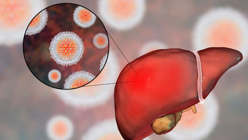
    Hepatitis means inflammation of the liver. Although hepatitis can be caused due to heavy alcohol use, drugs, and toxins, it is most commonly caused by a virus. Viral hepatitis is caused by a group of viruses, namely hepatitis A, B, C, D, and E. Hepatitis A virus (HAV) and Hepatitis E virus (HEV) are the most common causes of acute hepatitis worldwide.

    Hepatitis A

    Hepatitis A virus commonly spreads through contaminated food and water or direct contact with an infected person. Once inside the body, the virus attacks normal liver cells, causing inflammation and damage. Hepatitis A is generally acute or short-lived, though in rare cases, it can lead to severe infection or liver failure. The virus is highly contagious and can be transmitted in two main ways: person-to-person contact, such as through close physical contact like sexual activities or shared needles, and through the oral-fecal route, usually via contaminated food or water.

    Hepatitis E

    Hepatitis E, a member of the Hepeviridae family, spreads primarily through the fecal-oral route and is called enteric hepatitis. The infection usually resolves on its own within two to six weeks.

    Like Hepatitis A, Hepatitis E can be contracted by consuming contaminated food or water. Eating undercooked pork or organ meats from infected animals increases your chances of getting the virus from animal sources. Though less common, transmission can occur through blood transfusions, organ transplants from infected donors, or an infected mother to her child.

    People at risk of Hepatitis A and E

    Here are some groups of people who are at higher risk of getting Hepatitis A or Hepatitis E:

    1. Travelers

    If you travel to countries where these viruses are common, especially in areas with poor sanitation, you are more likely to be exposed and contract any of these viruses.

    2. People in Poor Sanitation

    Living in places without access to clean drinking water or proper waste management makes it easier for these viruses to spread.

    3. Drug Users

    Using drugs, whether injected or not, can increase your risk, especially if you’re in environments where hygiene and safety aren’t prioritized.

    4. Men Who Have Sex With Men

    You are at increased risk of getting Hepatitis A if you are a man who has sex with men.

    5. Close Contact With Infected Individuals

    Living with or caring for someone who has Hepatitis A puts you at higher risk, even before they start showing any symptoms.

    6. Food Handlers

    People who work with food are more likely to be exposed to Hepatitis A, especially if they come into contact with contaminated sources.

    7. Pregnant Women

    If you’re pregnant, there’s a higher risk of severe complications from Hepatitis E, particularly during the later stages of pregnancy.

    Symptoms of Hepatitis A and E

    Hepatitis A and Hepatitis E mostly present with similar symptoms; sometimes, the disease may develop without noticeable symptoms. The clinical presentation involves two phases: the prodromal phase followed by the icteric phase.

    Prodromal phase: This phase lasts 3 to 10 days and is primarily characterized by malaise and myalgia. Other symptoms during this phase may include:

    • Fever
    • Anorexia
    • Nausea and/or vomiting
    • Abdominal pain
    • Joint pain

    Icteric phase: The next phase is the icteric phase, which may last up to 3 weeks. Symptoms during the icteric phase include:

    • Yellowing of the skin and whites of the eyes
    • Anorexia, nausea, and vomiting
    • Dark urine and pale-colored stool
    • Skin irritation and/or rash
    • Abdominal pain and/or diarrhea

    People who are more likely to experience severe disease from Hepatitis A and E include:

    • Individuals with chronic liver disease, including Hepatitis B and C
    • People living with HIV
    • Pregnant women

    Diagnosis of Hepatitis A and E

    Initial Diagnostic Tests: Both Hepatitis A and E diagnoses involve blood tests to detect specific antibodies produced by your immune system in response to the viruses.

    IgM antibodies

    IgM antibodies are the first to emerge in response to Hepatitis A and E infections. They typically appear within the first week after infection, serving as an important marker for the body’s immune response. IgM antibodies indicate an acute infection and confirm recent exposure to either virus, helping healthcare providers diagnose the condition effectively.

    IgG antibodies

    IgG antibodies will develop as the infection progresses and your body starts to recover. These antibodies typically appear about 2 to 4 weeks after the onset of symptoms. IgG antibodies indicate that you have had a past infection or have developed immunity to the virus. This signifies that you are no longer infectious and have gained a certain level of protection against future infections.

    Viral RNA

    Detection of viral RNA is an important diagnostic tool for both Hepatitis A and E, complementing antibody testing. In Hepatitis A, the presence of Hepatitis A virus RNA can be identified using nucleic acid testing (NAT), which is particularly useful during the early stages of infection, often before antibodies are produced. Similarly, Hepatitis E virus RNA can also be detected through NAT, allowing for early diagnosis of the infection. This detection typically occurs shortly after infection, usually within the first week or two, before the immune response generates significant levels of antibodies. Identifying viral RNA is essential for diagnosing acute infections and can help distinguish Hepatitis A and E from other liver diseases. Viral  RNA is used to monitor the viral load in patients, assess the severity of the infection, and guide treatment decisions.

    Treatment of Hepatitis A and E

    Both hepatitis A and E mostly resolve on their own within 4-8 weeks. Almost all cases of HAV and HEV are self-limiting in immunocompetent patients.

    Supportive care rocks here. Both Hepatitis A and E typically require supportive care, as these infections are self-limiting and resolve on their own without specific antiviral medications. Adequate rest is crucial for helping your body recover from the infection. Drinking plenty of fluids can help prevent dehydration. Maintaining good hydration is essential, especially if symptoms like nausea and vomiting occur. You might need over-the-counter medications to alleviate specific symptoms, such as pain relievers for abdominal discomfort or fever reducers.

    Monitoring for complications and hospitalization  

    In most cases, both Hepatitis A and E do not lead to chronic infection, and patients can expect a full recovery. However, monitoring may be necessary for those with pre-existing liver conditions or weakened immune systems to manage any complications. Severe cases, including fulminant hepatitis, require hospitalization. Antiviral drugs like ribavirin and interferon may also be used in cases of chronic hepatitis E. Although very rare, more serious cases may also require liver transplantation.

    Complications of Hepatitis A and E

    Complications from hepatitis A and E are uncommon. Certain populations, such as individuals with pre-existing liver diseases like chronic hepatitis B or C, pregnant women, and those with weakened immune systems, are at higher risk for severe complications from Hepatitis A and E. Some of the possible complications include:

    1. Prolonged Symptoms

    While most individuals recover fully, some people may experience prolonged symptoms such as fatigue, weakness, and jaundice that can last for weeks or even months after the initial infection. This post-viral fatigue can significantly impact daily life and requires time for recovery.

    2. Chronic Liver Disease

    Although chronic infection is rare for both Hepatitis A and E, individuals with existing liver problems may experience worsening of their condition. This can lead to complications like cirrhosis or liver cancer over time.

    3. Acute Liver Failure

    In rare cases, both Hepatitis A and E can lead to acute liver failure, a severe condition where the liver suddenly loses its ability to function. This can result in symptoms such as jaundice, confusion, and bleeding tendencies. Acute liver failure requires immediate medical attention and often leads to hospitalization.

    4. Pregnancy Complications

    Pregnancy poses unique risks when it comes to Hepatitis E, as infected pregnant women are at a significantly higher risk of developing severe complications, including acute liver failure. This increased susceptibility is particularly concerning during the third trimester, with the greatest likelihood of severe outcomes. Infections during pregnancy can lead to adverse effects for both the mother and the fetus, including the risk of stillbirth, premature delivery, and maternal mortality. The transmission of the virus from the mother to the fetus can also occur, potentially resulting in neonatal infection.

    Prevention of Hepatitis A and E

    1. Good Hygiene Practices

    Both Hepatitis A and E are transmitted through the fecal-oral route, making good hygiene practices crucial. Regular handwashing with soap and water, especially after using the bathroom and before preparing or consuming food, can significantly reduce the risk of infection.

    2. Safe Food and Water

    Ensuring access to clean and safe drinking water is essential for preventing both Hepatitis A and E. Avoiding tap water in areas where the virus is common, drinking bottled or boiled water, and being cautious about ice made from unpurified water can help prevent infection. When eating, especially in areas where the viruses are endemic, opt for well-cooked foods and avoid raw or undercooked shellfish, fruits, and vegetables that cannot be peeled.

    3. Travel Precautions

    If you’re traveling to regions where Hepatitis A or E is common, take precautions by getting vaccinated ahead of time, practicing good hygiene, and being mindful of food and water safety. This is particularly important in developing countries where sanitation may be inadequate.

    4. Avoiding Risky Behaviors

    Reducing high-risk behaviors, such as sharing needles or engaging in unprotected sex with multiple partners, can help prevent Hepatitis A transmission, especially among men who have sex with men. Although Hepatitis E is less commonly transmitted through person-to-person contact, maintaining general safe practices is still advisable.

    5. Vaccination

    The most effective way to prevent Hepatitis A is through vaccination. The Hepatitis A vaccine is safe and effective and is recommended for all children aged 12-23 months. It is also recommended for individuals at higher risk, such as travelers to endemic areas, people with chronic liver diseases, and men who have sex with men. Although there is currently no vaccine for Hepatitis E, awareness of its prevention strategies remains important.

    Hepatitis A Vaccine Schedule and Its Side Effects

    In the US, there are now two registered single-antigen vaccinations. One year or older is the recommended age for the first dose of all inactivated hepatitis A vaccinations, authorized for intramuscular injection into the deltoid muscle in a two-dose regimen.

    A 6-to-18-month delay between dosages one and two is normally recommended, although this can be extended to 4 or 5 years. Over-18-year-olds can receive the combination vaccine containing both HAV and HBV antigens. Three doses are given during primary immunizations in the first, first, and sixth months. Common side effects of the vaccine include fever, headache, tiredness, and soreness at the injection site.

    Questions To Ask Your Doctor

    1. If I have been exposed to someone with Hepatitis A or E, what steps should I take?
    2. How long should I expect symptoms to last?
    3. Can I take over-the-counter drugs during hepatitis A and E?
    4. Are there any dietary restrictions?
    5. How often should I have follow-up appointments after recovering from Hepatitis A or E?

    References

    1. Webb GW, Kelly S, Dalton HR. Hepatitis A and Hepatitis E: Clinical and Epidemiological Features, Diagnosis, Treatment, and Prevention. Clin Microbiol Newsl 2020;42:171. https://doi.org/10.1016/J.CLINMICNEWS.2020.10.001.
    2. Van Damme P, Pintó RM, Feng Z, Cui F, Gentile A, Shouval D. Hepatitis A virus infection. Nat Rev Dis Primers 2023;9. https://doi.org/10.1038/s41572-023-00461-2.
    3. Jeong SH, Lee HS. Hepatitis a: Clinical manifestations and management. Intervirology 2010;53:15–9. https://doi.org/10.1159/000252779.
    4. Waqar S, Sharma B, Koirala J. Hepatitis E. Treatment and Management of Tropical Liver Disease 2023:1–7. https://doi.org/10.1016/B978-0-323-87031-3.00010-X.
    5. Migueres M, Lhomme S, Izopet J. Hepatitis A: Epidemiology, High-Risk Groups, Prevention and Research on Antiviral Treatment. Viruses 2021;13:1900. https://doi.org/10.3390/v13101900.
    6. Hepatitis E | CDC Yellow Book 2024 [Internet]. wwwnc.cdc.gov. Available from: https://wwwnc.cdc.gov/travel/yellowbook/2024/infections-diseases/hepatitis-e
    7. Hepatitis and reproduction. Fertil Steril 2008;90:S226–35. https://doi.org/10.1016/j.fertnstert.2008.08.040.
    8. Nelson NP, Link-Gelles R, Hofmeister MG, Romero JR, Moore KL, Ward JW, et al. Update: Recommendations of the Advisory Committee on Immunization Practices for Use of Hepatitis A Vaccine for Postexposure Prophylaxis and for Preexposure Prophylaxis for International Travel. MMWR Morb Mortal Wkly Rep 2018;67:1216–20. https://doi.org/10.15585/mmwr.mm6743a5.
    9. Mirazo S, Ramos N, Mainardi V, Gerona S, Arbiza J. Transmission, diagnosis, and management of hepatitis E: an update. Hepat Med 2014;6:45. https://doi.org/10.2147/HMER.S63417.
    10. Miller JM, Binnicker MJ, Campbell S, Carroll KC, Chapin KC, Gilligan PH, et al. A Guide to Utilization of the Microbiology Laboratory for Diagnosis of Infectious Diseases: 2018 Update by the Infectious Diseases Society of America and the American Society for Microbiologya. Clinical Infectious Diseases 2018;67:e1–94. https://doi.org/10.1093/cid/ciy381.
    11. Hepatitis E | CDC Yellow Book 2024 [Internet]. wwwnc.cdc.gov. Available from: https://wwwnc.cdc.gov/travel/yellowbook/2024/infections-diseases/hepatitis-e
    12. Workowski KA, Bachmann LH, Chan PA, Johnston CM, Muzny CA, Park I, et al. Sexually Transmitted Infections Treatment Guidelines, 2021. MMWR Recommendations and Reports 2021;70:1–187. https://doi.org/10.15585/mmwr.rr7004a1.
    13. Aslan AT, Balaban HY. Hepatitis E virus: Epidemiology, diagnosis, clinical manifestations, and treatment. World J Gastroenterol 2020;26:5543–60. https://doi.org/10.3748/wjg.v26.i37.5543.
    14. Abravanel F, Lhomme S, Marion O, Péron JM, Kamar N, Izopet J. Diagnostic and management strategies for chronic hepatitis E infection. Expert Rev Anti Infect Ther 2023;21:143–8. https://doi.org/10.1080/14787210.2023.2166932.
    15. Shingina A, Mukhtar N, Wakim-Fleming J, Alqahtani S, Wong RJ, Limketkai BN, et al. Acute Liver Failure Guidelines. American Journal of Gastroenterology 2023;118:1128–53. https://doi.org/10.14309/ajg.0000000000002340.
    16. Kamar N, Izopet J, Pavio N, Aggarwal R, Labrique A, Wedemeyer H, et al. Hepatitis E virus infection. Nat Rev Dis Primers 2017;3:17086. https://doi.org/10.1038/nrdp.2017.86.
    17. Sarkar M, Brady CW, Fleckenstein J, Forde KA, Khungar V, Molleston JP, et al. Reproductive Health and Liver Disease: Practice Guidance by the American Association for the Study of Liver Diseases. Hepatology 2021;73:318–65. https://doi.org/10.1002/hep.31559.
    18. Liang Y, Zhang J, Luo D, Cheng L, Wang Y. Deregulation of immune response contributing to fulminant hepatitis in HEV infected pregnant women. J Med Virol 2024;96. https://doi.org/10.1002/jmv.29639.
    19. Wu C, Wu X, Xia J. Hepatitis E virus infection during pregnancy. Virol J 2020;17:73. https://doi.org/10.1186/s12985-020-01343-9.
    20. Van Damme P, Pintó RM, Feng Z, Cui F, Gentile A, Shouval D. Hepatitis A virus infection. Nat Rev Dis Primers 2023;9:51. https://doi.org/10.1038/s41572-023-00461-2.
    21. Letafati A, Taghiabadi Z, Roushanzamir M, Memarpour B, Seyedi S, Farahani AV, et al. From discovery to treatment: tracing the path of hepatitis E virus. Virol J 2024;21:194. https://doi.org/10.1186/s12985-024-02470-3.
    22. Bhandari P, Brett C, Batool A, Sapra A. Hepatitis A Vaccine. Pediatric Vaccines and Vaccinations: A European Textbook: Second Edition 2023:115–25. https://doi.org/10.1007/978-3-030-77173-7_12.

  • Bulimia Nervosa – Types, Causes, Symptoms, Diagnosis and Impact on Fertility

    Bulimia Nervosa – Types, Causes, Symptoms, Diagnosis and Impact on Fertility

    Highlights Of Bulimia Nervosa

    1. Bulimia nervosa is a serious eating disorder characterized by cycles of binge eating followed by purging behaviors like vomiting or excessive exercise to avoid weight gain.
    2. People with bulimia often experience a sense of losing control during binge eating episodes. They may eat large amounts of food in a short period, which brings temporary relief, followed by intense guilt, shame, or fear of weight gain.
    3. The disorder can lead to electrolyte imbalances, severe dental issues, anxiety, and depression.
    4. Bulimia can affect anyone, but it is most commonly diagnosed in teenage girls and young women. However, men and older adults can also be affected, and the condition can go undetected due to stigma and secrecy.
    5. Recovery from bulimia is possible, but it requires early intervention and consistent support.

    What is Bulimia Nervosa?

    Bulimia nervosa is a significant eating disorder marked by recurrent episodes of eating unusually large amounts of food in a short time, followed by efforts to counteract the overeating through purging (vomiting), using laxatives, or engaging in extreme physical exercise. [1] Feelings of distress and loss of control accompany these episodes. The disorder can severely affect your physical health, causing electrolyte imbalances, damage to your gastrointestinal tract, and serious dental problems. It is estimated that bulimia nervosa affects 1-2% of young women worldwide, though the actual numbers might be higher due to underreporting. [2]

    Types of Bulimia Nervosa

    1. Purging type

    People in this category use self-induced vomiting or misuse laxatives, diuretics, or enemas to eliminate consumed food. The frequent vomiting exposes the body to harmful stomach acids, which can have lasting consequences, such as damage to the esophagus and tooth enamel. [3]

    2. Non-Purging type

    Instead of vomiting, individuals may engage in compensatory behaviors like excessive exercise, fasting, or strict dieting. These activities prevent weight gain and are just as harmful, putting the body under significant physical strain. [3]

    Who is at Risk of Bulimia Nervosa?

    Bulimia nervosa impacts more women than men, affecting up to 2% of women across all racial and ethnic backgrounds. [2] Teenage girls between 15 and 19, as well as young women in their early 20s, are the most vulnerable group. [2] However, there’s a rising trend of eating disorders occurring in older women too. A study found that 13% of American women over 50 had signs of an eating disorder. [2]

    Causes and Risk Factors of Bulimia Nervosa

    Bulimia nervosa is a complex eating disorder influenced by a combination of psychological, biological, and social factors. Understanding the causes and risk factors of bulimia is essential to identifying those at risk and providing effective support and treatment.

    1. Genetics and family history

    If you have a relative with an eating disorder, your risk is higher. Research shows that genetic variations may make some people more vulnerable to developing bulimia. [4]

    2. Psychological factors

    Mental health issues, including low self-esteem, perfectionism, impulsive behavior, and anxiety disorders, are closely associated with bulimia. These psychological struggles often worsen the cycle of binge eating and purging. [4]

    3. Cultural and societal pressure

    Living in a society that idealizes thinness can have a powerful impact. The constant exposure to body image standards through social media and advertisements can trigger unhealthy attitudes toward food and body shape. [5]

    4. Trauma and stressful life events

    Experiencing trauma, abuse, or high levels of stress can be a catalyst for developing bulimia. Coping with major life changes, like moving to a new place or dealing with the death of a loved one, can also increase the risk. [6]

    5. Dieting and body image obsession

    Dieting is a common trigger for bulimia. Restrictive eating can lead to a feeling of deprivation, making it more likely that you’ll binge eat. People who are overly focused on their body weight are especially vulnerable. [4]

    Signs and Symptoms of Bulimia Nervosa

    Recognizing the signs and symptoms of bulimia nervosa is crucial for early intervention and effective treatment. Understanding these warning signs can help support those struggling with this condition.

    1. Binge eating episodes

    People may consume large amounts of food in a short period, often feeling out of control during these binges. The food is typically high in calories and easy to eat quickly, like snacks or desserts. This behavior is usually done secretly, adding to feelings of shame and isolation. [7]

    2. Purging behaviors

    To compensate for the binges, they might induce vomiting, misuse laxatives, or engage in excessive exercise. These actions prevent weight gain but can severely damage the body over time. [7]

    3. Weight fluctuations

    People with bulimia often maintain a normal or near-normal weight, but frequent weight changes may still occur. This can make it difficult to detect and mask the severity of the disorder. [8]

    4. Preoccupation with body image

    A persistent fear of gaining weight and an obsession with body shape are common. They may feel extreme dissatisfaction with their body, which worsens the cycle of unhealthy eating behaviors. [7]

    5. Oral signs

    There might be swollen cheeks or jaw areas from enlarged salivary glands, chronic sore throat, and acid reflux. Teeth may show enamel erosion, leading to increased cavities and tooth sensitivity. [9]

    6. Calluses or scars on knuckles

    These are sometimes referred to as “Russell’s sign,” resulting from using hands to induce vomiting. The skin on the knuckles may become rough or scarred from repeated contact with teeth. [10]

    7. Gastrointestinal issues

    People may experience bloating, constipation, or abdominal pain due to disrupting normal digestion and using laxatives or other purging methods. These symptoms can become chronic and interfere with daily life. [11]

    8. Emotional and behavioral symptoms

    Mood swings, irritability, and withdrawal from friends or family can occur. They may also exhibit signs of anxiety, depression, or obsessive-compulsive behaviors, which can be both causes and consequences of bulimia. [7]

    Complications of Bulimia Nervosa

    Bulimia nervosa can lead to a range of severe complications that affect both physical and mental health. Recognizing these complications highlights the importance of timely treatment and support.

    1. Electrolyte imbalances

    Vomiting and laxative abuse can lower your potassium, sodium, and calcium levels. These imbalances can cause irregular heartbeats and even lead to heart failure or sudden death. [12]

    2. Gastrointestinal issues

    Repeated vomiting can cause chronic sore throats, inflammation of the esophagus, and a condition known as acid reflux. Laxative abuse can lead to long-term colon damage and constipation. [13]

    3. Tooth decay and oral health problems

    Stomach acid from vomiting can erode your teeth’s enamel, increasing your risk of cavities, tooth sensitivity, and gum disease. You might also experience swollen salivary glands, which can alter your face’s appearance. [9]

    4. Kidney damage

    The strain caused by severe dehydration and the constant disruption of electrolyte levels can impair kidney function, sometimes permanently. [12]

    5. Mental health complications

    Bulimia often coexists with mood disorders like depression and anxiety. Substance abuse is also more common in people with bulimia, further complicating treatment and recovery. [14]

    Diagnosis of Bulimia Nervosa

    The diagnosis of Bulimia Nervosa is entirely clinical. Some of the tests are done to assess the effect of the condition on the body.

    1. Physical examination

    Your doctor can look for physical signs of bulimia, such as swollen glands, dental issues, and calluses on your hands from induced vomiting.

    2. Psychological evaluation

    A mental health specialist will discuss your eating behaviors, attitudes toward body weight, and emotional state. This step is crucial to understanding the severity and root causes of the disorder. [14]

    3. Blood and urine tests

    These tests check for nutritional imbalances, dehydration, and electrolyte abnormalities. They also help assess the disorder’s overall impact on your organs. [15]

    4. Electrocardiogram (ECG)

    Since bulimia can affect heart health, an ECG may monitor your heart’s rhythm and function.  [15]

    5. Bone Density Scan

    A bone scan detects osteoporosis or bone thinning, especially if there is severe or long-standing malnutrition. [16]

    Treatment of Bulimia Nervosa

    Effective treatment for bulimia nervosa involves:

    1. Cognitive Behavioral Therapy (CBT)

    This therapy helps you change negative thought patterns related to food and body image. CBT teaches practical coping strategies and emphasizes the importance of developing a healthy relationship with food. [17]

    2. Nutritional rehabilitation and counseling

    Dietitians work with you to create a balanced meal plan that eliminates the cycle of bingeing and purging. They also educate you on the importance of proper nutrition for overall well-being. [18]

    3. Medication

    Antidepressants, particularly SSRIs like fluoxetine (Prozac), can be effective in reducing binge-purge cycles and managing co-occurring depression or anxiety. It is the only medication approved by the Food and Drug Administration (FDA) for treating bulimia in adults. However, these medications work best when combined with therapy. [19]

    4. Family-Based Therapy

    This approach involves family members in the treatment process, especially for adolescents. Families learn to support the individual without judgment and help them build healthier habits. [20]

    5. Hospitalization and inpatient programs

    In severe cases, hospitalization may be needed to address medical complications and stabilize weight. Inpatient programs offer intensive therapy and a structured environment for recovery. [21]

    Effect of Bulimia Nervosa on Fertility

    Bulimia nervosa can significantly impact fertility, as the disorder often disrupts normal reproductive function. Frequent bingeing and purging behaviors can lead to nutritional deficiencies, hormonal imbalances, and irregular menstrual cycles, which make it more difficult for someone to conceive. Chronic stress and extreme fluctuations in weight further impair reproductive health, affecting the body’s ability to support a healthy pregnancy. Additionally, the physical strain of bulimia may reduce ovarian function and affect the quality of eggs released during ovulation. [22]

    Questions to Ask Your Doctor

    1. What strategies can I use to manage my urge to binge eat?
    2. How can I ensure I’m getting the proper nutrients without feeling overwhelmed?
    3. Is there a support group or community resource you recommend for ongoing encouragement?
    4. What lifestyle changes should I make to support my treatment and recovery?
    5. How do I involve my friends and family in my recovery without feeling judged?
    6. Do I need any supplemental vitamins?

    References

    1. Jain A, Yilanli M. Bulimia Nervosa [Internet]. PubMed. Treasure Island (FL): StatPearls Publishing; 2023. Available from: https://www.ncbi.nlm.nih.gov/books/NBK562178/
    2.  Gagne DA, Von Holle A, Brownley KA, Runfola CD, Hofmeier S, Branch KE, et al. Eating disorder symptoms and weight and shape concerns in a large web-based convenience sample of women ages 50 and above: results of the Gender and Body Image (GABI) study. The International Journal of Eating Disorders [Internet]. 2012 Nov 1;45(7):832–44. Available from: https://www.ncbi.nlm.nih.gov/pubmed/22729743
    3. Jordan J, McIntosh VVW, Carter JD, Rowe S, Taylor K, Frampton CMA, et al. Bulimia nervosa-nonpurging subtype: Closer to the bulimia nervosa-purging subtype or to binge eating disorder? International Journal of Eating Disorders. 2013 Nov 26;47(3):231–8. Available from:https://pubmed.ncbi.nlm.nih.gov/24282157
    4. Keski-Rahkonen A. Eating disorders: etiology, risk factors, and suggestions for prevention. Current Opinion in Psychiatry [Internet]. 2024 Aug 22 [cited 2024 Oct 22];37(6):381–7. Available from: https://pubmed.ncbi.nlm.nih.gov/39239881/
    5. ‌Yamamiya Y, Stice E. Risk factors that predict future onset of anorexia nervosa, bulimia nervosa, binge eating disorder, and purging disorder in adolescent girls. Behavior Therapy [Internet]. 2023 Oct 31;55(4). Available from: https://www.sciencedirect.com/science/article/pii/S0005789423001235
    6. Pike KM, So M, Hilbert A, Maekawa H, Shimanouchi T, Wilfley D, et al. Risk factors for anorexia nervosa and bulimia nervosa in Japan and compared to a U.S. sample. International Journal of Eating Disorders. 2020 Dec 23;54(2):155–67. Available from: https://pubmed.ncbi.nlm.nih.gov/33355934
    7. Levinson CA, Zerwas S, Calebs B, Forbush K, Kordy H, Watson H, et al. The core symptoms of bulimia nervosa, anxiety, and depression: A network analysis. Journal of Abnormal Psychology [Internet]. 2017 Apr [cited 2019 Dec 13];126(3):340–54. Available from: https://www.ncbi.nlm.nih.gov/pmc/articles/PMC5378619/
    8. Keel PK, Bodell LP, Forney KJ, Appelbaum J, Williams D. Examining weight suppression as a transdiagnostic factor influencing illness trajectory in bulimic eating disorders. Physiology & Behavior. 2019 Sep;208:112565.Available from: https://pubmed.ncbi.nlm.nih.gov/31153878
    9. Kim IH, Hong WW, Mupparapu M. A Patient Diagnosed with Bulimia Reports to the Dental Office Seeking Cosmetic Dental Work. Dental Clinics of North America [Internet]. 2023 Jun 18 [cited 2024 Nov 6];67(4):699–702. Available from: https://pubmed.ncbi.nlm.nih.gov/37714628
    10. Stamu-Obrien C, Shivakumar S, Messas T, Kroumpouzos G. Through the Looking Glass: Skin Signs that Help Diagnose Eating Disorders. Clinics in Dermatology. 2023 Mar; Available from: https://pubmed.ncbi.nlm.nih.gov/36878454
    11. Riedlinger C, Schmidt G, Weiland A, Stengel A, Giel KE, Zipfel S, et al. Which Symptoms, Complaints and Complications of the Gastrointestinal Tract Occur in Patients With Eating Disorders? A Systematic Review and Quantitative Analysis. Frontiers in Psychiatry [Internet]. 2020 Apr 20;11(195). Available from:https://www.ncbi.nlm.nih.gov/pmc/articles/PMC7212454/
    12. Nitsch A, Dlugosz H, Gibson D, Mehler PS. Medical complications of bulimia nervosa. Cleveland Clinic Journal of Medicine. 2021 Jun;88(6):333–43. Available from: https://pubmed.ncbi.nlm.nih.gov/34078617
    13. Bern EM, Woods ER, Rodriguez L. Gastrointestinal Manifestations of Eating Disorders. Journal of Pediatric Gastroenterology & Nutrition. 2016 Nov;63(5):e77–85. Available from: https://pubmed.ncbi.nlm.nih.gov/27579693
    14. Himmerich H, Hotopf M, Shetty H, Schmidt U, Treasure J, Hayes RD, et al. Psychiatric comorbidity as a risk factor for the mortality of people with bulimia nervosa. Social Psychiatry and Psychiatric Epidemiology [Internet]. 2019 Feb 11 [cited 2019 Dec 19];54(7):813–21. Available from: https://link.springer.com/article/10.1007/s00127-019-01667-0
    15. Harrington. Initial evaluation, diagnosis, and treatment of anorexia nervosa and bulimia nervosa. American family physician [Internet]. 2015 [cited 2024 Nov 6];91(1). Available from: https://pubmed.ncbi.nlm.nih.gov/25591200/
    16. Robinson L, Aldridge V, Clark EM, Misra M, Micali N. A systematic review and meta-analysis of the association between eating disorders and bone density. Osteoporosis International. 2016 Jan 18;27(6):1953–66. Available from: https://pubmed.ncbi.nlm.nih.gov/26782684
    17. Glasofer DR, Devlin MJ. Cognitive behavioral therapy for bulimia nervosa. Psychotherapy. 2013;50(4):537–42. Available from: https://pubmed.ncbi.nlm.nih.gov/24295463
    18. Hackert AN, Kniskern MA, Beasley TM. Academy of Nutrition and Dietetics: Revised 2020 Standards of Practice and Standards of Professional Performance for Registered Dietitian Nutritionists (Competent, Proficient, and Expert) in Eating Disorders. Journal of the Academy of Nutrition and Dietetics. 2020 Nov;120(11):1902-1919.e54.Available from: https://pubmed.ncbi.nlm.nih.gov/33099403
    19. Aikaterini Argyrou, Lappas AS, Dimitra Rafailia Bakaloudi, Eirini Tsekitsidi, Elissavet Mathioudaki, Michou N, et al. Pharmacotherapy compared to placebo for people with Bulimia Nervosa: A systematic review and meta-analysis. Psychiatry Research-neuroimaging. 2023 Sep 1;327:115357–7. Available from: https://pubmed.ncbi.nlm.nih.gov/37562154
    20. Valenzuela F, Lock J, Le Grange D, Bohon C. Comorbid depressive symptoms and self-esteem improve after either cognitive-behavioural therapy or family-based treatment for adolescent bulimia nervosa. European Eating Disorders Review. 2018 Feb 15;26(3):253–8. Available from: https://pubmed.ncbi.nlm.nih.gov/29446174
    21. Hagan KE, Walsh BT. State of the Art: The Therapeutic Approaches to Bulimia Nervosa. Clinical Therapeutics [Internet]. 2021 Jan 1 [cited 2021 Jun 17];43(1):40–9. Available from: https://www.clinicaltherapeutics.com/article/S0149-2918(20)30483-5/fulltext
    22. Micali N, dos-Santos-Silva I, De Stavola B, Steenweg-de Graaf J, Jaddoe V, Hofman A, et al. Fertility treatment, twin births, and unplanned pregnancies in women with eating disorders: findings from a population-based birth cohort. BJOG: An International Journal of Obstetrics & Gynaecology. 2013 Oct 30;121(4):408–16. Available from: https://pubmed.ncbi.nlm.nih.gov/24206173

  • Melanoma Explained – Symptoms, Stages, Treatment & Prevention

    Melanoma Explained – Symptoms, Stages, Treatment & Prevention

    Highlights of Melanoma

    1. Melanoma is a type of skin cancer that is less common than other forms of skin cancer but is dangerous. This is due to its ability to spread quickly to other body parts.
    2. Fair skin, family history of melanoma, significant sun exposure or sunburns, a high number of moles, and male gender all increase your risk of developing melanoma.
    3. Key warning signs of melanoma include irregular or poorly defined borders, variegated colors, lesions larger than 6 mm, and any changes in shape or size over time.
    4. The earlier melanoma is diagnosed, the higher the chance of successful treatment.
    5. Protecting your skin from UV radiation is the most effective way to reduce your risk. Use broad-spectrum sunscreen, wear protective clothing, and avoid tanning beds.

    What is Melanoma?

    Melanoma is a type of skin cancer that is derived from melanocytes. Melanocytes are special skin cells that secrete melanin, the dark pigment responsible for giving the skin its characteristic color.

    Melanoma is less common in general populations when compared to other forms of skin cancers, but it is common in white populations. However, melanoma is dangerous, particularly because of its ability to quickly spread to other parts of the body. Fair-skinned and light-haired people with high UV exposure are at increased risk of developing melanoma.

    Risk Factors and Causes of Melanoma

    Melanoma has been linked to several potential causes, such as:

    1. Family History

    If someone in your family, like a parent or sibling, has had melanoma, you’re more likely to get it too. 5 to 10% of the cases of melanoma are known to have a family history, and if you have one family member with melanoma then you are 2.2 times more likely than general people to get it.

    2. Individual Features

    Blue eyes, red or blonde hair, fair skin tone, history of sunburns, and freckles are known high-risk factors for causing melanoma.

    3. Lifetime Sun Exposure

    Lifetime sun exposure is a key risk factor for melanoma, as the more time your skin is exposed to the sun, the more UV radiation it absorbs, leading to cumulative damage. Each time your skin is in the sun without protection, the UV rays can harm the DNA in your skin cells. Over the years, even without frequent sunburns, small, repeated exposures—like tanning or spending time outdoors without sunscreen add up, increasing your risk.

    4. Latitude

    If you live in places closer to the equator or at higher elevations, you’re exposed to stronger UV rays, which increases your risk of developing melanoma. At higher altitudes, the air is thinner, so UV rays can penetrate more easily, and near the equator, the sun’s rays are more direct.

    5. Moles

    If you have a lot of moles or unusual-looking moles, your risk of melanoma goes up. Changes in shape, size, or the color of the moles are considered risks for having melanoma.

    6. Socioeconomic Status

    At the time of diagnosis, a lower socioeconomic position may be associated with a more advanced condition. Patients of low socioeconomic class perceive a lower risk of melanoma, are less knowledgeable about the disease, and follow fewer preventative measures.

    Symptoms of Melanoma

    Melanoma can appear on any area of the body. Melanomas are more common on the trunk in cases of males and on lower limbs in cases of females. Melanoma can present over the skin in many different ways. The typical signs of melanoma are often described by the ABCDE mnemonic. The ABCDEs of melanoma include:

    1. A – Asymmetry

    One half of the mole or skin spot doesn’t match the other half. If you draw a line through the middle of a mole, both sides should look the same. In melanoma, one-half is often uneven or irregular in shape.

    2. B – Border

    Melanomas tend to have irregular, scalloped, or poorly defined edges. If the borders of your mole are jagged or blurred, it’s something to watch out for, as healthy moles usually have smooth and clear borders.

    3. C – Color

    Normal moles are usually a single shade of brown or black. Melanomas often have a variety of colors, including different shades of brown, black, tan, or even red, blue, or white. A mole with multiple colors or unusual tones is a warning sign.

    4. D – Diameter

    Melanomas are often larger than 6 millimeters in diameter (about the size of a pencil eraser), although they can be smaller when first detected.

    5. E – Evolving

    Look out for any mole that changes over time, whether in size, shape, color, or elevation—or any new symptoms like bleeding, itching, or crusting. Evolution is a key sign that something may be wrong.

    Other Warning Signs

    If a mole or spot becomes painful, itchy, or tender, it may indicate a problem. Bleeding, oozing, or crusting from a mole is also a red flag, as healthy moles don’t typically do this. Another sign is any new growth or mole that suddenly appears on your skin, especially if it looks unusual. Also, pay attention to changes in texture; if a mole becomes rough, scaly, or raised, it could be a sign of melanoma.

    Diagnosis of Melanoma

    Examining the area of skin under question is the most important aspect of diagnosing melanoma.

    1. Dermatoscopy

    Dermatoscopy is a non-invasive tool doctors use to examine your skin more closely. It’s like a special magnifying glass that helps your doctor see deeper into a mole or spot, looking for patterns, colors, and other details that aren’t visible to the naked eye. This tool helps in deciding whether a mole might be melanoma.

    2. Skin Biopsy and Histopathology

    Skin biopsy and histopathology are the key steps to confirm melanoma. If your doctor suspects melanoma after examining your skin, they’ll remove a small part (or all) of the mole in a biopsy. This sample is sent to a lab where specialists look at the cells under a microscope to check if they are cancerous. This is the most definitive way to diagnose melanoma.

    3. Imaging

    These tests might be used if there’s a concern that melanoma has spread to other parts of your body. Common imaging techniques include ultrasound, which uses sound waves to create images of soft tissues; CT scans, which provide detailed cross-sectional images of the body; and MRIs, which use magnetic fields to create detailed images of organs and tissues. These scans help doctors check if the cancer has affected organs like your lymph nodes, lungs, or liver.

    Treatment of Melanoma

    Treating involves several strategies depending on the stage and location of the cancer and patient-specific factors. These treatment options vary depending on the stage and specific characteristics of the disease. Various treatment options involve:

    Surgical

    Surgery is the primary treatment for early-stage melanoma. During this procedure, the surgeon removes the melanoma along with a margin of healthy skin surrounding it to ensure the complete removal of cancerous cells. If the melanoma is thicker or has spread to nearby lymph nodes, a wider excision may be necessary. Sometimes, a sentinel lymph node biopsy is performed to check if the cancer has spread. This involves removing and examining the nearest lymph node to the melanoma. Successful surgical intervention can lead to excellent outcomes, especially when the melanoma is diagnosed at an early stage.

    Immunotherapy

    Immunotherapy enhances the body’s natural immune response to recognize and fight melanoma cells. One of the most common types of immunotherapy is checkpoint inhibitors, which help to remove the “brakes” on the immune system, allowing it to attack cancer more effectively. Drugs such as pembrolizumab and nivolumab have shown significant success in treating advanced melanoma. Patients often experience durable responses, meaning that the benefits can last for a long time, even after treatment has stopped.

    Targeted Therapy

    This therapy focuses on specific genetic mutations that are present in melanoma cells. For instance, many melanomas have mutations in the BRAF gene, making them susceptible to drugs like vemurafenib and dabrafenib, which specifically target and inhibit the growth of these mutated cells. Targeted therapy is particularly beneficial for patients with advanced melanoma and can be combined with other treatments to enhance efficacy.

    Chemotherapy

    Chemotherapy involves the use of powerful drugs to kill rapidly dividing cells, including cancer cells. Although it is less commonly used for melanoma today due to the effectiveness of immunotherapy and targeted therapy, chemotherapy can still play a role in certain cases, particularly when the cancer has spread significantly. While chemotherapy can be effective, it may also come with significant side effects.

    Radiation Therapy

    Radiation therapy utilizes high-energy rays to target and destroy cancer cells, often used when surgery isn’t an option or to treat areas of the body where melanoma has recurred. This treatment can help shrink tumors or relieve pain caused by advanced melanoma. Radiation may be used as an adjunct therapy after surgery to eliminate remaining cancer cells, thereby reducing the risk of recurrence. Patients undergoing radiation therapy typically receive several sessions over a set period, and the side effects can vary, but many find that it effectively helps manage their disease.

    Prognosis of Melanoma

    The prognosis of melanoma depends primarily on the stage at diagnosis. Early-stage melanomas have a favorable prognosis, with five-year survival rates exceeding 90%. Prognosis worsens with regional spread and is poor for distant metastasis, though advancements in immunotherapy and targeted therapy have improved outcomes in advanced cases.

    Other factors influencing prognosis include tumor thickness and ulceration. Younger patients and females generally have a better prognosis than older patients and males. Overall, health and genetic factors have a role in prognosis. Similarly, the effectiveness of initial and subsequent treatments greatly influences the prognosis. Patients responding well to therapies such as immunotherapy or targeted therapy often experience prolonged survival and improved quality of life.

    Prevention of Melanoma

    Here are four effective strategies for melanoma prevention:

    1. Sun Protection

    Protecting your skin from the sun is crucial. When outdoors, use sunscreen with at least SPF 30, applying it generously and reapplying every two hours, especially after swimming or sweating. Wearing protective clothing, such as long sleeves and wide-brimmed hats, can also shield your skin from harmful UV rays. Additionally, seek shade during peak sun hours, typically between 10 AM and 4 PM, when the sun’s rays are the strongest.

    2. Avoid Tanning Beds

    Tanning beds can expose your skin to high levels of UV radiation, increasing your risk of developing melanoma and other skin cancers.

    3. Regular Skin Checks

    Performing regular self-examinations of your skin can help you spot any new or changing moles or spots early on. Familiarize yourself with the appearance of your moles and look for any changes in size, shape, color, or texture.

    4. Awareness of Risk Factors

    Being aware of the factors that increase your risk for melanoma, such as a family history of skin cancer, having many moles, or a history of sunburns, can help you take preventive measures.

    Questions To Ask Your Doctor

    1. Are there any genetic factors that could affect my prognosis or treatment?
    2. How often should I schedule follow-up visits after treatment?
    3. How likely is melanoma to recur after treatment?
    4. What are the signs and symptoms of melanoma recurrence I should watch for?
    5. How can I access support groups or resources for patients with melanoma?

    References

    1. Siegel RL, Miller KD, Jemal A. Cancer statistics, 2018. CA Cancer J Clin 2018;68:7–30. https://doi.org/10.3322/caac.21442.
    2. Papageorgiou C, Apalla Z, Manoli S-M, Lallas K, Vakirlis E, Lallas A. Melanoma: Staging and Follow-Up. Dermatol Pract Concept 2021;11:e2021162S-e2021162S. https://doi.org/10.5826/dpc.11S1a162S.
    3. Leiter U, Eigentler T, Garbe C. Epidemiology of Skin Cancer. Sunlight, Vitamin D and Skin Cancer, New York, NY: Springer New York; 2014, p. 120–40. https://doi.org/10.1007/978-1-4939-0437-2_7.
    4. Olsen CM, Pandeya N, Neale RE, Law MH, Whiteman DC. Phenotypic and genotypic risk factors for invasive melanoma by sex and body site. British Journal of Dermatology 2024. https://doi.org/10.1093/bjd/ljae297.
    5. Kwa M, Ravi M, Elhage K, Schultz L, Lim HW. The risk of ultraviolet exposure for melanoma in Fitzpatrick skin types I– <scp>IV</scp> : A 20‐year systematic review with meta‐analysis for sunburns. Journal of the European Academy of Dermatology and Venereology 2024. https://doi.org/10.1111/jdv.20316.
    6. Crombie IK. Variation of melanoma incidence with latitude in North America and Europe. Br J Cancer 1979;40:774–81. https://doi.org/10.1038/bjc.1979.260.
    7. Swetter SM, Tsao H, Bichakjian CK, Curiel-Lewandrowski C, Elder DE, Gershenwald JE, et al. Guidelines of care for the management of primary cutaneous melanoma. J Am Acad Dermatol 2019;80:208–50. https://doi.org/10.1016/j.jaad.2018.08.055.
    8. Longo C, Pampena R, Moscarella E, Chester J, Starace M, Cinotti E, et al. Dermoscopy of melanoma according to different body sites: Head and neck, trunk, limbs, nail, mucosal and acral. Journal of the European Academy of Dermatology and Venereology 2023;37:1718–30. https://doi.org/10.1111/jdv.19221.
    9. Xing Y, Bronstein Y, Ross MI, Askew RL, Lee JE, Gershenwald JE, et al. Contemporary Diagnostic Imaging Modalities for the Staging and Surveillance of Melanoma Patients: a Meta-analysis. JNCI Journal of the National Cancer Institute 2011;103:129–42. https://doi.org/10.1093/jnci/djq455.
    10. Rutkowski P, Zdzienicki M, Nowecki ZI, Van Akkooi ACJ. Surgery of primary melanomas. Cancers (Basel) 2010;2:824–41. https://doi.org/10.3390/cancers2020824.
    11. Long G V, Luke JJ, Khattak MA, de la Cruz Merino L, Del Vecchio M, Rutkowski P, et al. Pembrolizumab versus placebo as adjuvant therapy in resected stage IIB or IIC melanoma (KEYNOTE-716): distant metastasis-free survival results of a multicentre, double-blind, randomised, phase 3 trial. Lancet Oncol 2022;23:1378–88. https://doi.org/10.1016/s1470-2045(22)00559-9.
    12. Luke JJ, Rutkowski P, Queirolo P, Del Vecchio M, Mackiewicz J, Chiarion-Sileni V, et al. Pembrolizumab versus placebo as adjuvant therapy in completely resected stage IIB or IIC melanoma (KEYNOTE-716): a randomised, double-blind, phase 3 trial. The Lancet 2022;399:1718–29. https://doi.org/10.1016/s0140-6736(22)00562-1.
    13. Henderson MA, Burmeister BH, Ainslie J, Fisher R, Di Iulio J, Smithers BM, et al. Adjuvant lymph-node field radiotherapy versus observation only in patients with melanoma at high risk of further lymph-node field relapse after lymphadenectomy (ANZMTG 01.02/TROG 02.01): 6-year follow-up of a phase 3, randomised controlled trial. Lancet Oncol 2015;16:1049–60. https://doi.org/10.1016/s1470-2045(15)00187-4.
    14. Burmeister BH, Henderson MA, Ainslie J, Fisher R, Di Iulio J, Smithers BM, et al. Adjuvant radiotherapy versus observation alone for patients at risk of lymph-node field relapse after therapeutic lymphadenectomy for melanoma: a randomised trial. Lancet Oncol 2012;13:589–97. https://doi.org/10.1016/s1470-2045(12)70138-9.
    15. Goldinger SM, Buder-Bakhaya K, Lo SN, Forschner A, McKean M, Zimmer L, et al. Chemotherapy after immune checkpoint inhibitor failure in metastatic melanoma: a retrospective multicentre analysis. Eur J Cancer 2022;162:22–33. https://doi.org/10.1016/j.ejca.2021.11.022.
    16. Bliley R, Avant A, Medina TM, Lanning RM. Radiation and Melanoma: Where Are We Now? Curr Oncol Rep 2024;26:904–14. https://doi.org/10.1007/s11912-024-01557-y.
    17. An S, Kim K, Moon S, Ko K-P, Kim I, Lee JE, et al. Indoor Tanning and the Risk of Overall and Early-Onset Melanoma and Non-Melanoma Skin Cancer: Systematic Review and Meta-Analysis. Cancers (Basel) 2021;13:5940. https://doi.org/10.3390/cancers13235940.
  • Cushing’s Syndrome – Symptoms, Causes, and Treatment Options

    Cushing’s Syndrome – Symptoms, Causes, and Treatment Options

    Highlights on Cushing’s Syndrome

    1. Cushing’s syndrome is a hormonal disorder caused by excessive cortisol in the body, with the most common cause being prolonged use of corticosteroids.
    2. The two tell-tale signs are moon face and buffalo hump, which arise due to fat redistribution caused by high cortisol levels.
    3. Cushing’s syndrome can cause mood swings, irritability, anxiety, and depression. In severe cases, it may even lead to cognitive decline or psychosis.
    4. Cushing’s disease is a type of Cushing’s syndrome caused due to the excess production of cortisol because of a pituitary tumor.
    5. There is a strong link between Cushing’s syndrome and diabetes, osteoporosis, and metabolic syndrome.

    What is Cushing’s Syndrome?

    Cushing’s syndrome occurs when the body is exposed to high levels of the hormone cortisol for an extended period. This can result from prolonged use of corticosteroid medications or, less commonly, from the body producing excess cortisol due to a tumor in the pituitary gland (Cushing’s disease), adrenal glands, or other body parts.

    Typically, cortisol, a stress hormone, is produced from adrenal glands located on the top of each kidney. Cortisol is responsible for the flight and fight response. But when high, it causes Cushing’s syndrome. The annual incidence of Cushing’s syndrome ranges from 1.8 to 3.2 cases per million population.

    Causes of Cushing’s Syndrome

    Some of the common causes of cushing’s syndrome are:

    1. Long-Term Use of Steroid Medications

    This is the most common cause. If you take corticosteroid medications like prednisone for a long time, it can cause your body to have high levels of cortisol. These drugs often treat conditions like asthma, arthritis, or lupus. While they help control these illnesses, long-term use can lead to Cushing’s syndrome.

    2. Cushing’s Disease

    Sometimes, a small tumor can grow on your pituitary gland, which is located at the base of your brain. This tumor can cause your body to produce too much of a hormone called Adrenocorticotrophic Hormone (ACTH), which in turn causes your adrenal glands to produce more cortisol. This is the most common cause of Cushing’s syndrome when it happens naturally in the body and is called Cushing’s disease. It makes up about 60-70% of the cases.

    3. Adrenal Gland Tumors

    Your adrenal glands can develop a tumor that directly produces extra cortisol. These tumors are usually not cancerous, but they can still cause your cortisol levels to spike. These benign tumors account for 0.6 million of the yearly incidence of Cushing’s syndrome. 

    4. Ectopic ACTH Syndrome

    In rare cases, tumors in other parts of your body, like your lungs or pancreas, can produce ACTH. This is called ectopic ACTH syndrome, as it is made outside of the body’s normal control system. These tumors are usually cancerous and cause your adrenal glands to make more cortisol, causing Cushing’s syndrome.

    Symptoms of Cushing’s Syndrome

    The most common symptoms of cushing’s syndrome are:

    1. Weight Gain

    Too much cortisol in your body makes your body produce and store fat differently. You’ll notice that you gain weight mostly around your belly, face, and upper back, giving you a rounded face called moon face and a hump on your upper back called buffalo hump. You will probably start to see these changes slowly, with your face and trunk getting bigger first.

    2. Thin Skin

    Excess cortisol in your body can make your skin thin and make it easy to bruise. It stops cells from making collagen, weakens blood vessels, and delays the healing of wounds. If you notice that you bruise easily, along with other skin changes like purple stretch marks and thinning skin, it could be a sign of Cushing’s syndrome.

    3. Muscle Weakness

    In Cushing’s syndrome, your muscles weaken because high cortisol levels break down muscle proteins and prevent new ones from forming. This can even make it difficult for you to climb stairs, get up from chairs, or lift things easily.

    4. Bone Loss

    An increased risk of fractures results from bone loss. It may have an overall sense of being poorly or feeling exhausted all the time. According to the 50% prevalence of osteoporosis, between 30 and 50 percent of individuals with CS suffer fractures, especially at the spinal level.

    5. High Blood Pressure

    High cortisol levels in the body cause your kidneys to retain salt and water and raise the blood pressure.

    6. Mood Changes

    Many people with this condition experience mood swings, irritability, and anxiety. Depression is particularly common, often presenting as persistent sadness, lack of interest in activities, and feelings of hopelessness. Some individuals may develop more severe psychiatric conditions, such as memory problems or cognitive decline. Sleep disturbances are also common, which can worsen feelings of fatigue and emotional instability. In rare cases, high cortisol can lead to psychosis, where individuals may experience hallucinations or delusions.

    Diagnosis of Cushing’s Syndrome

    The diagnosis of cushing’s syndrome is made with the following tests:

    Laboratory Tests

    1. 24-Hour Urinary Free-Cortisol Tests

    In this test, you’ll collect your urine over a 24-hour period. The sample is then checked to see how much cortisol is in it. High cortisol levels in your urine can point to Cushing’s syndrome.

    2. Late-Night Salivary Cortisol Test

    This test measures cortisol in your saliva late in the evening. Normally, cortisol drops after you fall asleep, but in Cushing’s syndrome, it doesn’t. You can collect your saliva sample at home and send it to a lab.

    3. Dexamethasone Suppression Test

    This is an important tool used to diagnose Cushing’s syndrome, and there are two types.

    • Low-Dose Dexamethasone Suppression Test: This test confirms whether your body is producing too much cortisol. You’ll take a small dose of dexamethasone, a synthetic steroid, usually at night (1 mg around 11 PM). The next morning, your blood is tested to check your cortisol levels. In a normal response, dexamethasone suppresses the production of cortisol by the adrenal glands. If your cortisol levels remain high, it suggests that your body is not properly regulating cortisol, which can indicate Cushing’s syndrome.
    • High-Dose Dexamethasone Suppression Test: If the low-dose test indicates that you may have Cushing’s syndrome, the high-dose test is used to figure out the cause. For this test, you’ll take a higher dose of dexamethasone (8 mg) the night before, and your cortisol levels are measured the next morning. If the cortisol levels drop significantly, it usually means the cause is a pituitary tumor. If cortisol levels stay high, it might be caused by an adrenal tumor or ectopic ACTH-producing tumor.

    4. Imaging Tests

    If the other tests suggest Cushing’s syndrome, imaging tests like a CT scan of the chest or MRI of the brain may be used to look for tumors in your pituitary or adrenal glands, which could be causing the excess cortisol.

    Treatment of Cushing’s Syndrome

    Cushing’s syndrome treatment options:

    1. Reducing Corticosteroid Use

    If the syndrome is due to long-term medication use, your doctor will gradually lower your dosage or switch medications.

    2. Surgery

    If Cushing’s syndrome is caused by a tumor, the most effective treatment is often surgical removal of the tumor. For pituitary tumors, this typically involves transsphenoidal surgery, where the tumor is accessed through the nose. If the tumor is in the adrenal gland, it may require laparoscopic surgery. In cases of ectopic ACTH syndrome, the tumor causing excess ACTH secretion is removed when possible. Temporary or life-long cortisol replacement therapy may be needed after surgery.

    3. Radiation Therapy

    If surgery isn’t an option or is unsuccessful, radiation therapy may be used, either over several weeks or as a single high-dose treatment. This is more common for pituitary tumors. It can help shrink the tumor over time and lower cortisol production. Cortisol levels may take years to normalize after radiation so medications may be prescribed in the interim.

    4. Medications

    Several medications can help control cortisol production or control symptoms if surgery and radiation aren’t suitable.

    • Drugs Inhibiting Cortisol Production:  Several medications are available to manage Cushing’s syndrome, each targeting cortisol production in different ways. These include ketoconazole, metyrapone, osilodrostat, mitotane, and etomidate. Ketoconazole blocks enzymes involved in cortisol synthesis but may pose a risk of liver damage.Etomidate is used intravenously for rapid control of severe hypercortisolism during emergencies.
    • Drugs for The Pituitary Gland: Pasireotide targets pituitary tumors to reduce ACTH and cortisol levels. Cabergoline can effectively control ACTH secretion alone or in combination with other drugs.
    • Mifepristone: This medication blocks glucocorticoid receptors, helping to control high blood sugar and other metabolic issues of Cushing’s syndrome. While it doesn’t lower cortisol levels, it reduces the impact of cortisol on the body.

    5. Lifestyle Modifications

    Alongside medical treatments, lifestyle changes can help manage symptoms. This may include a healthy diet, regular exercise, salt restriction if you have high blood pressure, and stress management techniques to improve overall well-being and help combat the effects of high cortisol.

    Complications of Cushing’s Syndrome

    Cushing’s syndrome comes with several complications due to long-term high cortisol levels. These include:

    1. Type 2 Diabetes

    The link between Cushing’s syndrome and Type 2 Diabetes Mellitus lies in the role of cortisol in glucose metabolism. In Cushing’s Syndrome, elevated cortisol levels trigger increased glucose production, insulin resistance, and impaired insulin secretion.

    2. Metabolic Complications

    Cushing’s syndrome often causes metabolic syndrome, with symptoms like obesity, high blood pressure, abnormal cholesterol levels, and glucose problems, including diabetes. These issues raise the risk of heart problems like heart attacks and strokes.

    3. Musculoskeletal Problems

    Patients often have weak muscles and fragile bones, leading to a higher risk of fractures, especially in the spine.

    4. Cardiovascular Risks

    High blood pressure, which is common in Cushing’s syndrome, increases the chances of heart issues and death from heart-related problems. Cushing’s syndrome also raises the risk of blood clots, increasing the likelihood of strokes and other clot-related issues.

    5. Immune System Weakness

    Chronic high cortisol weakens the immune system, making people more prone to infections. This can lead to severe infections and even life-threatening conditions like sepsis.

    6. Osteoporosis

    In Cushing’s syndrome, excessive cortisol weakens your bones by slowing down bone formation, accelerating bone breakdown, disrupting calcium regulation, and causing hormonal imbalances. This can result in weaker bones and a higher risk of fractures despite appearing strong. Additionally, excess fat in bones and blood sugar issues further contribute to bone weakening.

    7. Excess Hair Growth

    Excessive hair growth in Cushing’s syndrome results from the overproduction of adrenal androgens, particularly in ACTH-dependent forms of the disease. This leads to hirsutism, a common manifestation in Cushing’s Syndrome patients.

    8. Reproductive and Sexual Issues

    High cortisol levels can disrupt menstrual cycles, cause infertility, impaired spermatogenesis, and lower sex drive by affecting reproductive organs and hormone levels.

    Questions To Ask Your Doctor

    1. What symptoms should I watch for that might indicate my condition is worsening?
    2. How will my treatment affect my daily life?
    3. What is the prognosis with treatment?
    4. How often will I need follow-up appointments, and what will they involve?
    5. If I have Cushing’s syndrome, is my child at risk of developing it?

    References

    1. Lindholm J, Juul S, Jørgensen JOL, Astrup J, Bjerre P, Feldt-Rasmussen U, et al. Incidence and Late Prognosis of Cushing’s Syndrome: A Population-Based Study 1. J Clin Endocrinol Metab 2001;86:117–23. https://doi.org/10.1210/jcem.86.1.7093
    2. Reincke M, Fleseriu M. Cushing Syndrome. JAMA 2023;330:170. https://doi.org/10.1001/jama.2023.11305
    3. Williams DM. Clinical Pharmacology of Corticosteroids. Respir Care 2018;63:655–70. https://doi.org/10.4187/respcare.06314
    4. Isales CM, Zaidi M, Blair HC. ACTH is a novel regulator of bone mass. Ann N Y Acad Sci 2010;1192:110–6. https://doi.org/10.1111/j.1749-6632.2009.05231.x.
    5. Steffensen C, Bak AM, Zøylner Rubeck K, Jørgensen JOL. Epidemiology of Cushing’s Syndrome. Neuroendocrinology 2010;92:1–5. https://doi.org/10.1159/000314297
    6. Bavaresco A, Mazzeo P, Lazzara M, Barbot M. Adipose tissue in cortisol excess: What Cushing’s syndrome can teach us? Biochem Pharmacol 2024;223:116137. https://doi.org/10.1016/j.bcp.2024.116137
    7. Stratakis CA. Skin manifestations of Cushing’s syndrome. Rev Endocr Metab Disord 2016;17:283–6. https://doi.org/10.1007/s11154-016-9399-3
    8. Minetto MA, Lanfranco F, Botter A, Motta G, Mengozzi G, Giordano R, et al. Do muscle fiber conduction slowing and decreased levels of circulating muscle proteins represent sensitive markers of steroid myopathy? A pilot study in Cushing’s disease. Eur J Endocrinol 2011;164:985–93. https://doi.org/10.1530/eje-10-1169
    9. Rahaman SH, Jyotsna VP, Kandasamy D, Shreenivas V, Gupta N, Tandon N. Bone Health in Patients with Cushing’s Syndrome. Indian J Endocrinol Metab 2018;22:766–9. https://doi.org/10.4103/ijem.IJEM_160_18
    10. Magiakou MA, Smyrnaki P, Chrousos GP. Hypertension in Cushing’s syndrome. Best Practice &amp; Research Clinical Endocrinology &amp; Metabolism 2006;20:467–82. https://doi.org/10.1016/j.beem.2006.07.006
    11. Santos A, Webb SM, Resmini E. Psychological complications of Cushing’s syndrome. Curr Opin Endocrinol Diabetes Obes 2021;28:325–9. https://doi.org/10.1097/MED.0000000000000633
    12. Savas M, Mehta S, Agrawal N, van Rossum EFC, Feelders RA. Approach to the Patient: Diagnosis of Cushing Syndrome. J Clin Endocrinol Metab 2022;107:3162–74. https://doi.org/10.1210/clinem/dgac492
    13. Wright K, van Rossum EFC, Zan E, Werner N, Harris A, Feelders RA, et al. Emerging diagnostic methods and imaging modalities in cushing’s syndrome. Front Endocrinol (Lausanne) 2023;14. https://doi.org/10.3389/fendo.2023.1230447
    14. Honegger J, Nasi‐Kordhishti I. Surgery and perioperative management of patients with Cushing’s disease. J Neuroendocrinol 2022;34. https://doi.org/10.1111/jne.13177
    15. Lian X, Xu Z, Sun S, Wang W, Zhu H, Lu L, et al. Intensity-modulated radiotherapy for cushing’s disease: single-center experience in 70 patients. Front Endocrinol (Lausanne) 2023;14. https://doi.org/10.3389/fendo.2023.1241669
    16. Carroll TB, Peppard WJ, Herrmann DJ, Javorsky BR, Wang TS, Patel H, et al. Continuous Etomidate Infusion for the Management of Severe Cushing Syndrome: Validation of a Standard Protocol. J Endocr Soc 2018;3:1–12. https://doi.org/10.1210/js.2018-00269
    17. Pivonello R, De Leo M, Cozzolino A, Colao A. The Treatment of Cushing’s Disease. Endocr Rev 2015;36:385–486. https://doi.org/10.1210/er.2013-1048
    18. Molitch ME. Glucocorticoid receptor blockers. Pituitary 2022;25:733–6. https://doi.org/10.1007/s11102-022-01227-x
    19. Varlamov E V, Langlois F, Vila G, Fleseriu M. MANAGEMENT OF ENDOCRINE DISEASE: Cardiovascular risk assessment, thromboembolism, and infection prevention in Cushing’s syndrome: a practical approach. Eur J Endocrinol 2021;184:R207–24. https://doi.org/10.1530/EJE-20-1309
    20. Pivonello R, Isidori AM, De Martino MC, Newell-Price J, Biller BMK, Colao A. Complications of Cushing’s syndrome: state of the art. Lancet Diabetes Endocrinol 2016;4:611–29. https://doi.org/10.1016/S2213-8587(16)00086-3
    21. Hasenmajer V, Sbardella E, Sciarra F, Minnetti M, Isidori AM, Venneri MA. The Immune System in Cushing’s Syndrome. Trends in Endocrinology & Metabolism 2020;31:655–69. https://doi.org/10.1016/j.tem.2020.04.004
    22. Godang K, Ueland T, Bollerslev J. Decreased bone area, bone mineral content, formative markers, and increased bone resorptive markers in endogenous Cushing’s syndrome. Eur J Endocrinol 1999:126–31. https://doi.org/10.1530/eje.0.1410126
    23. Kaltsas G, Makras P. Skeletal Diseases in Cushing’s Syndrome: Osteoporosis versus Arthropathy. Neuroendocrinology 2010;92:60–4. https://doi.org/10.1159/000314298
    24. Livia Gheorghiu M, Fleseriu M. Conundrums of Diagnosis and Management of Cushing’s Syndrome in Pregnancy. Endocrinol Metab Clin North Am 2024;53:421–35. https://doi.org/10.1016/j.ecl.2024.05.007

  • Addison’s Disease – Key Symptoms, and Proven Treatment Strategies

    Addison’s Disease – Key Symptoms, and Proven Treatment Strategies

    Key Highlights Of Addison’s Disease

    1. Addison’s disease is a rare condition where the adrenal glands do not produce enough cortisol and aldosterone. It is also known as adrenal insufficiency.
    2. Symptoms of adrenal insufficiency include extreme fatigue, weight loss, skin darkening, low blood pressure, salt cravings, low blood sugar, nausea, and vomiting.
    3. Addisonian Crisis is a life-threatening emergency triggered by stress, which causes very low blood pressure, severe pain, vomiting, and confusion. It requires an immediate medical attention.
    4. Treatment involves lifelong hormone replacement therapy with medications like hydrocortisone and fludrocortisone to replace cortisol and aldosterone.
    5. To prevent an Addisonian crisis, wear a medical alert bracelet, carry an emergency hydrocortisone injection kit, and increase medication during stress or illness.

    What is Addison’s Disease?

    Addison’s disease, also called primary adrenal insufficiency, occurs when the adrenal glands that lie on top of each kidney produce insufficient amounts of hormones. The gland is responsible for producing three hormones, namely, cortisol, aldosterone, and androgen. In adrenal insufficiency, the gland produces no or decreased amounts of cortisol and aldosterone, which are critical for maintaining balance in the body. Since cortisol regulates metabolism and the body’s response to stress, a deficiency can lead to fatigue, low blood sugar, and an impaired stress response. Similarly, a lack of aldosterone disrupts sodium and potassium balance, leading to low blood pressure and dehydration.

    Causes of Addison’s Disease  

    Addison’s disease occurs when the adrenal glands are damaged due to the following causes:

    1. Autoimmune Destruction

    The most common cause is autoimmune adrenalitis, an autoimmune disease where the immune system attacks the body’s cells and organs. In developed countries, autoimmune diseases cause eight or nine out of every 10 cases of Addison’s disease.

    2. Infection

    Tuberculosis (TB) used to be the most common cause, but better treatments have made TB a less frequent cause. People with HIV/AIDS are at risk because of their weakened immune systems.

    3. Genetic Conditions

    Genetic disorders affecting adrenal gland development or function. It can be part of genetic conditions like Autoimmune Polyglandular Syndrome Type 1 (APS1) and X-linked Adrenoleukodystrophy (X-ALD).

    4. Medications

    Certain medications can cause Addison’s disease by damaging the adrenal glands, blocking hormone production, or triggering an immune attack. Drugs like ipilimumab, nivolumab, pembrolizumab, ketoconazole, fluconazole, etomidate, rifampicin, and warfarin can all lead to adrenal insufficiency through various mechanisms.

    5. Other Causes

    Other less common causes of Addison’s disease include cancer cells in the adrenal glands, surgical removal of the adrenal glands, and bleeding into the adrenal glands. 

    Symptoms of Addison’s Disease

    The symptoms of Addison’s Disease can develop slowly and may include:

    1. Weakness

    Low cortisol reduces glucose production and storage, leading to low energy and blood pressure. You might feel very tired and weak, even if you sleep well. This tiredness can make it hard for you to do your daily activities and may leave you feeling worn out with little effort.

    2. Weight Loss

    Intentional weight loss is a hallmark of Addison’s disease. The body’s metabolism is disrupted due to insufficient cortisol, leading to reduced appetite and fat loss. In some cases, muscle mass may also decrease.

    3. Darkening of the Skin

    Your skin might start to look darker, especially in areas exposed to the sun, around scars, and on the creases of your hands. This happens because your body produces more melanin, the pigment that colors your skin.

    4. Low Blood Pressure

    Aldosterone regulates sodium and water balance in the body. Low aldosterone levels cause the body to lose sodium and water, leading to low blood pressure. This can result in dizziness, especially when standing up, and you may even feel lightheaded.

    5. Salt Craving

    You may find yourself wanting salty foods more than usual. This craving is a response of your body as the body attempts to replenish sodium levels, which are essential for maintaining blood pressure and fluid balance.

    6. Low Blood Sugar

    Cortisol helps maintain normal blood sugar levels, particularly during stress or fasting. A lack of cortisol leads to low blood sugar, which can cause symptoms such as shakiness, sweating, confusion, and fainting.

    7. Nausea and Vomiting

    You could feel sick to your stomach or even throw up, sometimes with stomach pain. These issues are common with Addison’s Disease and can make it hard to eat and stay nourished.

    Addisonian Crisis  

    An Addisonian crisis, or adrenal crisis, is a life-threatening condition that occurs when your body’s cortisol levels. It is often triggered by stress like infection, surgery, trauma, or severe vomiting and diarrhea. Symptoms of an adrenal crisis include severe weakness, low blood pressure, vomiting, diarrhea, dehydration, confusion, and loss of consciousness. If left untreated, it can lead to shock, organ failure, or even death.

    Diagnosis of Addison’s Disease

    Diagnosis of Addison’s disease involves several steps to confirm adrenal insufficiency and identify the underlying cause.

    Blood Tests

    Blood tests are crucial for diagnosing Addison’s disease and include checking for:

    • Cortisol levels: Cortisol is the primary hormone affected in Addison’s disease. Low cortisol levels, especially in the morning when levels are typically highest, can suggest adrenal insufficiency. 
    • Adrenocorticotropic hormone (ACTH): It is a hormone produced by the pituitary gland and stimulates the adrenal glands to produce cortisol. In Addison’s disease, ACTH levels are often elevated as the body tries to signal the adrenal glands to produce more cortisol.
    • Sodium and potassium levels: Due to aldosterone deficiency, low sodium, and high potassium levels are common in Addison’s disease.
    • Renin and aldosterone levels: Renin is a hormone the kidneys produce to regulate blood pressure. In Addison’s disease, renin levels may be elevated, while aldosterone levels are typically low, indicating a dysfunction in adrenal hormone production.

    ACTH Stimulation Test

    This is one of the most definitive tests for Addison’s disease. A synthetic form of ACTH is injected into the patient, and cortisol levels are measured before and after the injection. In healthy individuals, the adrenal glands should respond by producing more cortisol. In Addison’s disease, the cortisol levels either remain low or show only a minimal increase, indicating impaired adrenal function.

    Imaging Tests

    • Computed Tomography (CT): A CT scan of the abdomen can help identify if the adrenal glands are damaged, shrunken, or affected by other conditions like infections, tumors, or hemorrhage.
    • Magnetic Resonance Imaging (MRI): Sometimes, if the cause of adrenal insufficiency is suspected to be secondary due to pituitary dysfunction, an MRI scan of the brain may be done to assess the pituitary gland.

    Autoimmune Antibody Tests

    Since Addison’s disease is often caused by an autoimmune attack on the adrenal glands, testing for certain antibodies (such as anti-21-hydroxylase antibodies) can help confirm an autoimmune cause.

    Treatment of Addison’s Disease  

    Treatment for Addison’s Disease involves lifelong replacement of the hormones your adrenal glands aren’t making.

    Hormone Replacement Therapy

    1. Glucocorticoid Replacement

    The primary treatment for Addison’s disease is to replace the missing cortisol with a synthetic corticosteroid. Hydrocortisone is the most commonly prescribed medication, taken orally one to two times a day. It mimics your body’s natural cortisol production. Depending on the patient’s response and preferences, Prednisone or Dexamethasone may be used as an alternative.

    2. Aldosterone Replacement

    Fludrocortisone is the medication that helps maintain sodium balance, but you need to monitor your salt intake carefully.

    3. Adrenal Androgen Replacement

    Dehydroepiandrosterone (DHEA) supplementation may improve your quality of life.

    Stress Dosing for Illness or Surgery

    You need to adjust your steroid doses during physical stress, such as illness, injury, or surgery. The body naturally produces more cortisol during stress, but in Addison’s disease, you can’t respond adequately as other people. So, you might need to increase the hydrocortisone dose to prevent adrenal crisis. Many people are also advised to carry a self-injection kit of hydrocortisone in case of severe illness or injury.

    Managing an Adrenal Crisis

    The first step in the management of an adrenal crisis is intravenous hydrocortisone. It helps to replace the cortisol that your body cannot produce quickly. You also need intravenous fluids to treat dehydration, correct low blood pressure, and electrolyte replacement to address imbalances like low sodium and high potassium. Sometimes, your glucose level may become very low, which might need to be added to your fluid.

    Tips for People living with Addison’s Disease

    Living with Addison’s disease requires careful management, but with the right approach, individuals can lead healthy lives. Here are some essential tips:

    1. Medication Adherence

    Taking prescribed corticosteroid medications daily is crucial to replace missing hormones. Missing doses can lead to severe complications like adrenal crises.

    2. Carry Emergency Kit

    Always have an emergency hydrocortisone injection kit on hand, especially during travel or stressful situations. This can be life-saving during an adrenal crisis.

    3. Recognize Warning Signs

    You should learn to recognize early symptoms of an adrenal crisis, such as severe fatigue, dizziness, vomiting, or confusion, and seek immediate medical help.

    4. Stress Management

    Stress can trigger an adrenal crisis, so it’s important to manage it through relaxation techniques like meditation or yoga.

    5. Maintain Regular Check-Ups

    Regular doctor visits are necessary to monitor hormone levels and adjust medications as needed.

    6. Wear Medical Alert ID

    A medical alert bracelet or necklace informs healthcare providers of your condition in emergencies when you cannot communicate.

    Dietary Recommendations

    To manage Addison’s disease, focus on dietary adjustments that address your specific needs.

    1. Electrolyte Balance

    Increase your sodium intake to compensate for sodium loss due to aldosterone deficiency. This is especially important in hot weather or during heavy sweating. Monitor your potassium levels, as you may need to keep an eye on them, particularly if you have issues with high potassium.

    2. Prevention of Hypoglycemia

    Eat regular, balanced meals with complex carbohydrates, proteins, and healthy fats to keep your blood sugar stable overnight. You can try eating uncooked cornstarch before bed, which slowly releases glucose. You might also snack on fruits, glucose tablets, or drinks with glucose. Complex carbs with protein, like whole grain crackers with cheese or a peanut butter sandwich, help maintain steady glucose levels. Nuts like almonds or pistachios are also good for keeping your energy up.

    3. Bone Health

    Ensure you get enough calcium and vitamin D to support bone health, as you are at risk for osteoporosis. You can get these nutrients through your diet or supplements if needed.

    4. General Nutritional Support

    Maintain a balanced diet with plenty of fruits, vegetables, lean proteins, and whole grains to support overall health. Also, drink plenty of fluids to prevent dehydration, which can worsen your symptoms.

    Addison’s Disease and Vitiligo

    Addison’s disease and Vitiligo are often linked because both involve autoimmune attacks. In vitiligo, the immune system destroys melanocytes, causing skin to lose color. In Addison’s disease, it attacks the adrenal glands, leading to low levels of cortisol and aldosterone.

    If you have vitiligo, it’s important to watch for symptoms of Addison’s disease, such as fatigue, low blood pressure, and electrolyte imbalances. Early detection and treatment of Addison’s disease can help prevent severe complications

    President of the United States and Addison’s Disease

    John F. Kennedy, the youngest person ever elected President of the United States, was diagnosed with Addison’s disease in 1947 at the age of 30 while he was in London. Although he had this condition and other autoimmune diseases, JFK kept them a secret during his time in office. He was tragically assassinated on November 22, 1963, while campaigning for re-election, and his full medical history wasn’t revealed until years later. Today, Addison’s Disease Day is celebrated on May 29, President Kennedy’s birthday.

    Questions To Ask Your Doctor

    1. What activities can trigger my symptoms?
    2. How will I know if my condition is getting worse, and what should I do?
    3. What should I do if I miss a dose of my medication?
    4. Are there any support groups or resources for people living with Addison’s disease?
    5. How can I manage fatigue and low energy levels on a day-to-day basis?

    References

    1. Autoimmune Addison disease: MedlinePlus Genetics [Internet]. medlineplus.gov. Available from: https://medlineplus.gov/genetics/condition/autoimmune-addison-disease/
    2. Munir S, Waseem M. Addison disease [Internet]. National Library of Medicine. Treasure Island (FL): StatPearls Publishing; 2020. Available from: https://www.ncbi.nlm.nih.gov/books/NBK441994/
    3. Thau L, Gandhi J, Sharma S. Physiology, cortisol [Internet]. National Library of Medicine. StatPearls Publishing; 2023. Available from: https://www.ncbi.nlm.nih.gov/books/NBK538239/
    4. Scott JH, Menouar MA, Dunn RJ. Physiology, Aldosterone [Internet]. PubMed. Treasure Island (FL): StatPearls Publishing; 2020. Available from: https://www.ncbi.nlm.nih.gov/books/NBK470339/#:~:text=Aldosterone%20causes%20sodium%20to%20be
    5. Dutt M, Wehrle CJ, Jialal I. Physiology, Adrenal Gland [Internet]. PubMed. Treasure Island (FL): StatPearls Publishing; 2021. Available from: https://www.ncbi.nlm.nih.gov/books/NBK537260/#:~:text=The%20adrenal%20gland%20is%20made
    6. Husebye E, Løvås K. Pathogenesis of primary adrenal insufficiency. Best Practice & Research Clinical Endocrinology & Metabolism. 2009 Apr;23(2):147–57.
    7. Nassoro DD, Mkhoi ML, Sabi I, Meremo AJ, Lawala PS, Mwakyula IH. Adrenal Insufficiency: A Forgotten Diagnosis in HIV/AIDS Patients in Developing Countries. International Journal of Endocrinology [Internet]. 2019 Jun 23;2019:1–9. Available from: https://www.hindawi.com/journals/ije/2019/2342857/
    8. Alevritis EM, Sarubbi FA, Jordan RM, Peiris AN. Infectious Causes of Adrenal Insufficiency. Southern Medical Journal. 2003 Sep;96(9):888–90.
    9. Eriksson D, Røyrvik EC, Aranda-Guillén M, Berger AH, Landegren N, Artaza H, et al. GWAS for autoimmune Addison’s disease identifies multiple risk loci and highlights AIRE in disease susceptibility. Nature Communications. 2021 Feb 11;12(1).
    10. Barthel A, Benker G, Berens K, Diederich S, Manfras B, Gruber M, et al. An update on Addison’s disease. Experimental and Clinical Endocrinology & Diabetes [Internet]. 2019;127(02/03):165–75. Available from: https://www.thieme-connect.com/products/ejournals/html/10.1055/a-0804-2715
    11. Soule S. Addison’s disease in Africa teaching hospital experience. Clinical Endocrinology. 1999 Jan;50(1):115–20.
    12. Jakobi JM, Killinger DW, Wolfe BM, Mahon JL, Rice CL. Quadriceps muscle function and fatigue in women with Addison’s disease. Muscle & Nerve. 2001;24(8):1040–9.
    13. ADAMS R, KAYHINKEBEIN M, McQUILLEN M, SUTHERLAND S, ASYOUTY SE, LIPPMANN S. Prompt Differentiation of Addisonʼs Disease From Anorexia Nervosa During Weight Loss and Vomiting. Southern Medical Journal. 1998 Feb;91(2):208–11.
    14. Bornstein SR, Allolio B, Arlt W, Barthel A, Don-Wauchope A, Hammer GD, et al. Diagnosis and Treatment of Primary Adrenal Insufficiency: an Endocrine Society Clinical Practice Guideline. The Journal of Clinical Endocrinology & Metabolism. 2016 Feb;101(2):364–89.
    15. Liamis G, Milionis HJ, Elisaf M. Endocrine disorders: Causes of hyponatremia not to neglect. Annals of Medicine. 2010 Oct 22;43(3):179–87.
    16. Kemp WL, Koponen MA, Meyers SE. Addison Disease: The First Presentation of the Condition May be at Autopsy. Academic Forensic Pathology [Internet]. 2016 Jun 1 [cited 2020 May 8];6(2):249–57. Available from: https://www.ncbi.nlm.nih.gov/pubmed/31239896
    17. Meyer G, Hackemann A, Reusch J, Badenhoop K. Nocturnal Hypoglycemia Identified by a Continuous Glucose Monitoring System in Patients with Primary Adrenal Insufficiency (Addison’s Disease). Diabetes Technology & Therapeutics [Internet]. 2012 May 1 [cited 2021 Jan 19];14(5):386–8. Available from: https://www.ncbi.nlm.nih.gov/pmc/articles/PMC3338953/#__ffn_sectitle
    18. Meyer G, Badenhoop K. [Addisonian Crisis – Risk Assessment and Appropriate Treatment]. Deutsche Medizinische Wochenschrift (1946) [Internet]. 2018 Mar 1;143(6):392–6. Available from: https://pubmed.ncbi.nlm.nih.gov/29544234/
    19. Al-Aridi R, Abdelmannan D, Arafah B. Biochemical Diagnosis of Adrenal Insufficiency: The Added Value of Dehydroepiandrosterone Sulfate Measurements. Endocrine Practice. 2011 Mar;17(2):261–70.
    20. Ospina NS, Al Nofal A, Bancos I, Javed A, Benkhadra K, Kapoor E, et al. ACTH Stimulation Tests for the Diagnosis of Adrenal Insufficiency: Systematic Review and Meta-Analysis. J Clin Endocrinol Metab 2016;101:427–34. https://doi.org/10.1210/jc.2015-1700.
    21. Winters SJ, Vitaz T, Nowacki MR, Craddock DC, Silverman C, Lopez FA. Addison’s Disease and Pituitary Enlargement. Am J Med Sci 2015;349:526–9. https://doi.org/10.1097/MAJ.0000000000000305.
    22. Oelkers W, Diederich S, Bähr V. Diagnosis and therapy surveillance in Addison’s disease: rapid adrenocorticotropin (ACTH) test and measurement of plasma ACTH, renin activity, and aldosterone. The Journal of Clinical Endocrinology & Metabolism. 1992 Jul;75(1):259–64.
    23. ​​Bornstein SR, Allolio B, Arlt W, Barthel A, Don-Wauchope A, Hammer GD, et al. Diagnosis and Treatment of Primary Adrenal Insufficiency: An Endocrine Society Clinical Practice Guideline. J Clin Endocrinol Metab 2016;101:364–89. https://doi.org/10.1210/jc.2015-1710.
    24. Løvås K, Husebye ES. Replacement therapy in Addison’s disease. Expert Opin Pharmacother 2003;4:2145–9. https://doi.org/10.1517/14656566.4.12.2145
    25. Napier C, Pearce SHS. Current and emerging therapies for Addison’s disease. Curr Opin Endocrinol Diabetes Obes 2014;21:147–53. https://doi.org/10.1097/MED.0000000000000067.
    26. Schöfl C, Mayr B, Maison N, Beuschlein F, Meyer G, Badenhoop K, et al. Daily adjustment of glucocorticoids by patients with adrenal insufficiency. Clin Endocrinol (Oxf) 2019;91:256–62. https://doi.org/10.1111/cen.14004
    27. Prete A, Taylor AE, Bancos I, Smith DJ, Foster MA, Kohler S, et al. Prevention of Adrenal Crisis: Cortisol Responses to Major Stress Compared to Stress Dose Hydrocortisone Delivery. J Clin Endocrinol Metab 2020;105:2262–74. https://doi.org/10.1210/clinem/dgaa133.
    28. Petersen KS, Rushworth RL, Clifton PM, Torpy DJ. Recurrent nocturnal hypoglycaemia as a cause of morning fatigue in treated Addison’s disease – favourable response to dietary management: a case report. BMC Endocr Disord 2015;15:61. https://doi.org/10.1186/s12902-015-0058-6.
    29. Lee P, Greenfield JR. What is the optimal bone‐preserving strategy for patients with <scp>A</scp> ddison’s disease? Clin Endocrinol (Oxf) 2015;83:157–61. https://doi.org/10.1111/cen.12730.
    30. Kemp EH, Waterman EA, Weetman AP. Immunological pathomechanisms in vitiligo. Expert Rev Mol Med 2001;3:1–22. https://doi.org/10.1017/S1462399401003362.
    31. Boniface K, Seneschal J, Picardo M, Taïeb A. Vitiligo: Focus on Clinical Aspects, Immunopathogenesis, and Therapy. Clin Rev Allergy Immunol 2018;54:52–67. https://doi.org/10.1007/s12016-017-8622-7.

  • ACL Injury – Discover the Causes, Symptoms, and Effective Treatments

    ACL Injury – Discover the Causes, Symptoms, and Effective Treatments

    Highlights of Anterior Cruciate Ligament (ACL) Injury

    • Anterior Cruciate Ligament (ACL) injury is the most common injury of the knee ligament.
    • ACL injury is mainly seen among athletes and occurs in sports requiring sudden stops or changes in position, such as soccer, basketball, football, and gymnastics.
    • MRI of the knee is the best imaging test to diagnose ACL injuries.
    • Surgery is an option for an ACL injury, but not all ACL injuries require surgery.
    • Physiotherapy, including stretches and knee strengthening exercises, is one of the most essential parts of managing ACL injury.

    What is Anterior cruciate ligament injury (ACL)?

    The knee is a hinge joint which is held together by four ligaments. There are two ligaments on each side of the knee, called collateral ligaments. The other two ligaments are found deeper in the knee joint, which cross each other and are called cruciate ligaments. [1] These cruciate ligaments are known as anterior and posterior cruciate ligaments, depending on their location. These ligaments are attached to the thigh bone (femur) on one side and the shinbone (tibia) on the other. [2]

    Injury to these ligaments due to any cause, like trauma or surgery, can lead to the instability of the knee joint. Among the two cruciate ligaments, anterior cruciate ligament (ACL) injury is the most common. Most of these ACL injuries occur in athletes, especially during sports requiring sudden stopping or direction changes. It can also occur due to a direct strike to the knee from player-to-player contact, sometimes caused by sudden jumping or landing. [3] Sports likely to cause ACL injury include soccer, basketball, football, and gymnastics. ACL injuries are, however, also seen in cases of work injuries or road traffic accidents. [4]

    Risk Factors of ACL Injury

    Numerous factors may increase your risk of an ACL injury. The common risk factors are:

    • Female: ACL injuries are found to be more common among females. This could be due to the anatomical differences in the knee joint or the effect of hormones on bones.[5]
    • Athlete: Since ACL injuries occur during various sports activities, athletes are always at increased risk of this injury.
    • Higher BMI: Increased BMI has been found to have a two times higher risk of causing ACL injury than among those who are not obese.[6,7]
    • Knee Alignment: If your knee is positioned inward, it can increase the stress on the anterior cruciate ligament during movement, increasing your risk of injury.
    • Overtraining: Inadequate rest and excessive repeated moments with the knee can strain the ligaments, increasing the risk of injury.

    Symptoms of ACL Injury

    The symptoms can vary in intensity depending on the severity of the injury. Some of the common signs are:

    1. Popping Sensation

    Many people report hearing or feeling a pop at the moment the ACL is injured. The sound is often an indicator of a complete tear of the ligament.

    2. Severe Pain

    You may have severe and immediate pain after the injury. It can be intense and prevent you from continuing the activity, such as walking or running.

    3. Rapid Swelling

    Swelling due to an ACL injury develops within a few hours of the injury due to bleeding within the joint. Sometimes, swelling may appear almost immediately or within the first 24 hours.

    4. Giving Away

    Your knee might feel unstable or as though it could buckle, especially when trying to turn or put some weight on it while walking.

    5. Decreased Range of Motion

    After an ACL injury, you might feel stiffness or restricted ability to bend or straighten your knee fully. [8]

    Diagnosis of ACL Injury

    The ACL injury can be diagnosed by a history, physical examination, and imaging test.

    1. Provocative Maneuvers

    Your doctor might see the changes during the provocative maneuvers to help diagnose. During Lachman test, the ACL will allow the tibial bone to move forward more than normal, indicating ligament instability. With your knee bent at 90°, the tibia will have an excessive forward moment hinting towards a torn ACL during the anterior drawer test. The pivot shift test, done to assess the knee’s stability, will show shifting of the tibia during the rotation and flexion of the knee joint, indicating an ACL injury.

    2. X-rays

    Although X-rays cannot damage, they are often done to rule out fractures or other bone injuries that may have occurred during the incident.

    3. Magnetic Resonance Imaging (MRI)

    MRI is the most accurate imaging test for diagnosing ACL injuries. The test is 97.4% sensitive, with an accuracy of 92%. An MRI can show the extent of the ligament and any associated injuries, such as damage to the meniscus cartilage or other ligaments. [9,10]

    4. Functional Ultrasound (USG)

    In resource-limited situations where an ACL injury is suspected, functional USG can be used as the first investigative technique. It has a good sensitivity (81.65%) and specificity (89%) for detecting ACL injury.[11]

    5. Knee Arthroscopy

    A knee arthroscopy may be performed in cases where the diagnosis remains unclear after the clinical and imaging evaluation. This is a minimally invasive procedure where a tiny camera is inserted into the knee to visualize the injury directly. Sometimes, this can be used even to manage the case. 12]

    Treatment for ACL Injury

    Treatment of ACL injuries depends on the severity of the injury, your activity level, and long-term goals.

    Non-Surgical Treatment Of ACL Injury

    Non-surgical treatment is more suitable for less severe injuries.

    • First Aid: General care and first aid measures may be enough for minor injuries. Regular application of ice packs may help with swelling. Further, compression bandage also reduces swelling. Elevating your leg, staying off your feet, and using crutches are other first-aid measures.
    • Medications: Pain medications are the primary way to manage the condition. Your doctor might prescribe you non-steroid anti-inflammatory drugs or may even provide you with steroids to reduce the inflammation in the joint.
    • Physiotherapy: Physiotherapy is an essential component of the treatment as it helps improve knee strength, stability, and range of motion. Exercises that target strengthening the muscles around the knee, improving balance, and stretching can help restore the full range of motion.

    Surgical Treatment Of ACL Injury

    Anterior cruciate ligament reconstruction(ACLR) is the surgical technique often done where the ligament is reconstructed using a graft taken from your body or tissue from a donor. [13]

    ACL Reconstruction Surgery

    ACL reconstruction surgery is a procedure used to fix a torn anterior ligament. Reconstruction is like replacing a broken rope in your knee with a new one, strengthening and stabilizing your knee. The surgery is usually recommended if the ACL is completely torn, your knee feels unstable, making it hard to do sports, or you want to return to high-demand sports and physical activity that requires knee stability.

    During reconstruction, the surgeon makes a small cut around your knee, and a tiny camera called an arthroscope is inserted to see what is inside your knee. The torn ACL is then located and removed. The new replacement ligament harvested from yourself or the donor is placed where the old ligament was present. The surgery usually takes about 1 to 2 hours, and most patients can go home the same day. Arthroscopic reconstruction is more commonly used than open-knee surgery. [14 ]

    Recovering from a reconstruction surgery is another essential part of the treatment. You need to rest your knees and use only crutches for a few weeks before you can start walking. You also need to coordinate with your physical therapist to regain your knees’ strength, flexibility, and balance. For most people, returning to sports or intense physical activity takes around 6 to 9 months. [15,16]

    As with many other surgical procedures, ACLR can cause a number of potential complications, including infection, blood clots or hemorrhage at the site, stiffness and knee discomfort, and graft failure or poor healing.

    Reoccurrence of ACL injury

    Even after successful treatment or surgery, injuries can re-occur. Studies show that about one in five people experience re-injury. [17]  The risk of tearing the ACL in the opposite knee is also higher in people with an injury. [17]

    Returning to sports too soon, especially high-demand sports like soccer, basketball, or skiing, before the knee fully recovers increases the risk of re-injury. Poor rehabilitation or not completing a full rehabilitation program can leave the knee unstable, leading to a higher chance of re-injury.

    With proper rehabilitation, strength training, and a gradual return to activities, you can resume sports without reinjury.

    Five tips to prevent ACL injuries

    1. Wear proper protective gear and equipment.
    2. Stretch and warm up before sports.
    3. Wear shoes that fit you right.
    4. Rest and stretch after sports activities.
    5. Don’t ignore symptoms like pain and swelling.

    Questions To Ask Your Doctor

    1. Should I get surgery for my ACL injuries?
    2. When will I be able to return to playing sports again?
    3. Can I have an ACL tear again in the future?
    4. How do I know if my ACL is completely recovered?
    5. How can I prevent reinjury after I recover?

    References

    1. In brief: What are ligaments? 2018.Institute for Quality and Efficiency in Health Care (IQWiG); 2006-. In brief: What are ligaments? 2018 Jul 26. Available from: https://www.ncbi.nlm.nih.gov/books/NBK525790/
    2. Gupton M, Imonugo O, Black AC, Launico M V., Terreberry RR. Anatomy, Bony Pelvis and Lower Limb, Knee. StatPearls 2023.
    3. Alsubaie SF, Abdelbasset WK, Alkathiry AA, Alshehri WM, Azyabi MM, Alanazi BB, et al. Anterior cruciate ligament injury patterns and their relationship to fatigue and physical fitness levels – a cross-sectional study. Medicine 2021;100. https://doi.org/10.1097/MD.0000000000024171.
    4. LaBella CR, Hennrikus W, Hewett TE, Brenner JS, Brooks A, Demorest RA, et al. Anterior Cruciate Ligament Injuries: Diagnosis, Treatment, and Prevention. Pediatrics 2014;133:e1437–50. https://doi.org/10.1542/PEDS.2014-0623.
    5. Alsubaie SF, Abdelbasset WK, Alkathiry AA, Alshehri WM, Azyabi MM, Alanazi BB, et al. Anterior cruciate ligament injury patterns and their relationship to fatigue and physical fitness levels – a cross-sectional study. Medicine 2021;100. https://doi.org/10.1097/MD.0000000000024171.
    6. Weir CB, Jan A. BMI Classification Percentile And Cut Off Points. StatPearls 2019.
    7. Alsayed HN, Alkhateeb MA, Aldossary AA, Houbani KM, Aljamaan YM, Alrashidi YA. Risk of anterior cruciate ligament injury in population with elevated body mass index. Med Glas (Zenica) 2023;20:83–7. https://doi.org/10.17392/1517-22.
    8. Sokal PA, Norris R, Maddox TW, Oldershaw RA. The diagnostic accuracy of clinical tests for anterior cruciate ligament tears are comparable but the Lachman test has been previously overestimated: a systematic review and meta-analysis. Knee Surgery, Sports Traumatology, Arthroscopy 2022;30:3287. https://doi.org/10.1007/S00167-022-06898-4.
    9. Lukas S, Putman S, Delay C, Blairon A, Chazard E, Letartre R. Knee Ligament Sprains: Diagnosing Anterior Cruciate Ligament Injuries by Patient Interview. Development and Evaluation of the Anterior Cruciate Ligament Injury Score (ACLIS). Orthopaedics and Traumatology: Surgery and Research 2022;108. https://doi.org/10.1016/j.otsr.2022.103257.
    10. D’Ambrosi R, Di Maria F, Ursino C, Ursino N, Di Feo F, Formica M, et al. Magnetic resonance imaging shows low sensitivity but good specificity in detecting ramp lesions in children and adolescents with ACL injury: A systematic review. Journal of ISAKOS 2024;9:371–7. https://doi.org/10.1016/j.jisako.2023.12.005.
    11. Kumar S, Kumar A, Kumar S, Kumar P. Functional Ultrasonography in Diagnosing Anterior Cruciate Ligament Injury as Compared to Magnetic Resonance Imaging. Indian J Orthop 2018;52:638. https://doi.org/10.4103/ORTHO.IJORTHO_28_17.
    12. Laprade RF, Spalding T, Murray IR, Chahla J, Safran MR, Larson CM, et al. Knee arthroscopy: evidence for a targeted approach. Br J Sports Med 2021;55:707. https://doi.org/10.1136/BJSPORTS-2020-103742.
    13. Knee ligament surgery – NHS n.d. https://www.nhs.uk/conditions/knee-ligament-surgery/
    14. Macaulay AA, Perfetti DC, Levine WN. Anterior Cruciate Ligament Graft Choices. Sports Health 2012;4:63. https://doi.org/10.1177/1941738111409890.
    15. Brukner P, Clarsen B, Cook J, Cools A, Crossley K, Hutchinson M, et al. Brunkner & Khans’s Clinical Sports Medicine: Injuries. Brunkner & Khans’s Clinical Sports Medicine: Injuries 2017:287.
    16. Eitzen I, Holm I, Risberg MA. Preoperative quadriceps strength is a significant predictor of knee function two years after anterior cruciate ligament reconstruction. Br J Sports Med 2009;43:371–6. https://doi.org/10.1136/BJSM.2008.057059.
    17. Rigg JD, Panagodage Perera NK, Toohey LA, Cooke J, Hughes D. Anterior cruciate ligament injury occurrence, return to sport and subsequent injury in the Australian High Performance Sports System: A 5-year retrospective analysis. Physical Therapy in Sport 2023;64:140–6. https://doi.org/10.1016/J.PTSP.2023.10.001.

  • Reactive Arthritis – Symptoms, Diagnosis, Treatment, and Prevention

    Reactive Arthritis – Symptoms, Diagnosis, Treatment, and Prevention

    Highlights of Reactive Arthritis

    • Reactive Arthritis is joint pain that occurs when the immune system reacts to an infection elsewhere in the body, such as the intestines or urinary tract. It is commonly triggered by infections from bacteria like Salmonella, Chlamydia, and Campylobacter.
    • HLA-B 27 is the most common gene associated with reactive arthritis; however, not all people with this gene will have reactive arthritis.
    • Reactive arthritis’s symptoms are not limited to the joint and can cause high inflammation, urinary problems, and skin rashes.
    • An anti-inflammatory diet rich in omega-3 fatty acids (fish), fruits, vegetables, whole grains and low in processed foods can help manage symptoms and overall health.
    • Practicing good hygiene, protection during sexual activity, and treating infections with appropriate antibiotics can help prevent reactive arthritis.

    What is Reactive Arthritis?

    Reactive Arthritis is an autoimmune condition that occurs when the body reacts to an infection in another part of the body, like gastroenteritis or a sexually transmitted infection. The immune system mistakenly attacks joints, making them swollen and painful. It usually affects large joints, like knees, and starts about 2-4 weeks after the infection.[1,2]

    Causes of Reactive Arthritis 

    Common Causes and Triggers of Reactive Arthritis:

    • Infections: Salmonella, Shigella, Chlamydia, and Campylobacter are common bacteria that can trigger ReA. These bacteria leave behind parts of themselves in the joints, which can provoke the immune system.[3]Additionally, Human immunodeficiency virus (HIV) infection can increase the risk of reactive arthritis.
    • Genetics: Reactive arthritis is often linked to genetics, with the HLA-B27 gene being a major risk factor. Around 50-80% of people with reactive arthritis carry this gene, but not everyone with HLA-B27 develops the condition. A family history of HLA-B27-associated diseases also raises the risk, highlighting the genetic influence on reactive arthritis. [4]

    Symptoms of Reactive Arthritis

    Reactive arthritis usually shows up with a mix of joint-related and other symptoms:

    1. Joint Pain and Swelling

    Reactive arthritis often causes inflammation in a small number of joints, typically affecting the lower limbs like the knees, ankles, and feet the most. This is the main sign of reactive arthritis. Fingers or toes may swell up, giving them a “sausage digit” appearance. [5]

    2. Low Back Pain

    Lower back pain due to inflammation in the sacroiliac joints is also one of the symptoms of Reactive arthritis. This type of pain starts gradually, often with stiffness in the lower back in the morning that gets better with movement. Unlike regular back pain, it improves with activity and can disturb sleep, especially later in the night. [6]

    3. Pain at The Sites of Tendons

    People may experience pain and tenderness at the sites where tendons connect to bones. Achilles tendon and plantar fascia are the most common areas of pain affecting your daily activities and causing functional limitations. [7]

    4. Non-Joint-Related Symptoms

    Reactive arthritis can cause various non-joint-related symptoms affecting other body parts. Fatigue is a common issue, often accompanied by low-grade fever. This condition can also lead to eye problems such as dryness, inflammation, and sensitivity to light. [8] The lungs may be affected, causing interstitial lung disease or pleuritis, leading to shortness of breath or chest pain. It may cause skin nodules, particularly near the elbows or fingers.

    Diagnosis of Reactive Arthritis

    There is no single test for diagnosing reactive arthritis. Common tests include:

    Laboratory Tests

    1. Microbiological Test: Stool cultures find enteric pathogens like Salmonella, Shigella, Campylobacter, or Yersinia. Urogenital swabs are tested using nucleic acid amplification tests (NAAT) to detect Chlamydia trachomatis.
    2. Blood Test: Blood tests help identify signs of inflammation, genetic predispositions, and recent infections. Common tests include measuring inflammatory markers like C-reactive protein (CRP) and erythrocyte sedimentation rate (ESR), often elevated due to the body’s inflammatory response. Additionally, blood tests can detect antibodies indicating recent bacterial infections by pathogens such as Chlamydia trachomatis, Salmonella, Shigella, Yersinia, or Campylobacter. A complete blood count (CBC) may assess overall health and rule out other conditions.
    3. HLA-B27 Test: It detects the presence of the HLA-B27 gene, associated with an increased risk of developing reactive arthritis and other autoimmune conditions.  [4]
    4. Imaging: X-rays or MRIs may assess joint damage or inflammation, though these changes are often subtle or absent in early reactive arthritis.
    5. Joint Fluid Test: This test involves taking fluid from an affected joint to study its characteristics in a lab. It helps rule out other types of arthritis, like those caused by infections or gout. The tests look at inflammation levels, check for bacteria, and examine crystals that can indicate different kinds of arthritis. It rules out septic arthritis and crystalline arthritis. [9]

    Treatment of Reactive Arthritis

    Treatment of reactive arthritis includes:

    1. Nonsteroidal Anti-Inflammatory Drugs (NSAIDs)

    These are usually the first choices to manage the inflammation and pain. They help reduce swelling and inflammation.[10]

    2. Antibiotics

    If reactive arthritis is caused by an STI, antibiotics like doxycycline, erythromycin, and azithromycin are used. Treatment of sexual partners at the same time is important to prevent reinfection.[11]

    3. Corticosteroids

    If NSAIDs don’t help, corticosteroid injections into the joints can reduce inflammation. Your doctor might prescribe corticosteroids to reduce the symptoms for more severe cases.

    4. Disease-Modifying Antirheumatic Drugs (DMARDs)

    If you have had reactive arthritis for a long time, especially if it has been present for more than six months, disease-modifying drugs like sulfasalazine may be used. In severe cases, other medications, such as methotrexate and azathioprine, may be used.

    5. Biologic Agents

    Sometimes reactive arthritis gets so severe that they need stronger agents to control the symptoms. Biologic agents like etanercept and infliximab are commonly used effective groups of medications.[12]

    6. Physiotherapy

    Physiotherapy helps prevent muscles from weakening and keeps joints flexible. These therapies include exercises to help maintain range of motion, strength, and muscles and improve overall fitness. One common type of exercise, low blood-floor restricted resistance training, has been shown to be helpful in reactive arthritis.

    7. Lifestyle Modification

    Maintaining a healthy weight is important to avoid extra stress on the joints, and an anti-inflammatory diet rich in fruits, vegetables, whole grains, and omega-3 fatty acids can help reduce inflammation. Adequate rest is essential for recovery, but avoiding prolonged inactivity is key to preventing stiffness. Managing stress through mindfulness or yoga, quitting smoking, and reducing alcohol intake can also enhance overall well-being and treatment effectiveness. [13]

    8. Footwear

    Shoes designed for arthritis, whether custom-made or off-the-shelf, can significantly reduce foot pain and improve mobility by providing better support. They help distribute pressure evenly and lessen strain on your joints. Using special inserts in these supportive shoes can reduce pain and discomfort, making it easier to move feet and toes. [14]

    How to Prevent Reactive Arthritis?

    Preventing reactive arthritis primarily involves managing the risk of infections that can trigger the condition. Good hygiene, such as regular handwashing and safe food handling, helps prevent gastrointestinal infections from bacteria like Salmonella and Shigella.  Protecting yourself during sexual activity can reduce the risk of genitourinary infections, such as Chlamydia, which are linked to reactive arthritis. Prompt treatment of infections with appropriate antibiotics or medications is crucial to minimize the risk of developing reactive arthritis.

    Pregnancy and Reactive Arthritis

    During pregnancy, reactive arthritis should be carefully managed to minimize risks such as preterm delivery and low birth weight. Certain medications, like methotrexate and leflunomide, should be stopped before conception due to their potential harm to the baby. Safer alternatives, such as corticosteroids and other medications, are preferred. Biologic agents, like certolizumab, may be cautiously used, especially early in pregnancy. Regular monitoring is important to address maternal complications like hypertension and blood clotting, as well as fetal growth problems. [15–17]

    Questions To Ask Your Doctor

    1. What are the potential complications if reactive arthritis is left untreated?
    2. How would I know if my condition is worsening?
    3. When should I seek immediate medical attention?
    4. How can I manage flare-ups and prevent them from worsening?
    5. What is the long-term outlook for someone with reactive arthritis?

    References

    1. Hannu T, Inman R, Granfors K, Leirisalo-Repo M. Reactive arthritis or post-infectious arthritis? Best Pract Res Clin Rheumatol 2006;20:419–33. https://doi.org/10.1016/J.BERH.2006.02.003.
    2. Flores D, Marquez J, Garza M, Espinoza LR. Reactive arthritis: newer developments. Rheum Dis Clin North Am 2003;29:37–59. https://doi.org/10.1016/S0889-857X(02)00081-9.
    3. Keat A. Reactive arthritis. Adv Exp Med Biol 1999;455:201–6. https://doi.org/10.1007/978-1-4615-4857-7_29.
    4. Bentaleb I, Kawther &, Abdelghani B, Rostom S, Amine B, Laatar A, et al. Reactive Arthritis: Update n.d. https://doi.org/10.1542/PEDS.2014-0623
    5. Jubber A, Moorthy A. Reactive arthritis: a clinical review. J R Coll Physicians Edinb 2021;51:288–97. https://doi.org/10.4997/JRCPE.2021.319.
    6. Selmi C, Gershwin ME. Diagnosis and classification of reactive arthritis. Autoimmun Rev 2014;13:546–9. https://doi.org/10.1016/J.AUTREV.2014.01.005.
    7. D’Agostino MA, Olivieri I. Enthesitis. Best Pract Res Clin Rheumatol 2006;20:473–86. https://doi.org/10.1016/J.BERH.2006.03.007.
    8. Ngaruiya CM, Martin IBK. A case of reactive arthritis: a great masquerader. Am J Emerg Med 2013;31:266.e5-266.e7. https://doi.org/10.1016/J.AJEM.2012.04.019.
    9. Courtney P, Doherty M. Joint aspiration and injection and synovial fluid analysis. Best Pract Res Clin Rheumatol 2009;23:161–92. https://doi.org/10.1016/J.BERH.2009.01.003.
    10. Rudwaleit M, Braun J, Sieper J. Treatment of reactive arthritis: a practical guide. BioDrugs 2000;13:21–8. https://doi.org/10.2165/00063030-200013010-00003.
    11. Barber CE, Kim J, Inman RD, Esdaile JM, James MT. Antibiotics for treatment of reactive arthritis: a systematic review and metaanalysis. J Rheumatol 2013;40:916–28. https://doi.org/10.3899/JRHEUM.121192.
    12. Colmegna I, Espinoza LR. Recent advances in reactive arthritis. Curr Rheumatol Rep 2005;7:201–7. https://doi.org/10.1007/S11926-996-0040-4.
    13. Nikiphorou E, Philippou E. Nutrition and its role in prevention and management of rheumatoid arthritis. Autoimmun Rev 2023;22. https://doi.org/10.1016/J.AUTREV.2023.103333.
    14. Riskowski J, Dufour AB, Hannan MT. Arthritis, foot pain and shoe wear: current musculoskeletal research on feet. Curr Opin Rheumatol 2011;23:148–55. https://doi.org/10.1097/BOR.0B013E3283422CF5.
    15. Skorpen CG, Hoeltzenbein M, Tincani A, Fischer-Betz R, Elefant E, Chambers C, et al. The EULAR points to consider for use of antirheumatic drugs before pregnancy, and during pregnancy and lactation. Ann Rheum Dis 2016;75:795–810. https://doi.org/10.1136/ANNRHEUMDIS-2015-208840.
    16. Sammaritano LR, Bermas BL, Chakravarty EE, Chambers C, Clowse MEB, Lockshin MD, et al. 2020 American College of Rheumatology Guideline for the Management of Reproductive Health in Rheumatic and Musculoskeletal Diseases. Arthritis Care Res (Hoboken) 2020;72:461–88. https://doi.org/10.1002/ACR.24130.
    17. Nalli C, Galli J, Lini D, Merlini A, Piantoni S, Lazzaroni MG, et al. The Influence of Treatment of Inflammatory Arthritis During Pregnancy on the Long-Term Children’s Outcome. Front Pharmacol 2021;12. https://doi.org/10.3389/FPHAR.2021.626258.

  • Post-Streptococcal Glomerulonephritis – Causes, Symptoms and Treatment

    Post-Streptococcal Glomerulonephritis – Causes, Symptoms and Treatment

    Highlights of Post-Streptococcal Glomerulonephritis

    • Post-streptococcal glomerulonephritis(PSGN) is an inflammation of the kidneys that occurs after throat or skin infection, affecting primarily children aged 5-12.
    • Malnutrition and weakened immune systems increase the risk of PSGN. It can be more common in lower socioeconomic areas and certain ethnic groups.
    • Swelling that begins around the eyes and increased blood pressure are the most common symptom of this condition.
    • In severe cases of kidney damage, kidney replacement therapy with hemodialysis or peritoneal dialysis may be needed in the treatment of PSG
    • The outlook of post-streptococcal glomerulonephritis is generally good, especially for children, who usually have complete recovery.

    What is Post-Streptococcal Glomerulonephritis?

    Post-streptococcal glomerulonephritis (PSGN) is a kidney disorder that usually occurs as a throat or skin infection complication with specific strains of Group A streptococcus bacteria. This condition is a form of glomerulonephritis, where the tiny filtering units of the kidney, called glomeruli, become inflamed and damaged, leading to impaired kidney function.

    It is often seen in children one to two weeks after a throat infection or up to six weeks after a skin infection. [1] It is often seen worldwide but is more prevalent in developing countries and areas with limited access to healthcare, where streptococcal infections are more widespread.

    Causes and Risk Factors of PSGN

    The causes and factors predisposing to PSGN are:

    • Immune reaction: After an infection, the body’s immune system creates antibodies to fight off the strep infection. These antibodies are meant to target the bacteria, but because parts of the bacteria look similar to molecules in the kidney, the antibodies also end up attacking the kidney. This attack on the kidney leads to inflammation and damage, which causes the symptoms of PSGN. [2]
    • Infections: PSGN can also result from other infections, including bacterial (endocarditis, enterocolitis, pneumonia), viral (hepatitis B and C, HIV, CMV, Epstein Barr virus, parvovirus B19), fungal (coccidioidomycosis, histoplasmosis), and parasitic (malaria, leishmania, toxoplasmosis, schistosomiasis) sources. [3]
    • Age: PSGN primarily affects children aged 5-12 years, is rare in adults, but can affect the elderly. [4]
    • Immunity: PSGN can occur in older adults with weakened immune systems due to conditions like diabetes or cancer. Malnutrition can weaken the immune system, making children more prone to infections that trigger PSGN. [5]
    • Socioeconomic Status: PSGN is more prevalent in communities with lower socioeconomic status. Children from these backgrounds tend to experience higher rates of the disease. [6]
    • Genetic Factors: A family history of PSGN increases your risk of developing the condition. [5]
    • Geography and Ethnicity: PSGN is of significant concern in developing countries and rural areas, like northwestern Ontario, where Indigenous and rural communities are particularly affected. In places like New Zealand and Australia, children of Pacific Island, Māori, and Torres Strait Islander descent have higher rates compared to European or other populations.[7,8]

    Symptoms of PSGN

    Around 50% of patients with PSGN have no symptoms. Common symptoms include:

    1. Blood in urine

    This is the most common symptom of PSGN. The inflammation and damage can cause red blood cells to leak into your urine. This could cause your urine to appear tea-colored, cola-colored, smoky, or rusty. [9]  Blood may not always be visible to the naked eye but can be detected through lab tests.

    2. Swelling

    Swelling from PSGN typically starts one to two weeks after infection. It usually begins around the eyes, may spread to other body parts like ankles or belly, and is most noticeable in the morning but decreases by evening. In severe cases, fluid may build up in the lungs, leading to breathing problems. [3]

    3. High blood pressure

    In PSGN, when your kidneys cannot filter blood properly, your body can retain sodium and water, which may increase blood pressure and sometimes cause headaches. [10]

    4. Frothy urine

    In PSGN, damage to the kidney allows the protein to leak through, making the urine appear frothy and bubbly. [11]

    5. Low urine output

    When kidney function drops, urine output may fall below 400 mL daily in adults. In severe cases, it can be less than 100 mL per day.

    Diagnosis of PSGN

    The diagnoses of PSGN are:

    1. Urine test

    It is essential to the diagnosis and helps detect red blood cells and protein in your urine. A dipstick test can detect protein in the urine, and the levels are usually moderate but significantly higher in PSGN. [12]

    2. Blood tests

    • Kidney function test: This test is used to find the functioning of the kidney, which is done by measuring creatinine and blood urea nitrogen. Elevated levels of both can indicate reduced kidney function due to PSGN. [13]
    • Antibody levels: Antistreptolysin O is an antibody the immune system produces in response to an infection. A high ASO titer indicates a recent strep infection. Other antibody tests, like Anti-DNase B, support the diagnosis of a recent infection if the ASO titer is not elevated. [14]
    • Complement level: Complement proteins like C3 are part of the immune system and help fight infections. In PSGN, immune complexes deposit in the kidneys, leading to inflammation. This process consumes C3, often leading to low levels in the blood. [15]

    3. Kidney ultrasound

    This imaging test may be performed to evaluate the size and structure of the kidney and rule out other possible causes of kidney dysfunction. In PSGN, the kidneys may appear slightly enlarged due to inflammation. [11]

    4. Kidney biopsy

    A kidney biopsy is not routinely performed in PSGN. However, a biopsy might be done if the diagnosis is uncertain or if the patient has an atypical presentation, like severe symptoms in adults with rapidly worsening conditions. This involves taking a small piece of kidney tissue that would typically show the deposited immune complexes in the glomeruli, inflammation, and damage that is happening there. [3,10,16]

    Treatment of PSGN

    PSGN treatment focuses on eradicating streptococcal infection, managing symptoms, and preventing complications.

    1. Medications

    • Antibiotics: If there is an ongoing streptococcal infection at diagnosis, antibiotics like penicillin or amoxicillin are given to clear the infection and prevent its spread. However, antibiotics do not reverse the kidney damage already caused by PSGN. In some instances, prophylactic antibiotics may be considered to prevent recurrent streptococcal infections, particularly in high-risk populations. [16]
    • Blood Pressure Medications: Medications like captopril, enalapril, and valsartan may control blood pressure by relaxing blood vessels and reducing the pressure within glomeruli. [17]
    • Diuretics: Medications like furosemide help your body get rid of excess fluid, reduce swelling, and lower blood pressure.[16]
    • Anti-inflammatory agents: In most cases of PSGN, anti-inflammatory medications like corticosteroids are not used, as the inflammation in the kidneys typically resolves on its own. [16]However, your doctor might consider their use in rare, severe cases with persistent kidney inflammation.[16]

    2. Fluids and electrolytes

    Due to swelling, limiting fluid intake may be necessary and should be restricted to match the insensible loss and urine output. Reducing salt intake can help you manage fluid retention and high blood pressure, which are common issues in PSGN. [18]

    3. Immune Suppression Therapy

    In rare cases of severe, rapidly worsening kidney damage from PSGN, treatments like cyclophosphamide might be used. These medicines help calm the immune system but are used carefully and only if needed. [19]

    4. Kidney Replacement

    If severe acute kidney failure doesn’t improve with standard treatments, kidney replacement therapy like hemodialysis or peritoneal dialysis might be needed. This therapy helps manage fluid buildup and electrolyte imbalances when kidney function is very poor.[16]

    Complications of PSGN

    PSGN can cause various complications, both short-term and long-term. The most common include:

    • Kidney injury: PSGN can lead to mild kidney issues in some children, known as AKI, and in severe cases, it may lead to long-term kidney damage, needing a transplant. [16]
    • Blood Pressure: High blood pressure is a common complication and often needs treatment with diuretics or other medications. [20]
    • Effect on heart and lungs: Severe high blood pressure and fluid overload can cause congestive heart failure and pulmonary edema. In rare cases, OSGN can cause diffuse alveolar hemorrhage, leading to respiratory distress requiring aggressive management. [21]
    • Chronic Kidney Disease: PSGN has a good prognosis, but a small proportion of people may develop chronic kidney disease, with persistent proteinuria, hypertension, and impaired function of the kidneys. [21]

    PSGN and Recovery of Kidneys

    The outlook for post-streptococcal glomerulonephritis is usually good, especially in children who often recover fully with normal kidney function. [22]In the long term, most people do well and don’t develop chronic kidney disease. [21] In rare cases, PSGN can cause severe kidney injury and require ongoing treatment and monitoring. [22] Overall, PSGN is usually not life-threatening, and most people recover well. [22]

    PSGN and Malnutrition

    Malnutrition can worsen PSGN and affect its progression. Low levels of albumin in the blood, often from malnutrition, can further reduce kidney function in PSGN patients. Studies show that 37.3% of children with PSGN had low albumin levels, which is linked to worse kidney function. [13] Malnutrition weakens the immune system, making people more likely to get infections, including the streptococcal infections that cause PSGN. It can delay recovery and increase the risk of complications like persistent protein in the urine and high blood pressure.[20]

    Questions To Ask Your Doctor

    1. How long will it take for my kidneys to recover fully?
    2. How often will I need to test my kidney health?
    3. Will I need to take medications long-term?
    4. How would I know my condition is getting worse?
    5. Does  PSGN recur in the future?

    References

    1. Rawla P, Padala SA, Ludhwani D. Poststreptococcal Glomerulonephritis. Prim Care Update Ob Gyns 2022;10:24–8. https://doi.org/10.1016/S1068-607X(02)00141-5.
    2. Alhamoud MA, Salloot IZ, Mohiuddin SS, AlHarbi TM, Batouq F, Alfrayyan NY, et al. A Comprehensive Review Study on Glomerulonephritis Associated With Post-streptococcal Infection. Cureus 2021;13:e20212–e20212. https://doi.org/10.7759/cureus.20212.
    3. Iyengar A, Kamath N, Radhakrishnan J, Estebanez BT. Infection-Related Glomerulonephritis in Children and Adults. Semin Nephrol 2023;43:151469. https://doi.org/10.1016/j.semnephrol.2023.151469.
    4. Herthelius M, Berg U. Renal function during and after childhood acute poststreptococcal glomerulonephritis. Pediatric Nephrology 1999;13:907–11. https://doi.org/10.1007/s004670050725.
    5. Kambham N. Postinfectious Glomerulonephritis. Adv Anat Pathol 2012;19:338–47. https://doi.org/10.1097/PAP.0b013e31826663d9.
    6. Vogel AM, Lennon DR, van der Werf B, Diack M, Neutze JM, Horsfall M, et al. Post‐streptococcal glomerulonephritis: Some reduction in a disease of disparities. J Paediatr Child Health 2018;55:652–8. https://doi.org/10.1111/jpc.14263.
    7. Wong W, Morris MC, Zwi J. Outcome of severe acute post-streptococcal glomerulonephritis in New Zealand children. Pediatric Nephrology 2009;24:1021–6. https://doi.org/10.1007/s00467-008-1086-5.
    8. Chong HC, Hung T, Hohls A, Francis JR, Chaturvedi S. Clinical characteristics of hospitalised children with acute post‐streptococcal glomerulonephritis in the Top End of Australia. J Paediatr Child Health 2023;59:735–42. https://doi.org/10.1111/jpc.16386.
    9. Pais PJ, Kump T, Greenbaum LA. Delay in Diagnosis in Poststreptococcal Glomerulonephritis. J Pediatr 2008;153:560–4. https://doi.org/10.1016/j.jpeds.2008.04.021.
    10. Brant Pinheiro SV, de Freitas VB, de Castro GV, Rufino Madeiro BC, de Araújo SA, Silva Ribeiro TF, et al. Acute Post-Streptococcal Glomerulonephritis in Children: A Comprehensive Review. Curr Med Chem 2022;29:5543–59. https://doi.org/10.2174/0929867329666220613103316.
    11. Bajracharya P, Khadgi A, Shrestha S, Silwal R, Tandukar A. Acute Post-streptococcal Glomerulonephritis in a Pediatric Population: A Five-Year Retrospective Study. Cureus 2024. https://doi.org/10.7759/cureus.56082.
    12. Hamadah AM, Gharaibeh K, Mara KC, Thompson KA, Lieske JC, Said S, et al. Urinalysis for the diagnosis of glomerulonephritis: role of dysmorphic red blood cells. Nephrology Dialysis Transplantation 2018;33:1397–403. https://doi.org/10.1093/ndt/gfx274.
    13. Demircioglu Kılıc B, Akbalık Kara M, Buyukcelik M, Balat A. Pediatric post-streptococcal glomerulonephritis: Clinical and laboratory data. Pediatr Int 2018;60:645–50. https://doi.org/10.1111/PED.13587.
    14. Maness DL, Martin M, Mitchell G. Poststreptococcal Illness: Recognition and Management. Am Fam Physician 2018;97:517–22.
    15. Abugrain K, McCulloch MI, Muloiwa R, Luyckx VA, Buys H. A 6-year review of acute post-streptococcal glomerulonephritis at a public children’s hospital in Cape Town, South Africa. Pediatr Nephrol 2024;39:1809–16. https://doi.org/10.1007/s00467-023-06247-8.
    16. Beck LH, Ayoub I, Caster D, Choi MJ, Cobb J, Geetha D, et al. KDOQI US Commentary on the 2021 KDIGO Clinical Practice Guideline for the Management of Glomerular Diseases. American Journal of Kidney Diseases 2023;82:121–75. https://doi.org/10.1053/j.ajkd.2023.02.003.
    17. VanDeVoorde RG. Acute Poststreptococcal Glomerulonephritis: The Most Common Acute Glomerulonephritis. Pediatr Rev 2015;36:3–13. https://doi.org/10.1542/pir.36.1.3.
    18. Duong MD, Reidy KJ. Acute Postinfectious Glomerulonephritis. Pediatr Clin North Am 2022;69:1051–78. https://doi.org/10.1016/j.pcl.2022.08.001.
    19. Kanai H, Sawanobori E, Koizumi K, Ohashi R, Higashida K. Pediatric case of crescentic post-streptococcal glomerulonephritis with myeloperoxidase anti-neutrophil cytoplasmic antibody. Clin Nephrol 2014. https://doi.org/10.5414/cn108286.
    20. Ayoob RM, Schwaderer AL. Acute Kidney Injury and Atypical Features during Pediatric Poststreptococcal Glomerulonephritis. Int J Nephrol 2016;2016:1–5. https://doi.org/10.1155/2016/5163065.
    21. Dhakal AK, Shrestha D, Singh SK, Acharya S. Clinical profile of children with acute post-streptococcal glomerulonephritis. Pediatric Nephrology 2023;38:3327–36. https://doi.org/10.1007/s00467-023-05982-2.
    22. Skrzypczyk P, Ofiara A, Zacharzewska A, Pańczyk-Tomaszewska M. Acute post-streptococcal glomerulonephritis – immune-mediated acute kidney injury – case report and literature review. Central European Journal of Immunology 2021;46:516–23. https://doi.org/10.5114/ceji.2021.112244.

Sign Up to Our Newsletter

Be the first to know the latest updates