Blog

  • Understanding Nephrotic Syndrome: Causes, Symptoms, Diagnosis, Treatment, and Diet

    Understanding Nephrotic Syndrome: Causes, Symptoms, Diagnosis, Treatment, and Diet

    Highlights of Nephrotic Syndrome

    • Nephrotic Syndrome is a group of disorders with kidney damage and loss of proteins in urine.
    • Most people with nephrotic syndrome have swelling, particularly in the face or legs, frothy or bubbly urine, rapid weight gain, fatigue, and loss of appetite.
    • The diagnosis of nephrotic syndrome can be made with simple tests done in your urine, like a urine dipstick, 24-hour urine, and urine albumin-to-creatinine ratio.
    • Steroid is the most commonly used medication for the management of nephrotic syndrome.
    • A low-sodium diet is recommended to help manage symptoms like edema and increased blood pressure.

    What is Nephrotic Syndrome?

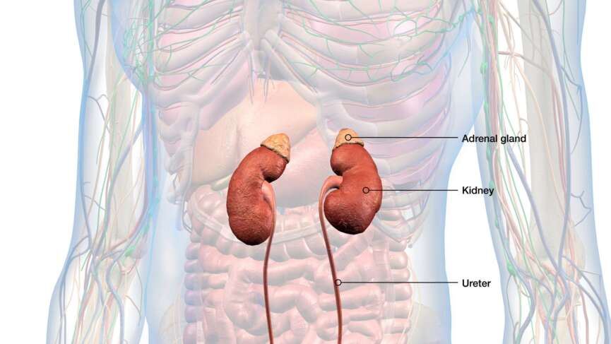
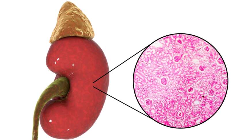
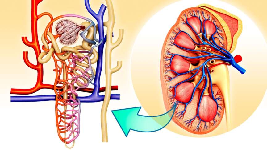
    A nephrotic syndrome is a group of disorders that damage the filtering units of your kidney, the nephrons.  Nephrons are responsible for filtering blood and producing urine. Each kidney contains thousands of nephrons, each consisting of a glomerulus and a tubule. As blood flows through your glomerulus, waste products and excess substances are filtered out of your bloodstream into the renal tubules. [1]

    Nephrons retain protein in the body, allowing less than 150 mg of protein to pass into the urine daily.  [1]However, when these filtering units are damaged in Nephrotic syndrome, three or more grams of protein are lost in the urine over a 24-hour period, a condition called proteinuria. [2]As more of these proteins are lost, the resulting osmotic imbalance can cause fluids to leak and accumulate in the spaces between cells, leading to swelling. [1]

    The estimated annual incidence rate of Nephrotic Syndrome is two to seven new cases per 100,000 in children of age below 18 years and 4.30 per 100,000 person-years in adults. [1,3] The condition affects children and adults of all ages, genders, or races.

    Causes of Nephrotic Syndrome

    Nephrotic syndrome can arise from various conditions that either solely impact the kidneys or those affecting multiple body parts.

    1. Diabetes

    In diabetic nephropathy, prolonged high blood sugar levels damage the small blood vessels in your kidneys, particularly the glomeruli. Additionally, if you have diabetes, you might have an increased risk of developing conditions like high blood pressure and cardiovascular disease, which can contribute to nephrotic syndrome. [4]

    2. Focal Segmental Glomerulosclerosis (FSGS)

    The glomerulus of your kidney becomes scarred, allowing proteins to leak into urine. It is the leading cause of nephrotic syndrome in African American adults. [1]

    3. Membranous Nephropathy

    Membranous nephropathy leads to protein build-up in your glomerulus, which causes the protein to leak into the urine. This is the predominant cause among white adults. [1]

    4. Minimal Change Disease (nil disease)

    It is the most common cause of nephrotic syndrome in children and accounts for a significant proportion of cases in adults as well. [5]

    5. Systemic Lupus Erythematous (SLE)

    SLE, commonly known as lupus, can cause nephrotic syndrome through a condition known as lupus nephritis. In lupus nephritis, the body’s immune system mistakenly attacks your kidneys, leading to inflammation and damage to the kidney tissue, particularly the glomeruli. [6]

    6. Amyloidosis

    Amyloidosis can cause nephrotic syndrome by depositing abnormal proteins called amyloids in your kidneys. These amyloid deposits interfere with the normal functioning of the glomeruli. [7]

    Symptoms of Nephrotic Syndrome

    Recognizing the symptoms early can lead to prompt treatment, which is critical in managing nephrotic syndrome.

    1. Swelling

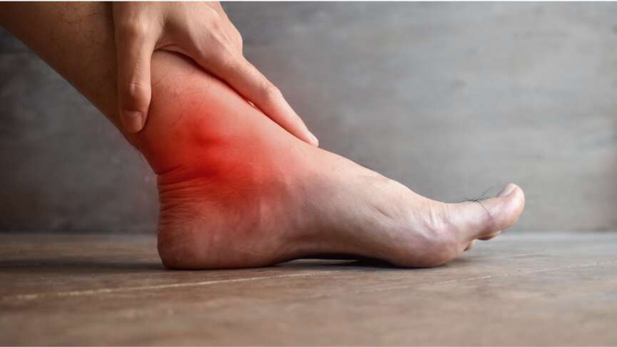
    Swelling is a prominent feature resulting from decreased oncotic pressure due to hypoalbuminemia. In children, swelling of the face or puffy eyelids is usually the earliest sign to appear. Later, this swelling extends to the rest of the body.  In adults, however, it typically appears in the legs, ankles, feet, lower abdomen, or other areas. [8]

    2. Frothy or bubbly urine

    It is one of the frequently reported symptoms and indicates the presence of protein in urine. [8]You can often notice this symptom right away. When it’s in the toilet bowl, it looks like it has bubbles or foam on the surface, more noticeable than the usual bubbles that may appear when urine hits the water.

    3. Weight gain

    Retention of fluid in the body causes people with nephrotic syndrome to experience a rapid increase in weight. This weight gain is not related to fat or muscle but is due to the build-up of excess tissue fluid. [2]

    4. Loss of appetite

    Many people with nephrotic syndrome experience a reduced appetite, which can be linked to the body’s overall inflammatory state, hypothalamic dysregulation, edema, or the general discomfort associated with the condition. [9]

    Complications of Nephrotic Syndrome

    Nephrotic syndrome can lead to several serious complications, primarily due to the loss of proteins in the urine and the body’s response to this imbalance. Some of the most common complications are:

    1. Infection

    Your body might be more susceptible to infections, including serious ones like spontaneous bacterial peritonitis, an infection of the lining of your abdomen. If the infection spreads throughout your body, you might develop sepsis. [10]

    2. High cholesterol and clogged arteries

    Nephrotic syndrome can lead to high cholesterol levels in your blood. The deposition of this cholesterol in your arterial wall might make your arteries narrow and stiff, which can cause heart problems. [10,11]

    3. Blood clotting

    The loss of protein like antithrombin III, which helps prevent blood clotting, combined with increased production of clotting factors by the liver, can lead to an increased risk of blood clots. These clots can form in the veins or arteries, leading to life-threatening conditions like deep vein thrombosis or pulmonary embolism. [10,12]

    4. High blood pressure

    The retention of fluid and salt can lead to high blood pressure, further damaging the kidneys and increasing the risk of cardiovascular problems. [13]

    5. Malnutrition

    The significant loss of proteins in the urine can lead to malnutrition, as the body loses essential proteins faster than they can be replaced. This can result in muscle wasting, weakness, and stunted growth in children. [10]

    Diagnosis of Nephrotic Syndrome

    Diagnosing nephrotic syndrome involves several tests:

    1. Urine Dipstick Test

    A chemically treated paper strip, or dipstick, is dipped into your urine sample to check for the presence of albumin. [8]

    2. 24-hour urine collection

    This measures the amount of protein lost in the urine over 24 hours. A protein loss greater than 3.5 grams per day indicates nephrotic syndrome.

    3. Urine albumin-to-creatinine ratio (UACR)

    Unlike the 24-hour urine collection, this test uses a single urine sample. It estimates the albumin lost in 24 hours by measuring albumin and creatinine, a byproduct of normal muscle breakdown. [14]

    4. Blood tests

    Blood tests for nephrotic syndrome check factors like protein levels, kidney function, cholesterol, electrolytes, lipid levels, and blood clotting parameters to diagnose and monitor the condition. [15]

    Kidney ultrasound is a noninvasive imaging test used to assess the size, structure, and presence of abnormalities such as cysts, tumors, or blockages. It also helps to rule out other causes of kidney disease.

    5. Kidney Biopsy

    A kidney biopsy is a diagnostic procedure where a small sample of your kidney tissue is taken using a needle, and the tissue is then sent for examination under a microscope. This procedure is performed to determine the underlying cause of the nephrotic syndrome and to assess the severity of your kidney damage. [14]

    Treatment of Nephrotic Syndrome

    The treatment of nephrotic syndrome is aimed at managing symptoms, addressing the underlying cause, and preventing complications. Here are common treatment options available:

    1. Corticosteroids

    Corticosteroids, such as Prednisone, play an important role in treating nephrotic syndrome, particularly in cases related to minimal change disease. These medications work by suppressing the immune system and reducing inflammation in the kidneys, which helps to restore the integrity of the glomeruli. This reduction in inflammation leads to decreased proteinuria. Initially, a high dose of steroids is prescribed daily for several weeks to achieve rapid control of symptoms. Once the condition improves, the dosage is gradually tapered down over several months. Steroids are one of the most effective treatments of nephrotic syndrome, but side effects such as weight gain, increased blood sugar levels, and increased risk of infections make monitoring essential. [16]

    2. Immunosuppressants

    These medications suppress the immune system. Drugs like cyclophosphamide, cyclosporin, or mycophenolate mofetil are commonly used, particularly in conditions like FSGS, lupus nephritis, or membranous nephropathy. In children, immunosuppressants are used if nephrotic syndrome recurs frequently or does not respond well to treatment. [16,17]

    3. Rituximab

    This is a monoclonal group of medications that are used in cases of steroid-resistant nephrotic syndrome and in patients with frequent relapses. [16,17]

    4. Diuretics

    Diuretics, often known as water pills, such as furosemide and spironolactone, help reduce fluid retention and swelling by increasing urine output. [18]

    5. Blood pressure medications

    Angiotensin-converting enzyme (ACE) inhibitor or Angiotensin II Receptor Blocker (ARB) can lower blood pressure and reduce proteinuria by decreasing the pressure in the glomeruli.

    6. Anticoagulants

    Blood thinners like warfarin or heparin may reduce the risk of blood clots, a common complication of nephrotic syndrome. [18]

    Vaccination in Nephrotic Syndrome

    Children with nephrotic syndrome should receive pneumococcal vaccination, including the 23-valent pneumococcal polysaccharide vaccine, to protect against severe pneumococcal infections. [19]Annual influenza vaccination is recommended for children with nephrotic syndrome and their household contacts, as it has been shown to reduce both influenza infections and relapses of nephrotic syndrome significantly. SARS-CoV-2 mRNA vaccines have proven safe and effective in these patients, though those on mycophenolate mofetil may exhibit lower antibody levels.[20] Inactivated, recombinant, and toxoid vaccines can be safely administered, with the varicella-zoster virus vaccine being given more frequently without significant side effects. [19,21]

    Live vaccines should be postponed until the child’s prednisone dose is below 1 mg/kg daily (less than 20 mg/day) or 2 mg/kg on alternate days (less than 40 mg on alternate days). [19] Live vaccines are contraindicated for children receiving corticosteroid-sparing immunosuppressive agents, and the child should also avoid close contact with the child vaccinated with live vaccine for 3-6 weeks. [22]

    Importance of Diet in Nephrotic Syndrome

    Dietary management for nephrotic syndrome consists of several key considerations. Protein intake depends on the type of nephrotic syndrome. A daily intake of 1.0-1.1 g/kg is recommended for minimal change nephrotic syndrome. Other types may require a lower intake of 0.8 g/kg/day to avoid exacerbating proteinuria and renal damage. [23,24]Sodium intake should be restricted to 5-7 grams daily to help manage edema and hypertension. [8]You should watch out for high-sodium seasonings and condiments that often get ignored to have the right amount of salt.

    Adequate caloric intake is also crucial to prevent malnutrition, with a recommended energy intake of over 35 kcal/kg/day to meet increased metabolic demands and prevent muscle wasting. [8]To manage hyperlipidemia, patients should limit saturated and trans fats, opting for a diet rich in monounsaturated and polyunsaturated fats. [18]

    How do You Restrict your Child’s Fluid Intake?

    It is often challenging to manage fluid intake and water balance in children. Fluid intake should be adjusted based on the patient’s edema and overall fluid balance, though strict fluid restriction is generally only necessary in cases of significant fluid overload. [25] A balanced diet that provides sufficient calories and protein while limiting salt and fluids is important for regulating fluid levels. [23]

    To help manage your child’s fluid intake and maintain their comfort, you start by determining the size of their favorite glass or cup to measure all the fluid they take properly. [25]You should remember all room-temperature liquids like milk, water, and juice should be counted as fluid intake. [25]

    You should avoid giving your child salty foods, which can increase thirst. [25]If your child feels thirsty, you can choose iced tea, lemonade, or frozen fruits, such as melon or berries, to avoid giving them too much fluid. [25]Offering chewing gum or hard candy can also help alleviate thirst, and rinsing the mouth with cold water (without swallowing) can provide relief. Sucking on a lemon wedge can further moisten the mouth. [25,26]

    Questions For Your Doctor

    1. How long does treatment for nephrotic syndrome usually take?
    2. How can we tell if my child’s nephrotic syndrome is worsening?
    3. How can I know if my child is responding to the treatment?
    4. What should we do if my child develops a fever or infection?
    5. Can my child participate in sports or physical activities? Are there any restrictions?

    References

    1. Tapia C, Bashir K. Nephrotic Syndrome [Internet]. Nih.gov. StatPearls Publishing; 2023. Available from: https://www.ncbi.nlm.nih.gov/books/NBK470444/
    2. Wang C, Greenbaum LA. Nephrotic Syndrome. Pediatr Clin North Am 2019;66:73–85. https://doi.org/10.1016/j.pcl.2018.08.006.
    3. Vestergaard SV, Birn H, Jensen SK, Sørensen HT, Nitsch D, Christiansen CF. Twenty-four-Year Trends in Incidence and Mortality of Nephrotic Syndrome: A Population-Based Cohort Study. Epidemiology 2023;34:411–20. https://doi.org/10.1097/EDE.0000000000001576.
    4. Samsu N. Diabetic Nephropathy: Challenges in Pathogenesis, Diagnosis, and Treatment. Biomed Res Int 2021;2021:1–17. https://doi.org/10.1155/2021/1497449.
    5. Vivarelli M, Massella L, Ruggiero B, Emma F. Minimal Change Disease. Clinical Journal of the American Society of Nephrology 2017;12:332–45. https://doi.org/10.2215/CJN.05000516.
    6. Nawata A, Hisano S, Shimajiri S, Wang K, Tanaka Y, Nakayama T. Podocyte and endothelial cell injury lead to nephrotic syndrome in proliferative lupus nephritis. Histopathology 2018;72:1084–92. https://doi.org/10.1111/his.13454.
    7. Karam S, Kaushal A, Abu Amer N, Royal V, KItchlu A. Non-Immunoglobulin Amyloidosis-Mediated Kidney Disease: Emerging Understanding of Underdiagnosed Entities. Advances in Kidney Disease and Health 2024;31:334–45. https://doi.org/10.1053/j.akdh.2024.02.001.
    8. Kodner C. Diagnosis and Management of Nephrotic Syndrome in Adults. American Family Physician [Internet]. 2016 Mar 15;93(6):479–85. Available from: https://www.aafp.org/pubs/afp/issues/2016/0315/p479.html
    9. Ueno H, Miyamoto T, Sanada K, Nakazono K, Tanaka K, Nishimura H, et al. Changes in gene expressions of hypothalamic neuropeptides controlling feeding behaviors in bilateral nephrectomized rats. Neurosci Lett 2019;711:134426. https://doi.org/10.1016/j.neulet.2019.134426 
    10. Claudio P, Gabriella M. Nephrotic syndrome: pathophysiology and consequences. J Nephrol 2023;36:2179–90. https://doi.org/10.1007/S40620-023-01697-7
    11. Agrawal S, Zaritsky JJ, Fornoni A, Smoyer WE. Dyslipidaemia in nephrotic syndrome: mechanisms and treatment. Nat Rev Nephrol 2018;14:57–70. https://doi.org/10.1038/nrneph.2017.155 
    12. Vestergaard SV, Birn H, Darvalics B, Nitsch D, Sørensen HT, Christiansen CF. Risk of Arterial Thromboembolism, Venous Thromboembolism, and Bleeding in Patients with Nephrotic Syndrome: A Population-Based Cohort Study. Am J Med 2022;135:615-625.e9. https://doi.org/10.1016/j.amjmed.2021.11.018 
    13. Ray EC, Rondon-Berrios H, Boyd CR, Kleyman TR. Sodium Retention and Volume Expansion in Nephrotic Syndrome: Implications for Hypertension. Adv Chronic Kidney Dis 2015;22:179–84. https://doi.org/10.1053/j.ackd.2014.11.006
    14. Kodner C. Nephrotic Syndrome in Adults: Diagnosis and Management. American Family Physician [Internet]. 2009 Nov 15;80(10):1129–34. Available from: https://www.aafp.org/pubs/afp/issues/2009/1115/p1129.html
    15. Stoycheff N, Stevens LA, Schmid CH, Tighiouart H, Lewis J, Atkins RC, et al. Nephrotic Syndrome in Diabetic Kidney Disease: An Evaluation and Update of the Definition. American Journal of Kidney Diseases 2009;54:840–9. https://doi.org/10.1053/j.ajkd.2009.04.016
    16. Vivarelli M, Gibson K, Sinha A, Boyer O. Childhood nephrotic syndrome. The Lancet 2023;402:809–24. https://doi.org/10.1016/S0140-6736(23)01051-6.
    17. Nagai K. Immunosuppressive Agent Options for Primary Nephrotic Syndrome: A Review of Network Meta-Analyses and Cost-Effectiveness Analysis. Medicina (B Aires) 2023;59:601. https://doi.org/10.3390/medicina59030601.
    18. Appel GB. Improved outcomes in nephrotic syndrome. Cleve Clin J Med 2006;73:161–7. https://doi.org/10.3949/ccjm.73.2.161.
    19. Kamei K, Miyairi I, Ishikura K, Ogura M, Shoji K, Funaki T, et al. Prospective Study of Live Attenuated Vaccines for Patients with Nephrotic Syndrome Receiving Immunosuppressive Agents. J Pediatr 2018;196:217-222.e1. https://doi.org/10.1016/j.jpeds.2017.12.061.
    20. Kamei K, Ogura M, Sato M, Nishi K, Shoji K, Funaki T, et al. Immunogenicity and safety of SARS-CoV-2 mRNA vaccine in patients with nephrotic syndrome receiving immunosuppressive agents. Pediatric Nephrology 2023;38:1099–106. https://doi.org/10.1007/s00467-022-05633-y.
    21. Angeletti A, Lugani F, La Porta E, Verrina E, Caridi G, Ghiggeri GM. Vaccines and nephrotic syndrome: efficacy and safety. Pediatric Nephrology 2023;38:2915–28. https://doi.org/10.1007/s00467-022-05835-4.
    22. NHS Choices. Nephrotic syndrome in children [Internet]. NHS. 2019. Available from: https://www.nhs.uk/conditions/nephrotic-syndrome/
    23. Lella G, Pecoraro L, Benetti E, Arnone OC, Piacentini G, Brugnara M, et al. Nutritional Management of Idiopathic Nephrotic Syndrome in Pediatric Age. Medical Sciences 2023;11:47. https://doi.org/10.3390/medsci11030047
    24. Polderman N, Cushing M, McFadyen K, Catapang M, Humphreys R, Mammen C, et al. Dietary intakes of children with nephrotic syndrome. Pediatric Nephrology 2021;36:2819–26. https://doi.org/10.1007/s00467-021-05055-2.
    25. National Kidney Foundation. KDOQI Clinical Practice Guideline for Nutrition in Children with CKD: 2008 Update. American Journal of Kidney Diseases 2009;53:S11–104. https://doi.org/10.1053/j.ajkd.2008.11.017.
    26. Nutrition and Nephrotic Syndrome [Internet]. www.stanfordchildrens.org. Available from: https://www.stanfordchildrens.org/en/topic/default?id=nutrition-and-nephrotic-syndrome-90-P03099

  • Osteoarthritis – Symptoms, Causes, Diagnosis, and Treatment

    Osteoarthritis – Symptoms, Causes, Diagnosis, and Treatment

    Highlights of Osteoarthritis

    1. Osteoarthritis is the most common joint condition, affecting the hands, knees, neck, hips, and lower back.
    2. Osteoarthritis is one of the major causes of disability in older people, but it can also occur in the young population.
    3. Osteoarthritis can cause symptoms such as pain, stiffness, limited range of motion, and swelling in a joint.
    4. Joint replacement surgery can be an option for individuals with severe joint pain, deformity, and dysfunction when conservative treatments are no longer effective.
    5. PRP injections are an emerging treatment for osteoarthritis, using the patient’s own blood to promote healing.

    What is Osteoarthritis (OA)?

    Osteoarthritis (OA) is the most common type of arthritis and occurs when the cartilage in joints breaks down, causing pain, stiffness, and difficulty moving. It usually affects the knees, hips, hands, and spine. [1] Worldwide, about 528 million people have OA in the year 2019, with about 60% of them being women. [2] In the United States, about 53 million adults have some form of arthritis, making it a major cause of disability in older people. [3]

    Types of Osteoarthritis

    Osteoarthritis can be classified into two types depending on the cause.

    1. Primary osteoarthritis

    It is the most common type of OA. It develops over time. It is believed to be caused due to wear and tear of the joint throughout your life. There is no predisposing trauma or disease in primary osteoarthritis.

    2. Secondary osteoarthritis

    This type of osteoarthritis occurs due to a preexisting joint disease. Predisposing conditions include congenital joint disorders, inflammatory arthritis, osteoporosis, metabolic disorders like hemochromatosis, injury, and trauma.

    Causes and Risk Factors of Osteoarthritis

    Although the exact cause of osteoarthritis is not yet known, certain risk factors increase your risk of developing it:

    • Aging: As people age, the cartilage that cushions joints naturally deteriorates, leading to OA. [4]
    • Gender: The cases of hip, knee, and hand OA are found to be higher in women than men, and the incidence of the condition increases around menopause. This has been attributed to the effect of decreasing levels of hormones in women. [4]
    • Joint injuries: Injuries from sports, accidents, or repetitive stress can damage the cartilage and increase the risk of OA. Among all the joints, the knee is one of the most commonly injured joints. Thus, the prevalence of OA is also higher among women. [4]
    • Obesity: Obesity that is defined by a body mass index of more than 30kg/m2 is strongly associated with knee OA. Excess body weight adds extra stress to weight-bearing joints like the knees and hips, accelerating cartilage breakdown. [4]
    • Genetics: A family history of OA can increase an individual’s risk, suggesting a genetic predisposition. It accounts for 60% of hand and hip OA, while it is the cause of 40% of cases of knee OA. [4]
    • Bone deformities: Some people are born with malformed joints, such as varus or valgus deformities or defective cartilage, which can lead to OA.

    Symptoms of Osteoarthritis

    OA symptoms can vary in severity and often develop gradually. Common symptoms include:

    1. Joint pain

    Pain in the affected joints during or after movement is a hallmark of osteoarthritis. This pain often worsens with activity and may improve with rest but can eventually become constant as the disease progresses. [5]

    2. Stiffness

    You may notice joint stiffness, especially upon waking up in the morning or after periods of inactivity, such as sitting for a long time. This stiffness typically eases within 30 minutes of movement but can recur throughout the day. [6]

    3. Loss of flexibility

    Reduced range of motion is common, making it difficult for you to move the joint fully. This loss of flexibility can interfere with simple movements like bending, reaching, or grasping objects. [7]

    4. Grating sensation

    You may feel a grating or hear a crackling sound (crepitus) when moving the joint. This occurs due to the rough surfaces of the cartilage and bone rubbing against each other. [7]

    5. Bone spurs

    Extra bits of bone, known as osteophytes or bone spurs, can form around the affected joint. These spurs can cause additional pain and limit joint movement.[6,7]

    6. Swelling

    You may see occasional swelling around the joint due to inflammation. This swelling can cause further discomfort and stiffness, and it may fluctuate depending on activity levels and other factors. [7]

    Diagnosis of Osteoarthritis

    Diagnosis of OA is primarily done with the help of presenting symptoms and imaging. Some of the diagnostic modalities for OA are:

    Imaging tests for osteoarthritis

    • X-rays: X-rays are commonly used to diagnose OA. They can reveal joint space narrowing, the presence of bone spurs, and other changes in the bone structure that are indicative of OA. [8]
    • Magnetic Resonance Imaging (MRI): MRI is not routinely performed for OA, but it helps by providing more detailed images of the joint’s soft tissues, including cartilage, tendons, and ligaments. It is particularly useful in early OA detection and in cases where the diagnosis is uncertain or more complex. [8,9]

    Laboratory tests for osteoarthritis

    • Blood tests: While there are no specific blood tests for OA, they can help rule out other types of arthritis, such as rheumatoid arthritis or gout, which can present similar symptoms. Inflammatory markers such as C-reactive protein (CRP) and erythrocyte sedimentation rate can also be used in severe cases, though OA is not related to systemic inflammation. [10]
    • Joint fluid analysis: In some cases, your doctor may extract a sample of synovial fluid from the affected joint using a needle. Analyzing this fluid can help exclude other inflammatory joint conditions, such as infection or gout. [11,12]

    Treatment of Osteoarthritis

    The principles of management and treatment of osteoarthritis include several key objectives: reducing pain and other symptoms, improving joint movement and function, stopping the progression of the disease, and maintaining quality of life. Here are the treatment modalities for OA:

    1. Pain medications

    You can use over-the-counter pain medication to relieve your pain. They can be either topical, like creams, ointments, and patches, or oral. Oral anti-inflammatory medications like NSAIDs can also be helpful. [13]

    2. Intra-articular medications

    Intra-articular injections of corticosteroids and hyaluronic acids can also be effective, especially for acute pain.[13]

    3. Exercise

    Exercise helps improve symptoms like pain and stiffness alongside increasing flexibility. Low-impact exercises are particularly helpful. Stretching and strengthening exercises are also equally beneficial. Your doctor may recommend a physiotherapist to help you find the best exercises. [14]

    4. Supportive devices

    Wearing aids, knee braces, or shoe inserts take pressure off affected joints. These devices, therefore, help with the symptoms. Simple steps such as wearing comfortable shoes can also make a considerable difference.[15]

    5. Heat or cold therapy

    Heat therapy uses methods like heating pads or warm baths, while cold therapy uses ice packs or cold compresses. Heat helps to relax and loosen tissues, increase blood flow, and alleviate stiffness and chronic pain. Cold therapy, on the other hand, reduces inflammation, numbs pain, and decreases swelling, especially during flare-ups. [16]

    6. Joint replacement surgery

    Joint replacement therapies offer a solution for individuals experiencing severe joint pain and dysfunction when conservative treatments are no longer effective. Total joint replacement, such as hip or knee arthroplasty, involves surgically removing the damaged joint and replacing it with an artificial implant. This procedure aims to relieve pain, restore function, and improve quality of life by providing a new, smooth surface for joint movement. Partial joint replacement, where only the damaged portion of the joint is replaced, is another option that may be suitable for some patients. These therapies can significantly reduce pain and enhance mobility, allowing you to return to daily activities and improve your overall well-being. However, candidates for joint replacement typically undergo a thorough evaluation to ensure the procedure is appropriate based on their overall health, activity level, and specific joint damage. [17,18]

    Prevention of Osteoarthritis

    It is not possible to prevent osteoarthritis altogether. However, a healthy lifestyle and certain other factors can reduce your likelihood of developing or progressing OA. Some preventive measures include:

    1. Maintain a healthy weight

    Excess weight puts added stress on weight-bearing joints, particularly the knees and hips. Achieving and maintaining a healthy weight can significantly reduce the risk of OA. [19]

    2. Regular exercise

    Engaging in low-impact exercises like swimming, cycling, and walking helps to strengthen the muscles around the joints, improve flexibility, and maintain overall joint function. Strengthening exercises can also support joint stability. [20]

    3. Proper joint care

    Using proper techniques during physical activities and sports can help prevent joint injuries. Employing correct posture and body mechanics in daily activities also contributes to joint health. [20]

    4. Healthy diet

    A balanced diet rich in nutrients, including omega-3 fatty acids, vitamins C and D, and antioxidants, supports joint health and reduces inflammation. [20]

    5. Avoiding repetitive stress

    Minimizing repetitive stress and overuse of joints, especially in occupations or activities that place high demands on specific joints, can help prevent OA.

    6. Education and self-management

    Educating patients about the risk factors for developing osteoarthritis can significantly reduce the risk. [21]

    Platelet-rich plasma (PRP) Injection for Osteoarthritis

    PRP injection is an emerging treatment for osteoarthritis, and it uses the patient’s own blood to promote healing. The process involves drawing blood, centrifuging it to concentrate the platelets, and then injecting the PRP into the affected joint. Platelets contain growth factors and proteins that support tissue repair and regeneration. [22]This treatment aims to reduce pain and improve joint function by leveraging the body’s natural healing mechanisms. PRP injections are considered a promising option for managing osteoarthritis symptoms, though more research is needed to understand their long-term efficacy. [22,23]

    Questions To Ask Your Doctor

    1. What exercises will benefit osteoarthritis?
    2. Will I have a normal life after developing osteoarthritis?
    3. Does surgery cure osteoarthritis?
    4. What natural therapies can I try to help with my joint function?
    5. What should I do if my symptoms suddenly worsen or if I experience new symptoms?

    References

    1. Hall M, van der Esch M, Hinman RS, Peat G, de Zwart A, Quicke JG, et al. How does hip osteoarthritis differ from knee osteoarthritis? Osteoarthritis Cartilage 2022;30:32–41. https://doi.org/10.1016/J.JOCA.2021.09.010.
    2. Osteoarthritis n.d. https://www.who.int/news-room/fact-sheets/detail/osteoarthritis (accessed July 29, 2024).
    3. Fallon EA, Boring MA, Foster AL, Stowe EW, Lites TD, Odom EL, et al. Prevalence of Diagnosed Arthritis — United States, 2019–2021. MMWR Morb Mortal Wkly Rep 2023;72:1101–7. https://doi.org/10.15585/MMWR.MM7241A1.
    4. Palazzo C, Nguyen C, Lefevre-Colau MM, Rannou F, Poiraudeau S. Risk factors and burden of osteoarthritis. Ann Phys Rehabil Med 2016;59:134–8. https://doi.org/10.1016/J.REHAB.2016.01.006.
    5. Perrot S, Anne-Priscille T. Pain in osteoarthritis from a symptom to a disease. Best Pract Res Clin Rheumatol 2023;37:101825. https://doi.org/10.1016/J.BERH.2023.101825.
    6. Ebell MH. Osteoarthritis: Rapid Evidence Review. Am Fam Physician 2018;97:523–6. https://www.aafp.org/pubs/afp/issues/2018/0415/p523.html
    7. Abhishek A, Doherty M. Diagnosis and Clinical Presentation of Osteoarthritis. Rheumatic Disease Clinics of North America 2013;39:45–66. https://doi.org/10.1016/J.RDC.2012.10.007.
    8. Mourad C, Vande Berg B. Osteoarthritis of the hip: is radiography still needed? Skeletal Radiol 2023;52:2259. https://doi.org/10.1007/S00256-022-04270-8.
    9. Wang X, Oo WM, Linklater JM. What is the role of imaging in the clinical diagnosis of osteoarthritis and disease management? Rheumatology 2018;57:iv51–60. https://doi.org/10.1093/RHEUMATOLOGY/KEX501.
    10. Woodell-May JE, Sommerfeld SD. Role of Inflammation and the Immune System in the Progression of Osteoarthritis. Journal of Orthopaedic Research® 2020;38:253–7. https://doi.org/10.1002/JOR.24457.
    11. Oliviero F, Scanu A, Galozzi P, Ramonda R. Synovial Fluid Analysis to Identify Osteoarthritis. Journal of Visualized Experiments 2022;2022. https://doi.org/10.3791/64351.
    12. Monibi F, Roller BL, Stoker A, Garner B, Bal S, Cook JL. Identification of Synovial Fluid Biomarkers for Knee Osteoarthritis and Correlation with Radiographic Assessment. Journal of Knee Surgery 2016;29:242–7. http://pubmed.ncbi.nlm.nih.gov/25927354/
    13. Brophy RH, Fillingham YA. AAOS Clinical Practice Guideline Summary: Management of Osteoarthritis of the Knee (Nonarthroplasty), Third Edition. Journal of the American Academy of Orthopaedic Surgeons 2022;30:e721–9. https://doi.org/10.5435/JAAOS-D-21-01233.
    14. Bannuru RR, Osani MC, Vaysbrot EE, Arden NK, Bennell K, Bierma-Zeinstra SMA, et al. OARSI guidelines for the non-surgical management of knee, hip, and polyarticular osteoarthritis. Osteoarthritis Cartilage 2019;27:1578–89. https://doi.org/10.1016/j.joca.2019.06.011.
    15. Kolasinski SL, Neogi T, Hochberg MC, Oatis C, Guyatt G, Block J, et al. 2019 American College of Rheumatology/Arthritis Foundation Guideline for the Management of Osteoarthritis of the Hand, Hip, and Knee. Arthritis & Rheumatology 2020;72:220–33. https://doi.org/10.1002/art.41142.
    16. Yıldırım N, Filiz Ulusoy M, Bodur H. The effect of heat application on pain, stiffness, physical function and quality of life in patients with knee osteoarthritis. J Clin Nurs 2010;19:1113–20. https://doi.org/10.1111/j.1365-2702.2009.03070.x.
    17. Lützner J, Kasten P, Günther K-P, Kirschner S. Surgical options for patients with osteoarthritis of the knee. Nat Rev Rheumatol 2009;5:309–16. https://doi.org/10.1038/nrrheum.2009.88.
    18. Grayson CW, Decker RC. Total Joint Arthroplasty for Persons With Osteoarthritis. PM&R 2012;4. https://doi.org/10.1016/j.pmrj.2012.02.018.
    19. Roos EM, Arden NK. Strategies for the prevention of knee osteoarthritis. Nat Rev Rheumatol 2016;12:92–101. https://doi.org/10.1038/nrrheum.2015.135.
    20. Marriott KA, Birmingham TB. Fundamentals of osteoarthritis. Rehabilitation: Exercise, diet, biomechanics, and physical therapist-delivered interventions. Osteoarthritis Cartilage 2023;31:1312–26. https://doi.org/10.1016/j.joca.2023.06.011.
    21. Whittaker JL, Runhaar J, Bierma-Zeinstra S, Roos EM. A lifespan approach to osteoarthritis prevention. Osteoarthritis Cartilage 2021;29:1638–53. https://doi.org/10.1016/j.joca.2021.06.015.
    22. Meheux CJ, McCulloch PC, Lintner DM, Varner KE, Harris JD. Efficacy of Intra-articular Platelet-Rich Plasma Injections in Knee Osteoarthritis: A Systematic Review. Arthroscopy: The Journal of Arthroscopic & Related Surgery 2016;32:495–505. https://doi.org/10.1016/j.arthro.2015.08.005.
    23. Xiong Y, Gong C, Peng X, Liu X, Su X, Tao X, et al. Efficacy and safety of platelet-rich plasma injections for the treatment of osteoarthritis: a systematic review and meta-analysis of randomized controlled trials. Front Med (Lausanne) 2023;10. https://doi.org/10.3389/fmed.2023.1204144.

  • Urinary Tract Infections – Causes, Symptoms, and Effective Management

    Urinary Tract Infections – Causes, Symptoms, and Effective Management

    Highlights Of Urinary Tract Infections

    • UTI is more common in women, affecting almost 50% of the female population at least once in their lifetime.
    • UTI typically presents as frequent urination, difficulty urinating, flank pain, fever, and chills.
    • Urine culture is a gold standard test to diagnose  UTI.
    • Overuse and misuse of antibiotics have led to antibiotic-resistant bacteria, making some UTIs harder to treat.
    • Cranberry juice, often used as a preventive measure for UTIs, has mixed scientific evidence supporting its effectiveness.

    What is Urinary Tract Infection (UTI)?

    A Urinary Tract Infection (UTI) occurs when bacteria enter the urinary system, which includes the kidneys, bladder, ureters, and urethra. It commonly causes symptoms like a burning sensation when you urinate, the need to urinate frequently, and cloudy or strong-smelling urine.

    UTIs can affect men and women of all age groups. However, the incidence is higher in females. About 50% of women report having UTIs at some point in their lives. [1]

    Types of Urinary Tract Infections

    There are various ways to classify UTI depending on various factors. On the basis of the onset of symptoms, it can be:

    • Acute UTI: An acute UTI is the acute onset of infection in the urinary tract. It can be caused by an infection in any part of the urinary tract and is usually treated with a short course of antibiotics.
    • Chronic UTI: A chronic UTI lasts for over two weeks and presents with severe symptoms.
    • Recurrent UTI: A urinary tract infection is termed recurrent when a person develops two or more infections in a 6-month period or three or more infections in a 12-month.

    Based on whether the infection is complicated or not, there are two types of UTIs, namely,

    1. Uncomplicated UTI

    This is usually less serious. A case of UTI is considered uncomplicated when there is no structural or functional abnormality of the urinary tract, and there are no significant comorbidities.[3]

    2. Complicated UTI

    UTI is classified as complicated under specific circumstances. These include if the patient is a child or pregnant, if there are structural or functional abnormalities in the urinary tract if the patient has underlying comorbidities such as diabetes, or if there has been recent instrumentation or surgery involving the urinary tract.[3]

    Causes of Urinary Tract Infection

    UTIs are primarily caused by bacteria, with Escherichia coli (E. coli) being the predominant pathogen. E.coli accounts for 70-95% of community-acquired UTIs.[4] Other common causative agents include Klebsiella pneumoniaeProteus mirabilis, and Enterococcus faecalis. [4]

    The most typical way these pathogens infect the urinary tract is through the ascending route, where bacteria travel from the urethra up to the bladder and potentially to the ureters and kidneys. They first colonize the areas around the urethra and then move upward. Less commonly, infections can spread through the bloodstream (hematogenous route) or lymphatic system. [5]

    Several host factors increase the risk of UTIs, such as being female, pregnancy, frequent or recent sexual activity, difficulties in urinating, and the use of spermicides. Recent antibiotic use and conditions like vesicoureteral reflux also elevate the risk. [2]

    Symptoms of Urinary Tract Infection

    UTIs present with various signs and symptoms, which vary depending on the location and severity of the infection. The primary symptoms include:

    1. Dysuria

    This is a burning sensation you have during urination. This symptom indicates inflammation or irritation in the urethra or bladder.[6]

    2. Increased Frequency and Urgency

    Many people experience an increased need to urinate, often producing only small amounts of urine each time. This symptom suggests bladder involvement.

    3. Hematuria

    Blood in the urine is a common symptom. It can range from microscopic hematuria, detectable only through a urine test, to gross hematuria, where blood is visible in the urine.[7]

    4. Fever and Chills

    In cases where the infection ascends to the kidneys (called as pyelonephritis), you might experience symptoms such as fever and chills.[8]

    5. Flank Pain

    Pain in the back or side, below the ribs, can indicate a kidney infection. This symptom is often severe and requires immediate medical attention. You may also experience nausea and vomiting. [9]

    Diagnosis of Urinary Tract Infection

    Your symptoms can easily suggest a UTI, and your doctor can order other tests to confirm the diagnosis. Here are some of the tests that you might require:

    1. Urinalysis

    This is the first step in diagnosing a UTI. A sample of urine is analyzed for the presence of white blood cells, red blood cells, and bacteria. The presence of nitrites and leukocyte esterase in the urine is a strong indicator of a UTI.[10]

    2. Urine Culture

    To confirm the diagnosis and identify the specific bacteria causing the infection, your doctor can ask for a urine culture. The sample is incubated to allow any bacteria present to grow. This test can also determine the sensitivity of the bacteria to various antibiotics, guiding effective treatment.[11]

    3. Complete Blood Count (CBC)

    A CBC can help identify the presence of an infection by showing elevated white blood cell counts, which is a sign of the body fighting off an infection. This is primarily done when you are suspected to have a kidney infection, as this can further help to figure out if your infection has subsided or not after the antibiotic therapy. [12]

    4. Blood Cultures

    In severe cases, especially when a kidney infection is suspected, blood cultures may also be taken to check if the infection has spread to the bloodstream.[12]

    5. Imaging Tests

    While not typically part of the initial laboratory diagnosis, imaging tests such as an ultrasound, cystoscopy, computed tomography scan, or magnetic resonance imaging can be used if there are concerns about structural abnormalities or complications like kidney stones or abscesses that might be responsible for your UTI. These tests may also be performed if you have recurrent UTIs. [13]

    Complications of Urinary Tract Infections

    Urinary tract infections can lead to a range of complications if not treated promptly and adequately. Some of such complications are:

    1. Urosepsis

    It is a severe systemic response to a UTI that spreads to the bloodstream, leading to sepsis. High-grade fever, chills, hypotension, and organ dysfunction are the symptoms.[14]

    2. Renal abscess

    An improperly treated UTI, especially pyelonephritis, can result in the formation of a renal abscess. Renal abscess is a localized collection of pus within the kidney tissue.[8]

    3. Chronic Kidney Disease (CKD)

    Recurrent or severe UTI can cause permanent renal damage and scarring. Over time, this leads to chronic kidney disease, reducing kidney function.[15]

    4. Complications in pregnancy

    UTIs in pregnancy increase the risk of complications such as preterm labor and low birth weight.[16]

    Management of Urinary Tract Infections

    The treatment options for urinary tract infections vary depending on the type and severity of the infection. They encompass both pharmacological and non-pharmacological approaches.

    1. Antibiotic Therapy

    The mainstay of treatment for UTI is antibiotic therapy. The first-line antibiotics often include trimethoprim-sulfamethoxazole, nitrofurantoin, and fosfomycin. These antibiotics are chosen for their effectiveness against common UTI pathogens. In cases where there is resistance or recurrent infections, culture-specific antibiotics are prescribed based on the sensitivity profile. The duration of treatment typically ranges from 3 to 7 days for uncomplicated UTIs and a longer duration of about seven to fourteen days for complicated ones to ensure complete eradication of the infection. [9]

    2. Pain Management

    To alleviate urinary discomfort, analgesics such as phenazopyridine can be used. This medication specifically targets urinary tract pain. Meanwhile, over-the-counter pain relievers like ibuprofen or acetaminophen can be used to manage general discomfort and reduce inflammation associated with the infection.[10]

    3. Hydration

    Drink plenty of fluids, as they are important for flushing bacteria from the urinary tract. Water is the best choice for hydration.[17]

    Prevention of Urinary Tract Infection

    Preventive measures for urinary tract infections are important, particularly for individuals prone to recurrent infections. Here are five preventive measures you can follow:

    1. Maintain Good Hydration

    Drink plenty of fluids, especially water, to help flush bacteria from the urinary tract.[17]

    2. Practice Proper Hygiene

    Wipe from front to back after using the toilet to prevent bacteria from the anal area from spreading to the urinary tract.[18]

    3. Urinate Frequently

    Do not hold urine for long periods. Emptying the bladder regularly can help prevent bacterial growth.[19]

    4. Urinate After Sexual Activity

    Urinating after intercourse can help flush out bacteria that may have entered the urinary tract.[19]

    5. Avoid Irritants

    Avoid using irritating feminine products such as deodorant sprays, douches, and powders in the genital area. These can cause irritation and increase the risk of infection.[19]

    Prophylactic Antibiotic Therapy for Recurrent UTIs

    Prophylactic antibiotic therapy is particularly important in those with recurrent urinary tract infections. Your doctor will prescribe antibiotics for daily use at a dose lower than required to treat the infection, which helps to reduce the frequency and severity of episodes.

    The antibiotics are the same as the ones used for treatment but at a lower dose, and the choice depends on the local antimicrobial resistance pattern.[20]

    Cranberry Juice and UTI

    Cranberry juice has long been suggested as a natural remedy for preventing UTIs. The primary mechanism behind this is the presence of proanthocyanidins in cranberries, which are believed to prevent bacteria, particularly E. coli, from adhering to the walls of the urinary tract. By inhibiting bacterial adhesion, cranberry juice may help reduce the likelihood of a developing infection. [21]

    However, while some studies support the potential benefits of cranberry juice in preventing UTIs, the evidence is not conclusive. It’s important to note that cranberry juice should not replace standard medical treatments and prevention strategies for UTIs. Those considering using cranberry juice as a preventive measure should consult with their doctor, especially if they have recurrent UTIs or other underlying health conditions.[21]

    Questions To Ask Your Doctor

    1. What are the signs that a UTI may be getting worse?
    2. How long will it take for antibiotics to start working?
    3. Are there any specific foods or drinks I should avoid during UTI?
    4. Does UTI during pregnancy affect my baby?
    5. Do I need a repeat urine test to find out if the infection has cleared?

    References

    1. Flores-Mireles AL, Walker JN, Caparon M, Hultgren SJ. Urinary tract infections: epidemiology, mechanisms of infection and treatment options. Nat Rev Microbiol 2015;13:269. https://doi.org/10.1038/NRMICRO3432.
    2. Alghoraibi H, Asidan A, Aljawaied R, Almukhayzim R, Alsaydan A, Alamer E, et al. Recurrent Urinary Tract Infection in Adult Patients, Risk Factors, and Efficacy of Low Dose Prophylactic Antibiotics Therapy. J Epidemiol Glob Health 2023;13:200–11. https://doi.org/10.1007/S44197-023-00105-4.
    3. Geerlings SE. Clinical Presentations and Epidemiology of Urinary Tract Infections. Microbiol Spectr 2016;4. https://doi.org/10.1128/MICROBIOLSPEC.UTI-0002-2012.
    4. Nielubowicz GR, Mobley HLT. Host-pathogen interactions in urinary tract infection. Nat Rev Urol 2010;7:430–41. https://doi.org/10.1038/NRUROL.2010.101.
    5. McLellan LK, Hunstad DA. Urinary Tract Infection: Pathogenesis and Outlook. Trends Mol Med 2016;22:946–57. https://doi.org/10.1016/J.MOLMED.2016.09.003.
    6. Urinary Tract Infections | National Kidney Foundation n.d. https://www.kidney.org/atoz/content/uti (accessed July 28, 2024).
    7. Lala V, Leslie SW, Minter DA. Acute Cystitis. 2024.
    8. Castaigne J, Georges B, Jouret F. [Acute pyelonephritis]. Rev Med Liege 2022;77:544–7.
    9. Al Lawati H, Blair BM, Larnard J. Urinary Tract Infections: Core Curriculum 2024. American Journal of Kidney Diseases 2024;83:90–100. https://doi.org/10.1053/j.ajkd.2023.08.009.
    10. Chu CM, Lowder JL. Diagnosis and treatment of urinary tract infections across age groups. Am J Obstet Gynecol 2018;219:40–51. https://doi.org/10.1016/J.AJOG.2017.12.231.
    11. Wojno KJ, Baunoch D, Luke N, Opel M, Korman H, Kelly C, et al. Multiplex PCR Based Urinary Tract Infection (UTI) Analysis Compared to Traditional Urine Culture in Identifying Significant Pathogens in Symptomatic Patients. Urology 2020;136:119–26. https://doi.org/10.1016/J.UROLOGY.2019.10.018.
    12. Gomi H, Goto Y, Laopaiboon M, Usui R, Mori R. Routine blood cultures in the management of pyelonephritis in pregnancy for improving outcomes. Cochrane Database Syst Rev 2015;2015. https://doi.org/10.1002/14651858.CD009216.PUB2.
    13. El-Ghar MA, Farg H, Doaa Elsayed S, El-Diasty T. CT and MRI in Urinary Tract Infections: A Spectrum of Different Imaging Findings. Medicina (B Aires) 2021;57:1–23. https://doi.org/10.3390/MEDICINA57010032.
    14. Wagenlehner FME, Pilatz A, Weidner W, Naber KG. Urosepsis: Overview of the Diagnostic and Treatment Challenges. Microbiol Spectr 2015;3. https://doi.org/10.1128/microbiolspec.UTI-0003-2012.
    15. Bandari B, Sindgikar SP, Kumar SS, Vijaya MS, Shankar R. Renal scarring following urinary tract infections in children. Sudan J Paediatr 2019;19:25. https://doi.org/10.24911/SJP.106-1554791193.
    16. Yan L, Jin Y, Hang H, Yan B. The association between urinary tract infection during pregnancy and preeclampsia: A meta-analysis. Medicine 2018;97. https://doi.org/10.1097/MD.0000000000012192.
    17. Urinary Tract Infection Basics | Urinary Tract Infection | CDC n.d. https://www.cdc.gov/uti/about/index.html (accessed July 28, 2024).
    18. Nkamedjie Pete PM, Mabvouna Biguioh R, Bita Izacar AG, Béchir Adogaye S Ben, Nguemo C. Genital hygiene behaviors and practices: A cross-sectional descriptive study among antenatal care attendees. J Public Health Afr 2019;10. https://doi.org/10.4081/JPHIA.2019.746.
    19. Pietrucha-Dilanchian P, Hooton TM. Diagnosis, Treatment, and Prevention of Urinary Tract Infection. Microbiol Spectr 2016;4. https://doi.org/10.1128/microbiolspec.UTI-0021-2015.
    20. Anger J, Lee U, Ackerman AL, Chou R, Chughtai B, Clemens JQ, et al. Recurrent Uncomplicated Urinary Tract Infections in Women: AUA/CUA/SUFU Guideline. J Urol 2019;202:282–9. https://doi.org/10.1097/JU.0000000000000296.
    21. Jepson RG, Williams G, Craig JC. Cranberries for preventing urinary tract infections. Cochrane Database Syst Rev 2012;2012. https://doi.org/10.1002/14651858.CD001321.PUB5.

  • Acute Respiratory Distress Syndrome -Symptoms, Diagnosis, Treatment and Recovery

    Acute Respiratory Distress Syndrome -Symptoms, Diagnosis, Treatment and Recovery

    Highlights of Acute Respiratory Distress Syndrome (ARDS)

    1. Acute Respiratory Distress Syndrome is a severe and life-threatening lung condition that causes low blood oxygen levels, leading to shortness of breath and bluish discoloration of the skin and lips.
    2. Sepsis is the leading cause of ARDS.
    3. Acute respiratory distress syndrome treatment focuses on supportive management and ventilator support to enhance oxygen levels and maintain lung function.
    4. A tracheostomy often becomes necessary as part of acute respiratory distress syndrome treatment for those who experience prolonged ARDS.
    5. While some individuals may recover fully within weeks to months, others may experience long-term lung damage and require ongoing medical care, and in severe cases, it can lead to death.

    What is Acute Respiratory Distress Syndrome (ARDS)?

    Acute respiratory distress syndrome (ARDS) is a severe and life-threatening lung condition that causes low blood oxygen levels and “stiff” lungs. It occurs when the tiny air sacs in your lungs, called alveoli, get damaged. These alveoli are essential because they allow oxygen and carbon dioxide to be exchanged. When they’re damaged, fluid leaks in, making it hard for your lungs to do their job. This damage also causes inflammation and breaks down a substance called surfactant, which keeps the alveoli open. Lung infections, inhalation of smoke, or other illnesses and injuries that cause lung inflammation can cause this damage.[1]

    Causes and Risk Factors of Acute Respiratory Distress Syndrome (ARDS)

    Risk factors for ARDS include:

    Risk factors of Acute Respiratory Distress Syndrome (ARDS)
    Several conditions such as infections, surgeries, and lung injuries can increase the risk of Acute Respiratory Distress Syndrome (ARDS).

    1. Infections

    Sepsis, which occurs when an infection spreads throughout the bloodstream, is the leading cause of acute respiratory distress syndrome. [2] Other common infections that can lead to ARDS include pneumonia and viral infections like the flu, respiratory syncytial virus, and COVID-19. [1]

    2. Heavy Alcohol Use

    If you drink too much alcohol, you’re more likely to develop ARDS. It harms your lungs by reducing an important antioxidant called glutathione, weakening lung walls and affecting your body’s ability to fight infections. [3]

    3. Smoking

    Smoking damages your lungs by irritating and inflaming the lung tissues, making them less effective at exchanging oxygen and carbon dioxide. Both active smoking and being exposed to smoke from others raise this risk. [4]

    4. Medical Conditions/Procedures

    Surgeries involving the heart or lungs and pancreatitis can be complicated with ARDS. [5]

    5. Medications

    Acute respiratory distress syndrome can be triggered by a variety of medications, particularly those that cause direct lung injury or provoke severe inflammatory responses. The common drugs known are chemotherapeutic agents like bleomycin and cyclophosphamide and antibiotics like nitrofurantoin and amphotericin B. [6]

    6. Major Trauma or Burns

    Big accidents or severe burns can harm your lungs directly or damage other parts of your body, leading to serious swelling and inflammation in your lungs. [1]

    Symptoms of Acute Respiratory Distress Syndrome (ARDS)

    The symptoms of acute respiratory distress syndrome typically develop quickly, within hours to a few days following the injury or illness. [7] Here are some of the symptoms to watch out for:

    Symptoms of Acute Respiratory Distress Syndrome (ARDS)
    Common symptoms of ARDS include severe shortness of breath, rapid breathing, and low oxygen levels in the blood.

    1. Shortness of Breath

    The first sign of ARDS is usually difficulty breathing, often reaching distressing levels, and feeling as though they are suffocating. The effort required to breathe significantly leads to rapid, shallow breaths as the body attempts to intake more oxygen. [1]

    2. Cough

    Patients with ARDS often have a dry cough that produces little to no mucus. This cough can be persistent and aggravating, causing discomfort. [1]

    3. Chest Pain

    Many people with ARDS report chest pain, particularly when taking deep breaths or coughing. This pain is often due to the inflammation and fluid buildup in the lungs, making breathing movements painful. [1]

    4. Fever

    If ARDS is caused by an underlying infection, such as pneumonia or sepsis, patients may experience fever. The body’s immune response to infection often includes elevated temperature. [1]

    5. Low Blood Pressure

    Low blood pressure can occur, particularly if ARDS is caused by conditions like sepsis or severe trauma. Hypotension further complicates the body’s ability to deliver oxygen to tissues and can lead to organ dysfunction. [8]

    Complications of Acute Respiratory Distress Syndrome (ARDS)

    Acute respiratory distress syndrome can lead to numerous complications, affecting both short-term and long-term health. Major complications are:

    Complications of Acute Respiratory Distress Syndrome (ARDS)
    ARDS can lead to serious complications such as lung damage, infections, and organ failure.

    • Pneumothorax: The use of mechanical ventilation, especially with high pressures, can cause air leaks in the lungs, filling air into the pleural cavity, a layer surrounding the lungs. [9]
    • Ventilator-Associated Pneumonia (VAP): Patients with ARDS often require mechanical ventilation, increasing the risk of developing pneumonia due to prolonged intubation. [10]
    • Sepsis: Acute respiratory distress syndrome can be both a cause and a consequence of sepsis, a life-threatening infection that can lead to widespread inflammation and organ failure. [10]
    • Arrhythmias: Electrolyte imbalances and hypoxemia associated with ARDS can lead to abnormal heart rhythms. [11]
    • Multi-organ failure: When organs don’t receive enough oxygen, they may not function properly or stop working. This can result in several organs failing simultaneously, posing a life-threatening situation. [2]
    • Pulmonary Fibrosis: Prolonged ARDS can lead to the scarring and thickening of lung tissue, resulting in chronic lung disease and persistent respiratory difficulties. [1]
    • Complications of hospital treatment: These can include blood clots from prolonged bed rest, muscle weakness, infections, stress ulcers, and mood disorders like depression. Long-term use of sedative medicines can also lead to problems with thinking and memory. [11,12]

    Diagnosis of Acute Respiratory Distress Syndrome (ARDS)

    The diagnosis of ARDS involves confirming the presence of ARDS and ruling out other potential causes of respiratory distress. Here are the key steps in diagnosing the condition:

    Diagnosis of Acute Respiratory Distress Syndrome (ARDS)
    Diagnosis of Acute Respiratory Distress Syndrome (ARDS) involves imaging studies and laboratory tests.

    1. Laboratory Tests

    • Arterial Blood Gas (ABG) Analysis: ABG analysis measures oxygen and carbon dioxide levels in the blood. Patients with acute respiratory distress syndrome usually have severely low levels of oxygen despite receiving supplemental oxygen. [13]
    • Blood Tests: Various blood tests can help identify underlying causes or associated conditions, such as infections, sepsis, or inflammation. Tests may include a complete blood count, blood cultures, and markers of infection or inflammation like C-reactive protein.

    2. Imaging Studies

    • Chest X-ray: A chest X-ray is the first imaging test used. It can show bilateral white lung areas, indicative of fluid accumulation, a hallmark of ARDS. These opacities are not due to heart failure or fluid overload. [14]
    • Computed tomography (CT) scan: A CT scan of the chest can provide more detailed images of the lungs, helping to identify the extent and pattern of lung involvement and rule out other conditions such as pulmonary edema or pneumonia. [15]
    • Echocardiogram: An echocardiogram may be performed to evaluate heart function and exclude cardiac causes of pulmonary edema, such as left heart failure or valvular heart disease. [11]

    Treatment of Acute Respiratory Distress Syndrome (ARDS)

    Acute respiratory distress syndrome is treated in an intensive care unit (ICU), with a focus on supporting the patient and addressing the condition’s underlying cause.

    Treatment of Acute Respiratory Distress Syndrome (ARDS)
    ARDS treatment often includes oxygen therapy and ventilator support.

    1. Ventilator Support

    All patients with ARDS will require extra oxygen support, but sometimes, oxygen alone isn’t enough and can even harm the lungs. That’s when ventilator support becomes necessary, as it helps open up closed airspaces and makes breathing easier. It’s connected to the patient through a face mask or a tube inserted into the windpipe. [8]

    2. Prone Positioning

    If oxygen and ventilator treatments are high and blood oxygen remains low, patients might be turned onto their stomachs or facedown to increase oxygen levels in the blood. This is called prone positioning and can temporarily boost oxygen levels. [8]

    3. Medications

    • Sedatives and muscle relaxants: Sedatives help keep patients calm and help them breathe better during adjustment. Muscle relaxants are given to prevent coughing or gagging while on a ventilator or to reduce the amount of oxygen the body requires. However, they have significant side effects and need continuous monitoring. [16]
    • Diuretics: Your doctor may prescribe diuretics to ARDS patients to increase urine production. This helps remove excess fluid from the body and prevents fluid buildup in the lungs. [17]
    • Antibiotics: Suitable antibiotics are given to prevent and treat infections that might have led to ARDS. [16]
    • Blood thinners: Blood thinners are given to prevent clot formation. Heparin is commonly used for adults with ARDS. [18]

    4. Extracorporeal membrane oxygenation (ECMO)

    ECMO is an advanced treatment where blood is taken out of your body, passed through a special membrane that adds oxygen, removes carbon dioxide, and then returns to your body. While this therapy can be effective, it’s not suitable for every ARDS patient and carries high risks. [8]

    5. Physical Therapy

    Physical therapy helps keep your muscles strong and prevents sores from developing. It can also shorten the time spent on a ventilator and help in post-hospital recovery. [8]

    6. Pulmonary Rehabilitation

    Pulmonary rehabilitation can increase your lung capacity, making it easier for you to breathe and improve your overall lung function. [19]

    ARDS and Need for Tracheostomy

    A tracheostomy often becomes necessary as part of their treatment for patients who experience prolonged ARDS. This procedure involves creating an opening in the neck to insert a tube directly into the windpipe, providing a stable airway and facilitating long-term mechanical ventilation. Tracheostomies are considered when conventional breathing support through an endotracheal tube is no longer sustainable or when prolonged ventilator support is anticipated. [20]

    Tracheostomy allows for better patient comfort, reduced risk of complications from prolonged intubation, and easier management of respiratory secretions. However, the decision to perform a tracheostomy in ARDS cases is complex, considering factors such as the patient’s overall condition, prognosis, and the potential for recovery. [20]

    Questions To Ask Your Doctor

    1. How long might my loved one need ventilator support for ARDS?
    2. What are the chances of complete recovery from ARDS?
    3. Can this happen again in the future?
    4. Will they require oxygen therapy after recovering from ARDS?
    5. How long will it take for someone to reach maximum recovery?

    References

    1. Matthay MA, Zemans RL, Zimmerman GA, Arabi YM, Beitler JR, Mercat A, et al. Acute respiratory distress syndrome. Nat Rev Dis Primers 2019;5(1):18. https://pubmed.ncbi.nlm.nih.gov/30872586/ 
    2. Xu Z, Huang Y, Mao P, Zhang J, Li Y. Sepsis and ARDS: The Dark Side of Histones. Mediators Inflamm 2015;2015:1–9.https://pmc.ncbi.nlm.nih.gov/articles/PMC4644547/
    3. Lassen MCH, Skaarup KG, Sengeløv M, Iversen K, Ulrik CS, Jensen JUS, et al. Alcohol Consumption and the Risk of Acute Respiratory Distress Syndrome in COVID-19. Annals of the American Thoracic Society. 2021 Jun;18(6):1074–6. https://pubmed.ncbi.nlm.nih.gov/33315543/
    4. Zhang L, Xu J, Li Y, Meng F, Wang W. Smoking on the risk of acute respiratory distress syndrome: a systematic review and meta-analysis. Critical care. 2024 Apr 14;28(1). https://pmc.ncbi.nlm.nih.gov/articles/PMC11017665/
    5. Zhou MT. Acute lung injury and ARDS in acute pancreatitis: Mechanisms and potential intervention. World J Gastroenterol 2010;16(17):2094. https://pmc.ncbi.nlm.nih.gov/articles/PMC2864834/
    6. Özbek AE, Divrikoğlu YS, Yılmaz S, Aytaş NÜ, Çelik E. Nonsteroidal anti-inflammatory drug-induced acute respiratory distress syndrome. Am J Emerg Med 2018;36(10):1929.e1-1929.e2. https://pubmed.ncbi.nlm.nih.gov/29983217/
    7. Saguil A, Fargo M V. Acute Respiratory Distress Syndrome: Diagnosis and Management. Am Fam Physician 2020;101(12):730–8. https://pubmed.ncbi.nlm.nih.gov/32538594/
    8. Griffiths MJD, McAuley DF, Perkins GD, Barrett N, Blackwood B, Boyle A, et al. Guidelines on the management of acute respiratory distress syndrome. BMJ Open Respir Res 2019;6(1):e000420. https://pubmed.ncbi.nlm.nih.gov/31258917/
    9. Teo YX, Geetha HS, Mishra AK, Lal A. Pneumomediastinum and pneumothorax in acute respiratory distress syndrome (ARDS) patients: a narrative review. Mediastinum 2024;8:3–3. https://pubmed.ncbi.nlm.nih.gov/38322185/
    10. Luyt CE, Bouadma L, Morris AC, Dhanani JA, Kollef M, Lipman J, et al. Pulmonary infections complicating ARDS. Intensive Care Med 2020;46(12):2168–83. https://pubmed.ncbi.nlm.nih.gov/33175277/
    11. Niehues P, Wegner FK, Wolfes J, Willy K, Ellermann C, Vollenberg R, et al. Incidence and predictors of cardiac arrhythmias in patients with COVID-19 induced ARDS. J Cardiol 2022;80(4):298–302. https://pubmed.ncbi.nlm.nih.gov/35589465/
    12. Frank P, Batty GD, Pentti J, Jokela M, Poole L, Ervasti J, et al. Association Between Depression and Physical Conditions Requiring Hospitalization. JAMA Psychiatry 2023;80(7):690. https://pubmed.ncbi.nlm.nih.gov/37133850/
    13. Balzanelli MG, Distratis P, Lazzaro R, Pham VH, Del Prete R, Dipalma G, et al. The importance of arterial blood gas analysis as a systemic diagnosis approach in assessing and preventing chronic diseases, from emergency medicine to the daily practice. Eur Rev Med Pharmacol Sci 2023;27(23):11653–63. https://pubmed.ncbi.nlm.nih.gov/38095412/
    14. Papazian L, Calfee CS, Chiumello D, Luyt CE, Meyer NJ, Sekiguchi H, et al. Diagnostic workup for ARDS patients. Intensive Care Med 2016;42(5):674–85. https://pubmed.ncbi.nlm.nih.gov/27007111/
    15. Pesenti A, Musch G, Lichtenstein D, Mojoli F, Amato MBP, Cinnella G, et al. Imaging in acute respiratory distress syndrome. Intensive Care Med 2016;42(5):686–98. https://www.researchgate.net/publication/299516646_Imaging_in_acute_respiratory_distress_syndrome
    16. Papazian L, Aubron C, Brochard L, Chiche JD, Combes A, Dreyfuss D, et al. Formal guidelines: management of acute respiratory distress syndrome. Ann Intensive Care 2019;9(1):69. https://pmc.ncbi.nlm.nih.gov/articles/PMC6565761/
    17. Vignon P, Evrard B, Asfar P, Busana M, Calfee CS, Coppola S, et al. Fluid administration and monitoring in ARDS: which management? Intensive Care Med 2020;46(12):2252–64. https://pubmed.ncbi.nlm.nih.gov/33169217/
    18. Stammers AH, Tesdahl EA, Barletti S, Mongero L, Patel K, Mosca M, et al. Anticoagulant Use During Extracorporeal Membrane Oxygenation Using Heparin and Direct Thrombin Inhibitors in COVID-19 and ARDS Patients. J Extra Corpor Technol 2022;54(3):223–34.  https://pubmed.ncbi.nlm.nih.gov/36742213/
    19. American Lung Association. ARDS Treatment and Recovery | American Lung Association [Internet]. Lung.org. 2019. Available from: https://www.lung.org/lung-health-diseases/lung-disease-lookup/ards/ards-treatment-and-recovery
    20. Battaglini D, Premraj L, White N, Sutt AL, Robba C, Cho SM, et al. Tracheostomy outcomes in critically ill patients with COVID-19: a systematic review, meta-analysis, and meta-regression. Br J Anaesth 2022;129(5):679–92. https://pubmed.ncbi.nlm.nih.gov/36182551/

  • Anorexia Nervosa: Symptoms, Types, Causes & Treatment (2026)

    Anorexia Nervosa: Symptoms, Types, Causes & Treatment (2026)

    | Highlights of Anorexia Nervosa

    1. Anorexia nervosa is a serious eating disorder where people lose a dangerous amount of weight due to fear of gaining weight.
    2. Anorexia nervosa is most commonly found among teenagers and has the highest mortality risk among mental illnesses and drug overdose.
    3. Dieting, excessive exercise, or the avoidance of food-related social situations may lead to behavioral changes, such as hiding food or wearing baggy clothes to conceal body shape.
    4. Cognitive Behavioral Therapy for anorexia nervosa focuses on changing distorted thinking patterns and behaviors related to food, weight, and body image.
    5. Refeeding syndrome poses a serious risk for severely malnourished individuals with anorexia nervosa when they start eating again, causing heart problems, breathing issues, seizures, and even death.

    Understanding Anorexia Nervosa

    Anorexia nervosa, commonly known as anorexia, is a serious eating disorder where people lose a dangerous amount of weight for their age and height out of fear of gaining weight. They avoid eating, severely limit their food intake, go on extreme diets, exercise excessively, or resort to other unhealthy methods to shed pounds. However, anorexia goes beyond starvation in the pursuit of thinness and involves psychological and medical influences. [1]

    It’s not just one of the most common illnesses among teenagers, but also the mental illness with the highest chance of death after a substance overdose. [2] The prevalence of anorexia nervosa varies across different studies, with lifetime prevalence ranging from 0.8 to 4% in females and 0.1% to 0.3% in males. [3]

    Types of Anorexia Nervosa

    Anorexia nervosa generally has two subtypes:

    1. Restricting Type

    It is the most well-known type of anorexia nervosa, where a person severely restricts their food consumption. This restriction can take many forms, including keeping a very low calorie count, avoiding certain types of food, or eating only one meal a day. People may also follow strict rules, like eating only food of a specific color. [4]

    2. Binge-eating/Purging type

    Here, the individuals restrict their food intake as previously described and engage in periodic binge-eating episodes. They try to eliminate what they have eaten in a short period by purging activities such as vomiting or abusing laxatives or diuretics. [5]

    3. Atypical Anorexia Nervosa

    Atypical anorexia nervosa is an eating disorder where individuals don’t fit the typical image of very low weight seen in anorexia nervosa. [6]

    Causes And Risk Factors of Anorexia Nervosa

    Anorexia nervosa causes and health risks include:

    Risk factors of Anorexia Nervosa

    1. Environmental and Social Causes

    People with anorexia often have a history of being bullied for their weight, along with abuse or familial problems. It is more likely to occur when they have been in circumstances where they feel pressured to conform to a certain appearance, such as in modeling, swimming, diving, wrestling, or ballet. In one study, it was found that approximately 13.7% of women with anorexia had symptoms of post-traumatic stress disorder (PTSD), with sexual trauma being the most predominant type of trauma experienced by them. [7]

    2. Psychological Causes

    Anorexia is often linked to how people feel about their bodies and a desire to look perfect. However, their perfectionism is rooted in self-doubt. They may have tried diets or other ways to manage their weight. Impulsive behavior, especially when feeling upset, can increase the risk of binge eating or purging. [8]

    3. Genetic Causes

    If a close family member has anorexia, your risk could be ten times greater than someone without a family history of the condition. Studies have shown that genetic factors are also responsible for causing anorexia nervosa, with the estimates ranging from 28% to 74%. [9]

    5. Age and Gender

    Women are 2 to 3 times more likely than men to have anorexia. It is more common in teens and young women in their early 20s than in other age groups. LGBT adults and adolescents have an elevated likelihood of experiencing eating disorders and disordered eating behaviors. [10]

    6. Mental Health Problems

    OCD, anxiety, depression, mood disorders, and other mental health disorders can increase your chances of getting anorexia. Nearly 1 in 10 individuals with autism spectrum disorder meet the diagnostic criteria for anorexia nervosa. [11]

    7. Medical Condition

    People with type 1 diabetes are at higher risk of developing eating disorders. Studies have shown that a significant number of people with type 1 diabetes, up to 39% of women and 15% of men, will develop an eating disorder. One common pattern is skipping insulin injections, a risky behavior known as diabulimia that has life-threatening consequences. [12]

    Symptoms of Anorexia Nervosa

    Some of the symptoms to watch out for in anorexia nervosa are:

    Symptoms of Anorexia Nervosa

    1. Significant Weight Loss

    This is one of the most evident symptoms of anorexia nervosa. People with anorexia can have rapid and excessive weight loss, leading to being significantly underweight for one’s age and height.

    2. Amenorrhea

    The nutritional deficiency and the hormonal changes that occur in anorexia nervosa can cause you to have irregular menstruation or absent menstruation for months.

    3. Hair and Skin Changes

    The skin of the anorexic individual is very dry, and fine, soft hair called lanugo grows on the face and body. There can be noticeable thinning of the hair and scalp.

    4. Distorted Body Image

    There is a significant distortion in the perception of one’s body size and shape, seeing oneself as overweight despite being underweight.

    5. Denial of Hunger

    Almost every person with anorexia denies being hungry or tries to stay away from situations where food will be served to maintain control over eating habits.

    6. Misuse of Pills

    Some of the anorexics might also make use of the pills like diuretics and laxatives to get the body weight they want. [13]

    Complications of Anorexia Nervosa

    Anorexia nervosa can lead to a wide range of severe health complications, affecting almost every organ system in the body. Here are the most significant complications:

    Complications of Anorexia Nervosa

    1. Heart and Blood

    People with anorexia nervosa are at high risk for cardiac complications, including sinus bradycardia, low blood pressure, and arrhythmia. [14]

    2. Hormonal Imbalances

    Long-term consequences of anorexia include irregular or absent periods, loss of libido, and infertility, especially if under 18. In younger women and girls, menstruation may not start or may stop before menopause. It causes low testosterone levels in men.[15]

    3. Hair and Skin

    Dry skin, hair loss from the scalp, or the growth of fine, downy hair on the body can occur as long-term symptoms of anorexia. Patients can also become intolerant to colds.

    4. Brain

    A person with anorexia nervosa could have headaches, fainting, dizziness, mood swings, anxiety, poor concentration, and depression due to their preoccupation with food and calories.[16]

    5. Bones and Muscles

    Anorexia can result in reduced bone density or brittle bones, known as osteopenia or osteoporosis. Excessive exercise may cause stress fractures.

    6. Intestines and Kidneys

    Dehydration, kidney failure, constipation, diarrhea, bloating, and abdominal pain are common complications associated with anorexia.

    Diagnosis of Anorexia Nervosa

    Diagnosis of anorexia is done clinically with the help of the symptoms that are present. Suppose someone has significant weight loss that affects health and well-being, an intense fear of gaining weight or becoming fat, a distorted perception of one’s body image, a negative impact of body weight and shape on self-esteem, and a failure to recognize the serious health consequences of their weight loss. In that case, they likely have anorexia nervosa. [17]

    Diagnosis of Anorexia Nervosa

    There is no single blood test to diagnose the condition. Your doctor will use various tests, such as a complete blood count, electrolyte panel, albumin, and liver function, to examine overall health and complications associated with anorexia. Tests include an electrocardiogram (EKG) to evaluate heart health and a bone density scan for bone strength. Hormone tests for pregnancy and menstrual problems, and testosterone levels may be performed by your primary care provider.

    Treatment Of Anorexia Nervosa

    The treatment of anorexia nervosa involves a comprehensive approach that addresses the physical, psychological, and nutritional aspects of the disorder. Here are the key components of the treatment:

    Treatment of Anorexia Nervosa

    1. Hospitalization

    Someone with severe anorexia may need to go to the hospital immediately. Hospitalization is necessary for stabilization and to treat possible complications like heart rhythm disturbances, electrolyte imbalances, dehydration, or malnutrition. People who refuse to eat may need to stay in mental health facilities. In certain cases, a tube may be inserted through the nose to provide nutrients directly despite refusal.

    2. Nutritional Counseling

    This involves educating individuals about their body’s nutritional needs and how much food they should eat to stay healthy based on age, size, and sex. It helps them recognize hunger cues, improve their relationship with food, address body image issues, and manage emotions. It gives them a strategy to eat more food and helps them understand that gaining weight is part of getting healthier. [18]

    3. Cognitive Behavioral Therapy (CBT)

    CBT is an integral part of treatment that helps gain weight after leaving the hospital. CBT uses techniques like establishing consistent eating habits and facing fears about certain foods. [19]

    4. Dialectical Behavioral Therapy (DBT)

    DBT is a form similar to CBT, where people learn to change their behavior by acquiring and practicing new skills. This therapy usually combines one-on-one and group sessions. For example, someone with a binge-eating disorder might learn to recognize when they feel anxious at work, which often leads to bingeing. Then, they can employ skills like mindfulness or self-soothing to manage their anxiety more healthily. [20]

    5. Family-based Treatment (FBT)

    FBT is an effective treatment for children and adolescents with anorexia nervosa who live with their families, spanning around a year and consisting of three stages. The first stage, re-feeding, involves families learning how to help the child eat normally again, with siblings providing important support and encouragement. The second stage, transitioning control, allows parents to guide their child to take more control over eating through trial periods. The final stage, returning to normal, focuses on the child building a healthy identity free from the eating disorder once stable eating habits are established.

    6. Medications

    While there are no medications specifically approved to treat anorexia, doctors may prescribe antidepressants like Prozac (fluoxetine), Celexa (citalopram), or Zoloft (sertraline) if symptoms don’t improve with therapy or nutritional rehabilitation. These medications can help with underlying symptoms of depression or anxiety in people with anorexia.[21]

    7. Experimental Treatments

    Art therapy, such as painting and sculpture, provides a creative outlet for individuals with anorexia to express emotions. For further exploration and understanding of their feelings, they can engage in psychodrama, which offers a group setting for role-playing and dramatic presentation.

    What is Refeeding Syndrome?

    Refeeding syndrome is a serious risk for severely malnourished patients with anorexia nervosa when they start eating again. This condition can cause dangerous shifts in the body’s fluids and electrolytes, leading to heart problems, breathing issues, seizures, and even death. Symptoms such as fatigue, confusion, and difficulty breathing require immediate medical attention. Those at risk for refeeding syndrome include individuals with a very low BMI, significant recent weight loss, prolonged periods of little to no food intake, and low levels of key electrolytes like phosphorus, potassium, and magnesium. [22]

    Can Anorexia Nervosa Be Prevented?

    Anorexia nervosa cannot always be prevented, but there are measures to reduce the likelihood of it occurring. Eating disorder prevention programs focus on self-esteem, healthy body image, and healthy food and exercise practices. They use interactive approaches to engage individuals and involve family and friends to foster a supportive environment. The programs are based on understanding the causes that contribute to eating disorders and how to prevent them, using age-appropriate materials, and addressing cultural impacts on body image and eating behaviors.

    Questions To Ask Your Doctor

    1. Will I ever eat normally again?
    2. Can I go on a diet again once I’m better?
    3. Will I gain weight during treatment?
    4. Why do I feel so bad about myself?
    5. Are there support groups or resources you recommend?

    References

    1. Moore CA, Bokor BR. Anorexia Nervosa . Nih.gov. StatPearls Publishing; 2022. Available from: https://www.ncbi.nlm.nih.gov/books/NBK459148/
    2. Edakubo S, Fushimi K. Mortality and risk assessment for anorexia nervosa in acute-care hospitals: A nationwide administrative database analysis. BMC Psychiatry. 2020 Jan 13;20(1). Available from: https://bmcpsychiatry.biomedcentral.com/articles/10.1186/s12888-020-2433-8
    3. van Eeden AE, van Hoeken D, Hoek HW. Incidence, prevalence and mortality of anorexia nervosa and bulimia nervosa. Curr Opin Psychiatry 2021;34(6):515–24. https://pubmed.ncbi.nlm.nih.gov/34419970/
    4. Miyata N, Hata T, Takakura S, Yoshihara K, Morita C, Mikami K, et al. Metabolomics profile of Japanese female patients with restricting-type anorexia nervosa. Physiol Behav 2021;228:113204 https://pubmed.ncbi.nlm.nih.gov/33053407/
    5. dfdf
    6. Matthews A, Lin J, Jhe G, Peters T, Sim L, Hebebrand J. Differentiating anorexia nervosa and atypical anorexia nervosa with absolute weight cut‐offs results in a skewed distribution for premorbid weight among youth. International Journal of Eating Disorders 2024;57(4):983–92.  https://pubmed.ncbi.nlm.nih.gov/38459568/
    7. Tagay S, Schlottbohm E, Reyes-Rodriguez ML, Repic N, Senf W. Eating Disorders, Trauma, PTSD, and Psychosocial Resources. Eating Disorders. 2013 Dec 23;22(1):33–49. https://pubmed.ncbi.nlm.nih.gov/24365526/
    8. Panero M, Longo P, De Bacco C, Abbate-Daga G, Martini M. Shame, Guilt, and Self-Consciousness in Anorexia Nervosa. J Clin Med 2022;11(22):6683. https://pubmed.ncbi.nlm.nih.gov/36431160/
    9. Zipfel S, Giel KE, Bulik CM, Hay P, Schmidt U. Anorexia nervosa: aetiology, assessment, and treatment. Lancet Psychiatry 2015;2(12):1099–111. https://pubmed.ncbi.nlm.nih.gov/26514083/
    10. Parker LL, Harriger JA. Eating disorders and disordered eating behaviors in the LGBT population: a review of the literature. Journal of Eating Disorders. 2020 Oct 16;8(1). https://pubmed.ncbi.nlm.nih.gov/33088566/
    11. McCrossin R. Finding the Proportion of Females with Autistic Spectrum Disorder Who Develop Anorexia Nervosa, the True Prevalence of Female ASD and Its Clinical Significance. Children. 2023 Jan 31;10(2):272. 
    12. Hanlan ME, Griffith J, Patel N, Jaser SS. Eating Disorders and Disordered Eating in Type 1 Diabetes: Prevalence, Screening, and Treatment Options. Current Diabetes Reports. 2013 Sep 12;13(6):909–16. Available from: https://www.ncbi.nlm.nih.gov/pmc/articles/PMC4002640/
    13. Lengvenyte A, Strumila R, Maimoun L, Seneque M, Olié E, Lefebvre P, et al. A specific association between laxative misuse and suicidal behaviours in patients with anorexia nervosa and bulimia nervosa. Eating and Weight Disorders – Studies on Anorexia, Bulimia and Obesity 2022;27(1):307–15.  https://pubmed.ncbi.nlm.nih.gov/33797033/
    14. Flamarique I, Vidal B, Plana MT, Andrés-Perpiñá S, Gárriz M, Sánchez P, et al. Long-term cardiac assessment in a sample of adolescent-onset anorexia nervosa. J Eat Disord 2022;10(1):12. https://pubmed.ncbi.nlm.nih.gov/35101147/
    15. Schorr M, Miller KK. The endocrine manifestations of anorexia nervosa: mechanisms and management. Nat Rev Endocrinol 2017;13(3):174–86. https://pubmed.ncbi.nlm.nih.gov/27811940/
    16. Olivo G, Gaudio S, Schiöth HB. Brain and Cognitive Development in Adolescents with Anorexia Nervosa: A Systematic Review of fMRI Studies. Nutrients 2019;11(8):1907. https://pubmed.ncbi.nlm.nih.gov/31443192/
    17. American Psychiatric Association. Diagnostic and statistical manual of mental disorders. Diagnostic and Statistical Manual of Mental Disorders, Fifth Edition, Text Revision (DSM-5-TR). 2022;5(5). Available from: https://dsm.psychiatryonline.org/doi/book/10.1176/appi.books.9780890425787
    18. Raatz S, Jahns L, Johnson L, Crosby R, Mitchell J, Crow S, et al. Nutritional Adequacy of Dietary Intake in Women with Anorexia Nervosa. Nutrients 2015;7(5):3652–65. https://pmc.ncbi.nlm.nih.gov/articles/PMC4446771/
    19. Monteleone AM, Pellegrino F, Croatto G, Carfagno M, Hilbert A, Trea sure J, et al. Treatment of eating disorders: A systematic meta-review of meta-analyses and network meta-analyses. Neurosci Biobehav Rev 2022;142:104857. https://pubmed.ncbi.nlm.nih.gov/36084848/
    20.  Rozakou-Soumalia N, Dârvariu Ş, Sjögren JM. Dialectical Behaviour Therapy Improves Emotion Dysregulation Mainly in Binge Eating Disorder and Bulimia Nervosa: A Systematic Review and Meta-Analysis. J Pers Med 2021;11(9):931. https://pubmed.ncbi.nlm.nih.gov/34575707/
    21. Harrington BC, Jimerson M, Haxton C, Jimerson DC. Initial evaluation, diagnosis, and treatment of anorexia nervosa and bulimia nervosa. Am Fam Physician 2015;91(1):46–52 
    22.  Mosuka EM, Muruga n A, Thakral A, Ngomo MC, Budhiraja S, St. Victor R. Clinical Outcomes of Refeeding Syndrome: A Systematic Review of High vs. Low-Calorie Diets for the Treatment of Anorexia Nervosa and Related Eating Disorders in Children and Adolescents. Cureus 2023; https://pubmed.ncbi.nlm.nih.gov/37351245/


  • Anticoagulants – Apixaban, Rivaroxaban, Warfarin, and Blood Thinner Medication

    Anticoagulants – Apixaban, Rivaroxaban, Warfarin, and Blood Thinner Medication

    Highlights on Anticoagulants 

    • Anticoagulants increase the risk of bleeding, which might show up as more bruising, nosebleeds, bleeding gums, pink or brown urine, or heavier menstrual periods.
    • Contact sports can increase your chance of bleeding, so wear protective sportswear to protect yourself from injuries while on anticoagulation.
    • Pregnancy is one of the conditions where you may require anticoagulants. However, it requires careful monitoring due to potential harm to the baby.
    • If you are on warfarin,  maintaining a consistent intake of vitamin K-rich foods is essential to avoid fluctuations in INR levels.
    • Newer anticoagulants like Eliquis, Xarelto, and Pradaxa do not need regular monitoring with INR.

    What are Anticoagulants?

    Blood thinner medication, also known as anticoagulants, helps slow down the clotting process in your blood. Although they’re sometimes called “blood thinners, ” they don’t really thin blood. Instead, these medications either break down existing clots or prevent new ones from forming in your heart, veins, and arteries.

    Anticoagulants are available in different forms. Some come in pills. Others are given to you as a shot or through the intravenous route. They’re often used to treat and prevent serious conditions caused by blood clots, like strokeheart attacks, and pulmonary embolisms. [1]

    Why Might Someone Need To Take Anticoagulants?

    When blood clots form at the right place to heal an injury, they are helpful. But if they move around in your bloodstream, they can be very dangerous. A large clot can block a small blood vessel, stopping blood flow to an organ, which can be life-threatening. Anticoagulants are often prescribed for the following health conditions:

    Reasons doctors prescribe anticoagulant medications

    1. Stroke

    A clot can block blood from flowing to your brain, damaging or killing brain cells, causing permanent damage or even death.

    2. Transient ischemic attack (TIA)

    These are like “mini-strokes” with similar symptoms to stroke but don’t last long, usually less than a day.

    3. Heart attack

    A clot can block a blood vessel in your heart, causing chest pain, and, in serious cases, death may result if your heart does not get enough oxygen.

    4. Deep vein thrombosis (DVT)

    Blood clots forming in your deep veins, often in the legs, can cause pain and swelling.

    5. Pulmonary embolism

    A blood clot blocking a blood vessel around your lungs can disrupt its blood supply and make you short of breath.

    If you’re at risk of these clot-related events due to clotting problems or diseases, anticoagulants are recommended.

    Additionally, they might be prescribed for individuals with:

    • History of blood clots: If you have had clots, your doctor might suggest anticoagulants after checking what caused them and analyzing your risk factors for developing further ones.
    • Post-surgery recovery: After hip or knee replacement surgery, you face a higher risk of clots forming in your leg veins. This condition, known as deep vein thrombosis, is a major cause of pulmonary embolism. To avoid this condition, anticoagulants are given prophylactically. [2]
    • Aortic valve replacement: When your valves are replaced, these new valves can be the site of clot formation. Anticoagulants help in preventing this clot formation.
    • Medical conditions: Certain conditions, like Factor V Leiden deficiency and antiphospholipid syndrome, affect blood clotting. Some of these conditions are genetic, which can be inherited from or passed to family members. These patients may require anticoagulant life long. [3]
    • Pregnancy: Anticoagulants might be recommended in pregnancy, as they can increase the risk of blood clots by five times, and this risk can rise even further, to twenty times or more, after childbirth. [4]

    How do Anticoagulants Work?

    When you get a cut or injury, your body must form a clot to stop the bleeding. Thrombin is a substance that helps in making these clots. However, too much thrombin can sometimes be a problem, leading to excessive clot formation. Anticoagulants help by reducing the production of thrombin or blocking its activity, preventing unwanted blood clotting.

    Some anticoagulants, like warfarin, work by interfering with vitamin K, which is needed for thrombin production. Others, like heparin, target a substance called factor Xa to prevent thrombin formation. Dabigatran is another anticoagulant that directly blocks thrombin formation. Overall, these medications help prevent the formation of dangerous blood clots, ultimately reducing your risk of having serious health problems like strokes or heart attacks.

    Types of Anticoagulants

    There are three main types of anticoagulant drugs:

    • Vitamin K antagonists
    • Direct Oral Anticoagulants (DOACs)
    • Low molecular weight heparins (LMWH)

    1. Vitamin K antagonist

    Warfarin, commonly known as Coumadin, is a pill you take once a day by mouth. It works by stopping an enzyme that activates vitamin K, which is important for making substances that help your blood clot.

    While warfarin can be very helpful for those who need it, it also raises the risk of bleeding. That is why it is important to have regular blood tests to make sure you are taking the right amount. It is not safe to take warfarin if you are pregnant because it can harm the baby. However, if you’re breastfeeding, you can usually take warfarin without any issues. Also, it won’t affect your ability to have children in the future.[5]

    2. Direct Oral Anticoagulants (DOACs)

    DOACs work faster than older anticoagulants like warfarin. Studies have shown that DOACs may lead to less bleeding than other anticoagulants like warfarin and are often more predictable. DOACs are easier for you to take because their dosing is simpler, and they are less affected by what you eat and other medications you might be taking. DOACs include:

    • Direct thrombin inhibitors: As their name suggests, these drugs interfere with thrombin directly. Although they are typically administered through injections, they are also available in pill form, like dabigatran (Pradaxa).
    • Direct factor Xa inhibitors: These anticoagulants prevent factor Xa, an important part of the clotting process. They come in pill form and include drugs like apixaban (Eliquis), edoxaban (Savaysa), dabigatran (Pradaxa), and rivaroxaban (Xarelto).

    With these medications, you don’t need regular blood tests to monitor your clotting function. Instead, you will have a blood test every 6 to 12 months to monitor your kidney function. [6] If you forget to take a dose, take it as soon as you remember. Then, continue with your regular schedule the next day. But if it is nearly time for your next dose, do not take the missed one and continue with your normal schedule. You must avoid taking two doses simultaneously to compensate for the missed one.

    3. Low Molecular Weight Heparin (LMWH)

    LMWH is an injection commonly used temporarily alongside oral anticoagulants like warfarin. It’s important to let your doctor know if you’re taking any nonsteroidal anti-inflammatory drugs (NSAIDs) like aspirin, ibuprofen, or naproxen, as they can increase your risk of bleeding when taken with heparin.

    LMWH stays in your body longer, so you won’t need to visit the hospital as frequently for injections, and you won’t need regular blood tests. However, it may be more expensive, and getting the injections might be uncomfortable, especially if you’re afraid of needles. Also, it can be harder to reverse its effects if needed. There are no specific food restrictions with LMWH, but your doctor might recommend calcium and vitamin D supplements to protect your bones if you’re using them for an extended period.

    Side Effects of Anticoagulants

    Knowing when to seek emergency help is crucial for anyone taking blood thinners. Common side effects of anticoagulants are:

    Side effects of blood thinner medication including bruising and bleeding risks

    1. Bleeding

    Any anticoagulants can put you at a greater risk of bleeding. You can have minor bleeding like bruising, nosebleeds that last longer than usual, bleeding gums, or prolonged bleeding from cuts. Sometimes, the bleeding can be more serious and include internal bleeding called major bleeding. This includes gastrointestinal bleeding, intracranial bleeding, and severe bleeding that requires medical attention or hospitalization. [7]

    2. Warfarin

    Apart from the risk of bleeding, warfarin can cause other side effects, like skin necrosis. It is one of the rare but serious complications that typically occur within the first few days of starting the therapy. It appears as painful, reddish-purple patches on the skin and can rapidly progress to blackened, necrotic tissue areas. This is due to the temporary imbalance between procoagulant and anticoagulant factors, as warfarin inhibits vitamin-K-dependent clotting factors. Other side effects of warfarin include purple toe syndrome, calciphylaxis, stomach pain, and changes in your sense of taste. [8]

    3. LMWH

    It can cause a serious condition called heparin-induced thrombocytopenia (HIT). HIT is a severe immune-mediated condition that leads to a paradoxical increase in clotting risk due to lower platelet count. HIT usually occurs after five days of starting heparin therapy and can lead to dangerous blood clots, increasing your risk of having complications such as pulmonary embolism, deep vein thrombosis, stroke, heart attack, and blockages in your major arteries. With heparin, you may experience injection-site reactions such as redness, swelling, or pain. Long-term use of LMWH can lead to other side effects like high potassium levels in your blood, hair loss, and weakening of your bones (osteoporosis). [9]

    When To Seek Emergency Help When You’re on Anticoagulants?

    Anticoagulants, commonly known as blood thinners, are vital for preventing blood clots. However, they can also pose significant risks. Knowing when to seek emergency help is crucial for anyone taking these medications. Here’s a comprehensive guide to help you understand when to take action.

    When to avoid anticoagulant medications due to bleeding risk and medical conditions

    When taking anticoagulants, it is important to seek medical help immediately if you experience signs of serious bleeding, such as prolonged or heavy bleeding from cuts, unusual or unexplained bruising, blood in urine or stool, blood in your cough, or vomit that looks like coffee grounds. Additionally, symptoms of internal bleeding, like severe headache, dizziness, weakness, unusual pain or swelling, and shortness of breath, require urgent medical attention. You should seek help if you have signs of a stroke, including sudden weakness, especially on one side of the body, difficulty speaking, confusion, or sudden vision problems. Also, report reactions such as itching,  rash, swelling, trouble breathing, or severe dizziness.

    International Normalized Ratio (INR)- What does it Mean?

    INR is a test to check how well the blood-thinning medication warfarin (Coumadin®) works. It’s calculated from another test called the Prothrombin Time (PT). These tests help doctors see if warfarin is preventing blood clots effectively. The longer it takes the blood to clot in the assay, the higher the PT and INR. The target INR range depends on your specific medical situation. Usually, the target range is between 2 and 3, but sometimes different ranges may be chosen depending on special circumstances. If someone is not taking warfarin, the INR typically averages around one. [10]

    If your INR is below the target range, you’re not getting enough blood thinning effect, and there’s a higher risk of clotting. If your INR is above the target range, it means you’re getting too much blood thinning effect, and there’s a higher risk of bleeding.

    Monitoring of Anticoagulants

    Monitoring anticoagulants is essential to ensure their effectiveness while decreasing the risk of adverse effects, especially bleeding. Regular blood tests to measure the INR are important for patients on warfarin, with the target INR range varying depending on the condition being treated. Patients on LMWH may require periodic monitoring of renal function and platelet counts to detect HIT and adjust dosages appropriately, especially in those with renal impairment. Newer oral anticoagulants, DOACs, generally require less frequent monitoring but still require periodic kidney and liver function tests. [11]

    Tips for Preventing Excessive Bleeding while Taking Anticoagulants

    To prevent bleeding while taking anticoagulants, it’s important to be cautious during activities that could lead to injury, cuts, or bruises. Here are some tips to help:

    How to prevent bleeding while taking anticoagulant medications

    • Avoid contact sports like football, rugby, hockey, and horse riding.
    • Use gloves when handling sharp objects such as scissors, knives, or gardening tools.
    • Instead of wet shaving or waxing, use an electric razor or hair-removing cream to prevent cuts.
    • Take out dentures or retainers for a few hours daily to rest your gums and ensure they fit well to avoid irritation.
    • Before any medical or dental procedures, including vaccinations and routine dental appointments, let your doctor, dentist, or nurse know you are taking anticoagulants. [12]

    Foods, Supplements, and Drugs That Interact with Anticoagulants

    When taking anticoagulants, it’s important to inform your healthcare team about any other medications or foods you’re taking or eating, such as supplements, natural medicines, vitamins, or alternative therapies. These can all affect how well your medication works.

    1. Foods

    Some foods, especially those high in vitamin K (broccoli, spinach, kale, and Brussels sprouts), cranberries, alcohol, soya beans, and canola oil, can impact how well your anticoagulant (such as warfarin) works. If you are on warfarin,  maintaining a consistent intake of vitamin K-rich foods is essential to avoid fluctuations in INR levels. Alcohol, cranberry juice, and grapefruit juice should be limited due to their potential to increase bleeding risk. But if you take newer anticoagulants like rivaroxaban, dabigatran, apixaban, or edoxaban, you have fewer dietary interactions but still require careful monitoring. [13]

    2. Drugs

    Medications can significantly interact with anticoagulants, affecting their efficacy and safety. NSAIDs, such as ibuprofen and aspirin, can increase the risk of gastrointestinal bleeding when taken with anticoagulants. Certain antibiotics, like ciprofloxacin and metronidazole, can enhance the effects of warfarin, raising the risk of bleeding and necessitating close INR monitoring and potential dosage adjustments. Selective serotonin reuptake inhibitors (SSRIs) and other antidepressants can also increase bleeding risk. [14]

    3. Supplements

    Herbal supplements like ginkgo biloba, garlic, and St. John’s wort may alter the anticoagulant’s effects, either by increasing bleeding risk or reducing efficacy. Fish oil supplements can enhance the anticoagulant effect, increasing the risk of bleeding. Vitamin E supplements may have a similar effect, potentiating bleeding risk. [13]

    When Should Anticoagulants be Avoided?

    Anticoagulants should be avoided or used with extreme caution in certain situations to prevent potential complications. Contraindications to anticoagulant therapy include

    • Active bleeding disorders,
    • Recent major surgery,
    • Severe liver disease,
    • Severe thrombocytopenia and
    • Uncontrolled hypertension.

    Additionally, individuals with a history of intracranial bleeding, recent stroke, or certain types of gastrointestinal bleeding may not be suitable candidates for anticoagulant therapy. Pregnancy and breastfeeding also require careful consideration, as some anticoagulants can pose risks to the developing fetus or nursing infant. Patients with a known hypersensitivity to anticoagulant medications or any of their components should avoid their use. However, if the benefit of using anticoagulants outweighs the risk, your doctor may prescribe them.

    Pregnancy and Anticoagulants

    Pregnancy is known as a state of hypercoagulability, and women are likely to develop blood clots both during and after giving birth. If you need to take anticoagulants during pregnancy, your doctor will prescribe them very cautiously because these medications can harm the baby. However,  it is very challenging to determine the right dosage during pregnancy. In addition, if you are someone who takes these medications regularly and wants to have a baby, you’ll need advice on how to keep the baby safe from the medicine’s effects and what to do during childbirth. [15]

    Questions To Ask Your Doctor

    1. Will I require regular blood tests while on the anticoagulant you’re prescribing?
    2. What should I do if I have another clotting event while taking this medication?
    3. Can you explain how home monitoring for Warfarin works?
    4. Is it true that anticoagulants can make me more prone to sunburn?
    5. Can anticoagulants make me feel colder?
    6. Do I need to change my birth control while taking anticoagulants?

    References

    1. Heestermans M, Poenou G, Hamzeh-Cognasse H, Cognasse F, Bertoletti L. Anticoagulants: A Short History, Their Mechanism of Action, Pharmacology, and Indications. Cells. 2022 Oct 13;11(20):3214. doi: 10.3390/cells11203214. PMID: 36291080; PMCID: PMC9600347. https://pubmed.ncbi.nlm.nih.gov/36291080/
    2. Jones A, Al-Horani RA. Venous Thromboembolism Prophylaxis in Major Orthopedic Surgeries and Factor XIa Inhibitors. Medical Sciences 2023;11(3):49. https://pubmed.ncbi.nlm.nih.gov/37606428/
    3. Kakkos SK, Gohel M, Baekgaard N, Bauersachs R, Bellmunt-Montoya S, Black SA, et al. Editor’s Choice – European Society for Vascular Surgery (ESVS) 2021 Clinical Practice Guidelines on the Management of Venous Thrombosis. European Journal of Vascular and Endovascular Surgery 2021;61(1):9–82. https://pubmed.ncbi.nlm.nih.gov/33334670/
    4. Marshall AL. Diagnosis, treatment, and prevention of venous thromboembolism in pregnancy. Postgrad Med. 2014 Nov;126(7):25-34. doi: 10.3810/pgm.2014.11.2830. PMID: 25387211. https://pubmed.ncbi.nlm.nih.gov/25387211/
    5. Bates SM, Rajasekhar A, Middeldorp S, McLintock C, Rodger MA, James AH, et al. American Society of Hematology 2018 guidelines for management of venous thromboembolism: venous thromboembolism in the context of pregnancy. Blood Adv 2018;2(22):3317–59. https://pmc.ncbi.nlm.nih.gov/articles/PMC6258928/
    6. Burnett AE, Mahan CE, Vazquez SR, Oertel LB, Garcia DA, Ansell J. Guidance for the practical management of the direct oral anticoagulants (DOACs) in VTE treatment. J Thromb Thrombolysis. 2016 Jan;41(1):206-32. doi: 10.1007/s11239-015-1310-7. PMID: 26780747; PMCID: PMC4715848. https://pubmed.ncbi.nlm.nih.gov/26780747/
    7. Piran S, Schulman S. Treatment of bleeding complications in patients on anticoagulant therapy. Blood 2019;133(5):425–35. https://pubmed.ncbi.nlm.nih.gov/30559261/
    8. Ageno W, Gallus AS, Wittkowsky A, Crowther M, Hylek EM, Palareti G. Oral Anticoagulant Therapy. Chest 2012;141(2):e44S-e88S. https://journal.chestnet.org/article/S0012-3692(12)60119-6/fulltext
    9. Warnock LB, Huang D. Heparin [Internet]. PubMed. Treasure Island (FL): StatPearls Publishing; 2023. Available from: https://www.ncbi.nlm.nih.gov/books/NBK538247/
    10. Papala M, Gillard D, Hardman J, Romano T, Rein LE. Extending INR testing intervals in warfarin patients at a multi-center anticoagulation clinic. J Thromb Thrombolysis 2022;53(3):626–32. https://pubmed.ncbi.nlm.nih.gov/34622376/
    11. Conway SE, Hwang AY, Ponte CD, Gums JG. Laboratory and Clinical Monitoring of Direct Acting Oral Anticoagulants: What Clinicians Need to Know. Pharmacotherapy: The Journal of Human Pharmacology and Drug Therapy 2017;37(2):236–48. https://pubmed.ncbi.nlm.nih.gov/27983747/
    12. Anticoagulants [Internet]. Heart and Stroke Foundation of Canada. Available from: https://www.heartandstroke.ca/heart-disease/treatments/medications/anticoagulants
    13. Tan CSS, Lee SWH. Warfarin and food, herbal or dietary supplement interactions: A systematic review. Br J Clin Pharmacol 2021;87(2):352–74. https://pubmed.ncbi.nlm.nih.gov/32478963/
    14. Mar PL, Gopinathannair R, Gengler BE, Chung MK, Perez A, Dukes J, et al. Drug Interactions Affecting Oral Anticoagulant Use. Circ Arrhythm Electrophysiol 2022;15(6). https://pubmed.ncbi.nlm.nih.gov/35622425/
    15. Alshawabkeh L, Economy KE, Valente AM. Anticoagulation During Pregnancy. Journal of the American College of Cardiology [Internet]. 2016 Oct [cited 2020 Jan 15];68(16):1804–13. Available from: http://www.onlinejacc.org/content/68/16/1804

  • Crohn’s Disease – Symptoms, Causes, Diagnosis and Treatment

    Crohn’s Disease – Symptoms, Causes, Diagnosis and Treatment

    | Highlights of Crohn’s Disease

    1. Crohn’s disease is an inflammatory bowel disease that can affect any part of the digestive tract, with the small intestine and colon being the most commonly affected areas.
    2. Crohn’s disease persists throughout life and follows a pattern of relapses and periods of improvement.
    3. While the exact cause of inflammatory bowel disease remains unclear, evidence suggests it stems from an abnormal immune response in the gut triggered by environmental factors like drugs, toxins, infections, or gut microbes in individuals with certain genetic susceptibilities.
    4. Smokers have a higher risk of developing Crohn’s disease than nonsmokers.
    5. To better manage Crohn’s disease, try increasing fluid intake, eating smaller, more frequent meals, and maintaining a food diary to identify possible triggers.

    What is Crohn’s Disease?

    Crohn’s disease is one component of inflammatory bowel disease (IBD), which is characterized by immune system-driven inflammation in the gastrointestinal tract.Crohns disease signs and symptoms can range from mild to severe — and knowing what to look for could make all the difference in getting diagnosed early. In Crohn’s disease, the inflammation affects the entire bowel wall, from the inner mucosa to the outer serosa layer. This inflammation in various parts of the bowel and the systemic impact of the disease lead to a range of clinical presentations and long-term risks.

    In the US, 721 out of every 100,000 people are living with IBD, which includes both Crohn’s disease and Ulcerative Colitis. [1]

    Causes and Risk Factors of Crohn’s Disease

    The exact cause of Crohn’s disease remains uncertain, but here are some factors that might be involved:

    1. Immune system

    Your immune system might mistakenly attack healthy cells when certain gut bacteria trigger a response, causing inflammation. This imbalance in gut bacteria weakens the gut’s immune system. [2]

    2. Genes

    Crohn’s disease sometimes runs in families, so if you have a close relative with it, you might be more likely to get it, too.

    3. Age

    Crohn’s disease can affect people of any age. It is, however, most commonly diagnosed in young adults. Most cases are identified in individuals between the ages of 15 and 35.

    4. Family history

    If one of your parents has Crohn’s disease, your chances of developing it are around 7 to 10 percent. However, if both of your parents have the condition, your risk increases significantly to about 35 percent. [3]

    5. Race and Ethnicity

    Ashkenazi Jews are about four times more likely to get Crohn’s disease compared to non-Jews in the same area. While most people with IBD worldwide are not Jewish, the Jewish community is significantly more affected by these conditions. [4]

    6. Other factors

    Certain things like smoking, taking certain pain relievers like aspirin or ibuprofen, antibiotics, and birth control pills might slightly increase your chances of getting Crohn’s disease. A diet high in fat could play a role in Chron’s disease. [5]

    Symptoms of Crohn’s Disease

    Patients often go through a cycle of active symptoms, known as flares, followed by remission, when symptoms lessen or disappear. These fluctuating symptoms can go on from weeks to years and range from mild to severe. The five most common symptoms are:

    crohns disease signs and symptoms

    1. Chronic Diarrhea

    During an IBD flare, the intestine lining swells up, making it hard to absorb all the liquid. This can lead to loose, watery, or even completely liquid stools. This is sometimes accompanied by the urgency to pass stool when the bowels are empty. Sometimes, you might also notice blood, mucus, pus, and loose stool.

    2. Abdominal pain

    Pain and cramping are frequent symptoms of Crohn’s disease. Many patients feel abdominal pain on the lower right side or around their navel, usually about 1 to 2 hours after eating. The pain tends to worsen during a flare-up. While pain in the small intestine is common, cramps can occur regardless of which part of the intestine is affected.

    3. Weight loss

    Inflammation makes it harder to absorb nutrients, leaving you malnourished. Furthermore, dehydration and a reduced appetite caused by diarrhea and cramps may prevent the body from getting enough energy and essential nutrients it needs and cause unintentional weight loss.

    4. Fatigue

    Fatigue is a common experience for people with Crohn’s and Colitis, affecting around 72% of individuals during a flare-up and about 30% during remission. In a study, individuals described their fatigue as “brain fog,” feeling completely wiped out, or being “very, very weary.” This fatigue doesn’t improve with rest. [6]

    5. Blood in stool

    Some people with Crohn’s disease might not have any bleeding. Unlike ulcerative colitis, bleeding is less common in Crohn’s. But if the lower colon and rectum are affected, you might see blood in your stool during flare-ups. [7]

    Diagnosis of Crohn’s Disease 

    No single test exists to diagnose Crohn’s disease. There are a variety of tests to confirm the diagnosis of Crohn’s disease, which may include:

    1. Lab Tests

    • Blood tests: Different blood tests are performed to diagnose Chron’s disease. A complete blood cell count can show changes in your red and white blood cells. Fewer or smaller red blood cells may indicate anemia, while a higher white blood cell count could mean inflammation or infection. Specific antibodies like Perinuclear Anti-Neutrophil Cytoplasmic Antibodies (pANCA), Anti-Saccharomyces Cerevisiae Antibodies (ASCA), and Outer Membrane Porin C (Omp C) can help diagnose Crohn’s disease. [8]
    • Stool test: When you take a stool test, your doctor will give you a container to collect a stool sample. They will tell you where to send or take the kit for analysis. A stool test helps rule out other causes of digestive diseases, including tests like fecal calprotectin, which detect inflammation in the intestines. It helps distinguish between IBD and Irritable Bowel Syndrome.

    2. Intestinal Endoscopy

    Intestinal endoscopy is the best way to diagnose Crohn’s disease and check for other issues. It shows how your digestive tract looks and checks for any fissures, ulcers, or changes caused by inflammation. [9]

    • Colonoscopy: This procedure uses a thin tube with a camera to visualize the rectum and colon. If Crohn’s disease is suspected, biopsies or tissue samples might be taken.
    • Upper GI endoscopy: During an upper GI endoscopy, a doctor guides the endoscope down your throat to see inside your stomach and duodenum.
    • Enteroscopy: Your small intestine is checked using a longer scope, which might involve different techniques, such as push enteroscopy or capsule endoscopy.

    3. Upper GI Series

    An upper GI series involves your doctor using X-rays, fluoroscopy, and a chalky liquid called barium to examine your upper digestive system.

    4. Computed Tomography (CT) Scan

    This imaging mode helps to see a super detailed picture of your insides, including your digestive tract.  CT scans help determine if you have Crohn’s disease and spot any problems it might be causing in your body, like inflammation, abnormal connections (fistula), or narrowing (strictures) in your intestines.

    5. Magnetic Resonance Imaging (MRI) Scan

    An MRI scanner obtains a detailed picture of your organs and tissues using the help of a magnetic field and radio waves. Doctors use MRI to check for fistulas near your anus (called pelvic MRI) or in your small intestine (MR enterography). They may also use MRI to monitor Crohn’s disease without exposing younger people to radiation. [10]

    Treatment of Crohn’s Disease

    Currently, there is no cure for this disease, and treatments aren’t one-size-fits-all, either. The goals of treatment are to lessen inflammation, prevent symptoms from worsening, and keep the disease under control. Crohn’s disease can be controlled with medications, periods of bowel rest, and, in some cases, surgery.

    1. Medications

    Lots of people with Crohn’s disease require medication. The specific medications you need depend on your symptoms.

    • Aminosalicylates: Aminosalicylates contain 5-aminosalicylic acid (5-ASA), which helps manage inflammation. They’re often prescribed for people with newly diagnosed Crohn’s disease and with mild symptoms, though contradictory findings have been shown regarding its efficacy in remission. [11] Examples include balsalazide, mesalamine, olsalazine, and sulfasalazine. When you’re under this medication, you might experience side effects such as diarrhea, headaches, heartburn, nausea, vomiting, and abdominal pain.
    • Corticosteroids: Corticosteroids provide quick relief from inflammation and symptoms. However, long-term use is not advisable because of potential side effects like acne, bone loss, high blood glucose, high blood pressure, increased infection risk, mood swings, and weight gain. Examples include budesonide, hydrocortisone, methylprednisolone, and prednisone. [12]
    • Immunomodulators: Immunomodulators reduce the activity of your immune system, which decreases inflammation in your digestive tract. Immunomodulators, such as 6-mercaptopurine (6-MP), azathioprine, cyclosporine, and methotrexate, help you go into remission or if you don’t respond to other treatments. However, they may have side effects such as low white blood cell count, fatigue, nausea, vomiting, and pancreatitis. [13]
    • Biologic therapies: Biologic therapies target proteins your immune system produces to reduce inflammation in the intestines and achieve remission, especially if other medications haven’t been effective. Examples include anti-tumor necrosis factor-alpha therapies like adalimumab, certolizumab, and infliximab, as well as anti-integrin therapies like natalizumab and vedolizumab and anti-interleukin-12 and interleukin-23 therapy such as ustekinumab. However, one might have a toxic reaction to the medication and an increased risk of infections, particularly tuberculosis. [13]
    • Other medications: Doctors may prescribe acetaminophen for mild pain relief, antibiotics for infection-related complications like abscesses and fistulas, and loperamide to alleviate severe diarrhea, though its use is usually short-term due to the risk of megacolon. Metronidazole doesn’t work in Crohn’s disease. [14]

    2. Bowel Rest

    If your disease is bad, your doctor might suggest resting your bowel. This means you’ll only drink certain liquids or nothing for a few days or weeks. During this time, your doctor might have you drink a special liquid with nutrients or give you nutrients through a tube in your stomach or vein. You might need to stay in the hospital, but sometimes you can do this at home. Most people’s intestines get better during bowel rest.

    3. Surgery

    Even with medications, many people with Crohn’s disease end up needing surgery. A study found that almost 60 percent of people had surgery within 20 years of being diagnosed. While surgery won’t cure Crohn’s disease, it can help with complications and improve symptoms. Doctors often suggest surgery for conditions like fistulas, life-threatening bleeding, intestinal blockages, and when medicines aren’t working well enough. [15]

    • Small bowel resection: Small bowel resection is a surgical procedure where part of the small intestine is removed. It is often necessary for severe forms of disease, and it can be done either laparoscopically with small incisions and a camera or through open surgery with a larger incision.
    • Subtotal colectomy: A subtotal colectomy, also known as a large bowel resection, is a procedure that removes a piece of the large intestine to treat severe Crohn’s disease. This procedure can be done with either laparoscopic colectomy, which uses small incisions and a camera, or open surgery, which uses a single bigger incision.
    • Proctocolectomy and ileostomy: Proctocolectomy takes out your colon and rectum, while ileostomy creates a small opening in your belly called a stoma, usually from part of your ileum. The ostomy pouch, connected to the stoma, gathers stool outside the body. Since the stoma doesn’t have muscle control, stool passes through the stoma whenever it happens instead of the anus. If you have this surgery, you’ll have the ileostomy for life.

    Complications of Crohn’s Disease

    Crohn’s disease is a systemic condition with various complications affecting the intestines and other parts of the body. Some complications associated with Crohn’s disease include:

    1. Strictures in the intestines
    2. Formation of fistulas and abscesses
    3. Increased risk of colorectal carcinoma
    4. Ankylosing spondylitis, causing joint pain
    5. Eye conditions like episcleritis and iritis cause eye redness or blurry vision
    6. Skin changes such as erythema nodosum and pyoderma gangrenosum
    7. Anemia due to chronic inflammation
    8. Weakening of bones leads to osteoporosis [16]

    Living with Crohn’s Disease

    If you have Crohn’s disease, it’s important to take care of yourself to feel your best. Here are some things you can do:

    1. Daily physical activity

    Even a short walk, can help you feel better and reduce stress linked with Crohn’s disease.

    2. Quit smoking

    If you smoke, stopping can help you manage your Crohn’s better and reduce the chances of complications.

    3. Stress management

    Stress management techniques, such as mindfulness and relaxation exercises, are important to prevent symptom flare-ups. Social and emotional support from family, friends, and support groups can provide significant relief and encouragement.

    Nutrition in Crohn’s Disease

    Nutrition plays a crucial role in managing Crohn’s disease, as it can help reduce symptoms and promote overall health. Individuals with Crohn’s disease often experience difficulty absorbing nutrients due to inflammation in the digestive tract, making it essential to focus on a nutrient-dense diet. This includes eating foods like fruits, vegetables, lean proteins, and whole grains while avoiding foods that can trigger flare-ups, such as high-fat, high-fiber, or spicy foods. Small, frequent meals and adequate hydration are recommended to ease digestion and maintain energy levels. Keeping a food diary can help you identify and eliminate specific dietary triggers. Supplements may be necessary to address deficiencies in vitamins and minerals such as vitamin D, calcium, iron, and B12. [17]

    Questions To Ask Your Doctor

    1. Is there a possibility that my child will develop Crohn’s disease?
    2. Will I require lifelong therapy?
    3. Can preventive measures help in preventing Crohn’s disease?
    4. How could being diagnosed with IBD impact my ability to conceive?
    5. Will Crohn’s disease affect my lifespan?

    References

    1. Lewis JD, Parlett LE, Jonsson-Funk M, Brensinger CM, Pate V, Wu Q, et al. Incidence, Prevalence and Racial and Ethnic Distribution of Inflammatory Bowel Disease in the United States. Gastroenterology. 2023 Jul 1;165(5). https://pubmed.ncbi.nlm.nih.gov/37481117/
    2. Brown K, DeCoffe D, Molcan E, et al. Diet-induced dysbiosis of the intestinal microbiota and the effects on immunity and disease. Nutrients. 2012;4(8):1095–119.‌ https://pubmed.ncbi.nlm.nih.gov/23016134/
    3. Crohn’s Disease [Internet]. American College of Gastroenterology. Available from: https://gi.org/topics/crohns-disease/
    4. Borren NZ, Conway G, Garber JJ, Khalili H, Budree S, Mallick H, et al. Differences in Clinical Course, Genetics, and the Microbiome Between Familial and Sporadic Inflammatory Bowel Diseases. J Crohns Colitis 2018;12(5):525–31. https://pubmed.ncbi.nlm.nih.gov/29145572/
    5. Ko Y, Butcher R, Leong RW. Epidemiological studies of migration and environmental risk factors in the inflammatory bowel diseases. World J Gastroenterol. 2014 Feb 7;20(5):1238-47. doi: 10.3748/wjg.v20.i5.1238. PMID: 24574798; PMCID: PMC3921506. https://pmc.ncbi.nlm.nih.gov/articles/PMC3921506/
    6. Włodarczyk M, Makaro A, Prusisz M, Włodarczyk J, Nowocień M, Maryńczak K, et al. The Role of Chronic Fatigue in Patients with Crohn’s Disease. Life 2023;13(8):1692. https://pubmed.ncbi.nlm.nih.gov/37629549/
    7. Matsuoka K, Kobayashi T, Ueno F, Matsui T, Hirai F, Inoue N, et al. Evidence-based clinical practice guidelines for inflammatory bowel disease. J Gastroenterol 2018;53(3):305–53. https://pubmed.ncbi.nlm.nih.gov/29429045/
    8. Mitsuyama K. Antibody markers in the diagnosis of inflammatory bowel disease. World J Gastroenterol 2016;22(3):1304. https://pubmed.ncbi.nlm.nih.gov/26811667/
    9. Spiceland CM, Lodhia N. Endoscopy in inflammatory bowel disease: Role in diagnosis, management, and treatment. World J Gastroenterol 2018;24(35):4014–20. https://pubmed.ncbi.nlm.nih.gov/30254405/
    10. Ahmad R, Ajlan AM, Eskander AA, Alhazmi TA, Khashoggi K, Wazzan MA, et al. Magnetic resonance imaging in the management of Crohn’s disease: a systematic review and meta-analysis. Insights Imaging 2021;12(1):118. https://pubmed.ncbi.nlm.nih.gov/34406519/
    11. Honap S, Sharma E, Samaan MA. 5-ASAs in Crohn’s Disease: Time to Stop the Salicylate? Dig Dis Sci 2022;67(7):2699–700. https://pubmed.ncbi.nlm.nih.gov/34797441/
    12. Farraj KL, Pellegrini JR, Munshi RF, Russe‐Russe J, Kaliounji A, Tiwana MS, et al. Chronic steroid use: An overlooked impact on patients with inflammatory bowel disease. JGH Open 2022;6(12):910–4. https://pubmed.ncbi.nlm.nih.gov/36514507/
    13. Rodríguez-Lago I, Gisbert JP. The Role of Immunomodulators and Biologics in the Medical Management of Stricturing Crohn’s Disease. J Crohns Colitis 2020;14(4):557–66. https://pubmed.ncbi.nlm.nih.gov/31541235/
    14. Bernstein CN, Eliakim A, Fedail S, Fried M, Gearry R, Goh KL, et al. World Gastroenterology Organisation Global Guidelines Inflammatory Bowel Disease. Journal of Clinical Gastroenterology. 2016;50(10):803–18. https://pubmed.ncbi.nlm.nih.gov/27741097/
    15. Peyrin-Biroulet L, Harmsen WS, Tremaine WJ, Zinsmeister AR, Sandborn WJ, Loftus EV. Surgery in a population-based cohort of Crohn’s disease from Olmsted County, Minnesota (1970-2004). The American journal of gastroenterology [Internet]. 2012 [cited 2019 Dec 10];107(11):1693–701. Available from: https://www.ncbi.nlm.nih.gov/pubmed/22945286
    16. Ranasinghe IR, Hsu R. Crohn Disease [Internet]. PubMed. Treasure Island (FL): StatPearls Publishing; 2023. Available from: https://www.ncbi.nlm.nih.gov/books/NBK436021/
    17. Caio G, Lungaro L, Caputo F, Zoli E, Giancola F, Chiarioni G, et al. Nutritional Treatment in Crohn’s Disease. Nutrients 2021;13(5):1628. https://pubmed.ncbi.nlm.nih.gov/34066229/

  • ED Solutions – Top Causes, Signs, and Treatments for Erectile Dysfunction

    ED Solutions – Top Causes, Signs, and Treatments for Erectile Dysfunction

    | Highlights – Erectile Dysfunction

    • Erectile dysfunction (ED) is the inability to achieve or maintain an erection sufficient for satisfactory sexual performance.
    • While ED is more common in older men, it is not an inevitable part of aging. Younger men can experience ED, often due to psychological factors or lifestyle choices.
    • ED can be caused by diabetes, high blood pressure, heart disease, and obesity, along with psychological conditions such as anxiety, stress, and depression.
    • Lack of sexual attraction to your partner can be one of the causes of your erectile dysfunction.
    • Treatment of ED consists of lifestyle changes, oral medications, counseling or therapy for psychological causes, vacuum erection devices, and surgical options like penile implants.

    What is Erectile Dysfunction(ED)?

    Erectile dysfunction (ED) is commonly known as impotence. It is the inability of an individual to achieve or maintain a sufficient erection for satisfactory sexual activity. Remember,  It is normal for someone to have erectile issues sometimes, and it is not usually a matter of concern; the persistence of the issue problem, however, needs to be taken seriously. It is one of the most common sexual disorders in men and affects about 30 million men in the United States [1]. The Massachusetts Male Aging Study (MMAS), a seminal study on male sexual health, found that about 52% of men aged 40-70 years experience some form of ED. [1]

    Causes and Risk factors of Erectile dysfunction

    ED involves various mechanisms disrupting the normal process of achieving and maintaining an erection, including vascular, neurogenic, hormonal, and psychological factors. [3]

    1. Vascular cause

    Vascular issues such as atherosclerosis and endothelial dysfunction reduce blood flow, while venous leak prevents adequate blood retention in the penis leading to erectile dysfunction. [1]

    2. Neurogenic cause

    Normally, sensory and psychological stimuli activate the brain signals sent through the spinal cord to penile nerves. This enhances the release of Nitric Oxide (NO), which helps cause vasodilation and maintain an erection. Neurological disorders such as multiple sclerosis, spinal injuries, and pelvic surgeries can disrupt the nerve signals required for erection. [3]

    3. Hormonal cause

    Hormones, particularly testosterone, are crucial for libido and penile tissue health. It influences the synthesis of NO synthesis; low testosterone due to hypogonadism and endocrine disorders like diabetes impair these functions. [3]

    4. Psychological cause

    Various psychological factors like stress, anxiety, depression, and performance anxiety inhibit the brain’s ability to initiate erections. [1]

    5. Other causes

    Medications like antihypertensives and antidepressants, along with substances such as tobacco,  alcohol, and illicit drugs, can interfere with neurovascular mechanisms, leading to ED. [3]

    Signs Of ED

    ED is not just about erection; it extends beyond the inability to achieve or sustain an erection, manifesting in a spectrum of associated symptoms [1]. The most common symptoms of ED include:

    1. Difficulty Achieving an Erection

    The most common symptom of ED is difficulty in starting an erection when desired. This can happen occasionally or more frequently and varies in severity from person to person.[1]

    2. Difficulty Maintaining an Erection

    It is another critical symptom where an individual can achieve an erection but has difficulty in maintaining it to complete the sexual intercourse. This can cause frustration, anxiety, and decreased sexual satisfaction. [1]

    3. Reduced Sexual Desire

    Men with ED often experience a reduced interest in sexual activity. This decrease in libido can be a result of psychological factors, hormonal imbalances, or a response to recurring issues with erectile function. [3]

    4. Premature Ejaculation

    Some men with ED may also experience premature ejaculation, where ejaculation occurs sooner than desired, either shortly after penetration or even before it. [3]

    5. Delayed Ejaculation

    In contrast to premature ejaculation, some men might find it difficult to ejaculate despite having a sufficient erection. The ejaculation gets delayed, leaving the person tired and frustrated. [3]

    6. Emotional and Psychological Distress

    The inability to perform sexually can lead to significant emotional distress, including anxiety, depression, low self-esteem, and relationship issues. These emotional responses can, in turn, exacerbate the symptoms of ED. [1]

    Diagnosis of Erectile Dysfunction

    The proper diagnosis method of erectile dysfunction is given below:

    1. Medical examination

    This includes an examination to check for the signs of systemic conditions that might contribute to ED, like hypertension and peripheral arterial disease. Your doctor will do a focused exam of the genitals to identify any anatomical abnormalities, such as curvature of the penis, penile plaque, or testicular atrophy, which all could be the cause of ED. [3]

    2. Blood Tests

    Blood tests are important for diagnosing ED. Key blood tests include measuring hormone levels, such as testosterone, luteinizing hormone (LH), and prolactin, to detect hormonal imbalances affecting sexual function. Assessing fasting blood glucose and HbA1c levels is important to identify diabetes, a common cause of ED. A lipid profile test evaluates cholesterol levels, which helps assess your cardiovascular risk of developing ED. [3]

    3. Psychological Assessment

    In some cases, psychological factors may contribute to ED. Your doctor may inquire about your mental health, stress, and emotional well-being. [1]

    4. Doppler Ultrasound

    This imaging test assesses blood flow to the penis. It can identify vascular problems such as arterial insufficiency or venous leak. [3]

    5. Nocturnal Penile Tumescence (NPT) Test

    This is a special kind of test that measures erections during sleep. This test is based on the natural physiological phenomenon wherein men experience erections during certain stages of sleep, particularly during rapid eye movement (REM) sleep. This test involves placing a small device called a nocturnal penile tumescence monitor around the penis before bedtime. The monitor records changes in penile circumference or rigidity throughout the night. Normal results typically show multiple episodes of nighttime erections, which are indicative of healthy erectile function. If these erections are absent or insufficient, it may suggest an underlying physical cause of erectile dysfunction, such as vascular problems or nerve damage. Conversely, if nocturnal erections occur regularly, it suggests that the ED may have a psychological origin, such as stress, anxiety, or relationship issues. The NPT test can help differentiate between physical and psychological causes of ED. Average results suggest a psychological cause, while abnormal results indicate a physical cause. [3]

    6. Intracavernosal Injection Test

    This test uses an injectable vasodilator to induce an erection. The response can help determine if there is a vascular issue causing ED. [3]

    ED Treatment

    Treatment for ED can range from lifestyle changes medications, or other therapies, depending on the cause and severity of the condition.

    1. Medications

    An oral medication like sildenafil (Viagra), vardenafil (Levitra, Staxyn), tadalafil (Cialis), or avanafil (Stendra) are commonly used for ED. These medications can come in various forms: oral, intravenous, or as pellets that can be placed in the urethra.  It relaxes the smooth muscle present in your penis and dilates blood vessels there, increasing the blood flow and improving erectile function. [1]

    However, there is one major side effect of these medications. These medications can cause the erection to last too long, causing you to have pain. This condition is known as priapism, and it needs immediate medical attention. A small percentage of people taking this medication may experience headaches or visual changes. [3]

    2. Injectable medications

    Alprostadil is a synthetic form of a naturally occurring prostaglandin E1 (PGE1) hormone. It has the same mechanism as other oral medications like Viagra. It can be injected directly into the base of the penis using a fine needle. This medication can produce an erection within 5 to 20 minutes, and the erection can last for up to an hour. It is an effective option for men who do not respond to oral medications. [1]

    3. Device

    This is also known as a penis pump, and it draws blood into your penis. To use this device, first of all, you place a tube part of the device around your penis. The pump then creates a vacuum within the tub that pulls the blood into your penis. Once you get an erection, you slide an elastic ring to the base of the penis to hold the blood and keep it firm. The ring is removed after the intercourse. [1]

    4. Implanted devices

    Many men with ED might benefit from implanted prosthetics in getting an erection. A urologist places these implants surgically. There are two main types of implants: malleable and inflatable. The malleable implants are rods that let you manually modify the position of your penis, and inflatable implants use a pump in the scrotum to make your penis longer and broader. [3]

    Complications of Erectile Dysfunction

    Some of the potential complications of erectile dysfunction include:

    1. Emotional Impact

    ED can lead to feelings of frustration, embarrassment, and lowered self-esteem. Anxiety, depression, and stress can also result from ED, creating a negative cycle that further exacerbates the condition. [1]

    2. Relationship Problems

    Sexuality forms an important part of an intimate relationship. ED can lead to reduced intimacy, emotional distance, and overall dissatisfaction. [1]

    3. Performance Anxiety

    ED may lead to performance anxiety, where a person becomes overly concerned about their ability to perform sexually, making it even more challenging to achieve an erection. [3]

    4. Infertility

    Infertility can be an issue when you have ED. ED can prevent you from having sexual intercourse, indirectly impacting your ability to conceive. [1]

    Tips To Prevent Erectile Dysfunction

    Here are five tips for you to follow to prevent ED:

    1. Healthy diet

    It would be best if you focused on eating a balanced diet rich in fruits and vegetables. Studies have also shown that the Mediterranean diet can help to improve cardiovascular health and prevent ED [2].

    2. Quit smoking

    Smoking is linked to various diseases of the heart and blood vessels, which can lead to ED. Hence, stopping smoking habit can help you to keep your sexual health on point. [2]

    3. Exercise

    Exercise is an integral part of sexual health. You can participate in physical activity such as brisk walking, running, swimming, or strength training. [2]

    4. Manage stress

    Chronic stress and anxiety can lead to ED. You can reduce your stress with the help of techniques such as mindfulness, yoga, meditation, and deep breathing exercises.

    5. Healthy sexual relationship

    You should maintain open communication with your partner about sexual preferences and concerns. This would help you to understand each other better and keep your sexual relationship healthy.

    When To See Your Doctor For ED?

    It is normal for someone to have ED sometimes, and it may not indicate a significant problem. However, if you are having new onset ED, have them frequently, or it began after starting a new drug, it is advisable to seek medical advice from your doctor. In some cases, the dysfunction can be a sign of underlying health conditions such as diabetes, cardiovascular disease, hypertension, or hormonal imbalances. You should talk to your doctor to find out the cause and get an appropriate treatment.

    Viagra and Erectile Dysfunction

    Viagra, also widely known as the “blue pill,” is a commonly prescribed medication for ED. It increases the blood flow to the penis, thereby helping with the erection. It is typically taken in doses of 25 mg, 50 mg, or 100 mg and taken about 30 to 60 minutes before sex and works for about four to five hours. The common side effects of the drug included headache, flushing, and indigestion. It sometimes can lead to a serious side effect of priapism where the erection does not settle down on its own and sudden onset of vision or hearing loss. Remember, you should not use Viagra with nitrates (your heart medication) and those with severe cardiovascular conditions.

    Vaping and Erectile Dysfunction

    Vaping is commonly linked to erectile dysfunction in men. The association has been found regardless of age, cardiovascular health, or other common risk factors for ED. Studies have found that men who use e-cigarettes are more likely to report ED compared to those who do not vape. The primary culprit appears to be nicotine, which causes the constriction of blood vessels reducing blood flow to the penis, thereby causing ED.

    Questions For Your Doctor

    1. How can I communicate with my partner about my erectile dysfunction?
    2. How soon should I expect to see results from the treatment?
    3. What would be the complication if I did not treat my erectile dysfunction?
    4. Are there any dietary supplements or herbal remedies that could help with erectile dysfunction?
    5. Can I engage in sexual activity while I am taking medications for erectile dysfunction?

    References

    1. Cleveland Clinic. (n.d.). Erectile dysfunction. Cleveland Clinic https://my.clevelandclinic.org/health/diseases/10035-erectile-dysfunction
    2. National Institute of Diabetes and Digestive and Kidney Diseases. (n.d.). Erectile dysfunction: Prevention. NIDDK https://www.niddk.nih.gov/health-information/urologic-diseases/erectile-dysfunction/prevention
    3. Mayo Clinic. (n.d.). Erectile dysfunction: Diagnosis and treatment. Mayo Clinic. https://www.mayoclinic.org/diseases-conditions/erectile-dysfunction/in-depth/erectile-dysfunction/art-20047821
    4. UH Hospitals. (2022, March). Can vaping cause erectile dysfunction in men? UH Hospitals https://www.uhhospitals.org/blog/articles/2022/03/can-vaping-cause-erectile-dysfunction-in-men

  • PCOS Fertility Treatment – How to Get Pregnant, Manage Symptoms, and Lose Weight

    PCOS Fertility Treatment – How to Get Pregnant, Manage Symptoms, and Lose Weight

    PCOS Highlights

        • PCOS involves a hormonal imbalance, with high levels of androgens, leading to symptoms like irregular menstrual cycles, hirsutism, acne, and hair loss.

        • Women with PCOS commonly experience irregular or absent menstrual periods due to chronic anovulation, impacting fertility and increasing the risk of endometrial cancer.

        • Many individuals with PCOS exhibit insulin resistance, resulting in elevated insulin levels, weight gain, and a heightened risk of type 2 diabetes and metabolic syndrome.

        • Polycystic ovaries can be identified through pelvic ultrasound as small follicles, although their presence alone does not confirm a PCOS diagnosis.

        • Lifestyle modifications, including a balanced diet, regular exercise, and weight loss, are key components of PCOS management.

      What is Polycystic Ovarian Syndrome(PCOS)?

      Polycystic ovary syndrome (PCOS) is a hormonal disorder affecting females of reproductive age. Understanding PCOS fertility treatment options is key to managing symptoms and improving your chances of conception. It occurs when your ovaries produce an excess of hormones called androgens. These androgens create an imbalance in your other reproductive hormones, leading you to have various symptoms like irregularity in menstruation and hirsutism, along with the development of cysts in your ovaries. It is estimated to affect up to 10% of women worldwide, making it one of the most prevalent endocrine disorders in this population.

      Causes and Risk Factors of PCOS

      Numerous risk factors elevate the likelihood of developing polycystic ovary syndrome. These factors include genetic predisposition, obesity, insulin resistance, and hormonal imbalances. Understanding these influences is crucial for detecting and effectively managing this complex condition.

      1. Genetics

      Family history plays a significant role in developing polycystic ovary syndrome. If you have someone with a first-degree relative, such as a mother or sister, who has PCOS, then you are at higher risk of developing the condition itself. This suggests a genetic predisposition to polycystic ovary syndrome.

      2. Hormonal Imbalances

      Polycystic ovary syndrome is marked by elevated levels of androgens, such as testosterone, in the blood. These high levels of androgens can impair the functioning of the ovaries and lead to the development of cysts.

      3. Insulin Resistance

      Insulin resistance in PCOS is closely linked to increased insulin in the blood and worsening hormonal imbalance. Increased insulin, in turn, stimulates the ovaries to produce more androgens, disrupting the normal hormonal balance and contributing to the symptoms of polycystic ovary syndrome.

      4. Obesity

      Obesity is strongly associated with PCOS. Excess body fat, particularly abdominal fat, can exacerbate insulin resistance and increase the production of androgens, further contributing to the hormonal imbalances seen in polycystic ovary syndrome.

      5. Ethnicity

      PCOS affects women of all ethnicities, but some groups may be at higher risk than others. Studies have found that women of South Asian, Middle Eastern, and Hispanic descent may have a higher prevalence of polycystic ovary syndrome compared to women of other ethnic backgrounds.

      Polycystic Ovary Syndrome Symptoms

      The most common signs and symptoms of polycystic ovary syndrome are:

          • Irregular menstrual periods, including absent periods or heavy or prolonged bleeding.

          • Excess growth of hair on the face and chest.

          • Thinning or loss of the scalp hair.

          • Severe acne particularly occurs in adulthood and is difficult to treat.

          • Raised, velvety discoloration called acanthosis nigricans is mostly present on the nape of the neck, underarms, groin, and underneath breasts.

          • Weight gain or difficulty losing weight.

        PCOS and Health Conditions

        Polycystic ovary syndrome is not just limited to its symptoms. It is also linked with an increased risk of developing other health conditions. Here are some of the disease conditions commonly associated with PCOS.

        1. Diabetes

        Insulin resistance is common among numerous PCOS-afflicted women, causing decreased sensitivity to insulin. Consequently, this condition can elevate blood sugar levels and heighten the likelihood of developing type 2 diabetes before reaching the age of 40.

        2. Hypertension

        Women diagnosed with polycystic ovary syndrome face a heightened likelihood of experiencing high blood pressure compared to those of similar age without the condition. High blood pressure represents a significant contributor to heart disease and stroke among women living with PCOS.

        3. Cholesterol problem

        Women with PCOS often have higher LDL (bad) cholesterol and low HDL (good) cholesterol levels. High cholesterol also raises your risk of heart disease and stroke.

        4. Sleep apnea

        Many women with polycystic ovary syndrome are overweight or obese, which can cause repeated momentary stops in breathing while sleeping. It raises your risk of heart disease and diabetes.

        5. Endometrial cancer

        Women with PCOS have been found to have an increased risk of developing endometrium cancer (lining of the uterus or womb) due to problems with ovulation, obesity, insulin resistance, and diabetes.

        Diagnosing PCOS

        A single test cannot specifically diagnose PCOS. Your doctor might recommend the following to establish the diagnosis:

        1. Pelvic examination

        This involves a physical examination of the reproductive organs to identify abnormalities and rule out other conditions. During this exam, a healthcare provider checks for enlarged ovaries and signs of other gynecological issues, such as ovarian tumors or endometriosis.

        2. Ultrasonography

        Transvaginal ultrasound is the most reliable method for detecting polycystic ovaries. It involves placing a probe into the vagina to obtain a detailed image of the ovaries and uterus. It also helps to count the number of follicles present in the ovary. The presence of 12 or more small follicles (2-9 mm in diameter) in each ovary or increased ovarian volume (>10 mL) is a key indicator of PCOS. In the case of young or sexually inactive women, transabdominal ultrasound can be used to look for polycystic ovaries.

        3. Blood tests

        A blood test is used to measure the level of hormones, and it can help exclude the possible causes of menstrual problems or androgen excess that mimics polycystic ovary syndrome. The hormones that are measured are:

            • Androgens: Androgens are the major culprit in PCOS. There is an elevated level of androgens, such as testosterone and androstenedione.

            • LH and FSH: The ratio of luteinizing hormone (LH) to follicle-stimulating hormone (FSH) is often altered in polycystic ovary syndrome, with an increased LH to FSH ratio being a common finding.

            • Prolactin and Thyroid Hormones: These are measured to rule out other causes of irregular menstruation or symptoms similar to polycystic ovary syndrome, such as hyperprolactinemia or thyroid dysfunction or Cushing’s syndrome.

            • Sex Hormone-Binding Globulin (SHBG): This protein binds to androgens and estrogens; lower levels can indicate higher levels of free androgens.

          4. Other tests

          Other tests include tests to look for glucose intolerance, cholesterol, and triglyceride levels and screening for depression, anxiety, and sleep apnea.

          PCOS Fertility Treatment and Symptom Management

          There is no cure for polycystic ovary syndrome. Treating PCOS involves addressing its diverse symptoms and underlying metabolic issues. Here are the polycystic ovary syndrome treatment options:

          1. Lifestyle measures

          Weight loss is one of the most essential factors in managing PCOS. Dietary modifications with balanced nutrition and low glycemic index foods can help manage weight and improve insulin sensitivity. Regular physician activity and combining aerobic and resistance training can also help with weight reduction and reduce the risk of cardiovascular disease.

          2. Medications

          There are various medications prescribed for PCOS, and here is the list.

              • Combined oral contraceptives: These agents contain both estrogen and progestin. They help decrease androgen production and regulate the level of estrogen. This helps regulate the menstrual cycle and helps with acne and hirsutism.

              • Antiandrogens: These consist of drugs like spironolactone and finasteride. They block the effects of androgen and help with excessive hair growth and acne. Spironolactone is known to cause birth defects, so you need to follow effective birth control methods while taking it.

              • Metformin: It improves the ability of insulin to lower your blood sugar. It is known to reduce both insulin and androgen levels. Metformin may also help restart ovulation after a few months of use. How does metformin help PCOS fertility? It works by lowering insulin and androgen levels, which can regulate the menstrual cycle and improve the chances of natural conception.

              • Fertility treatments: These ovulation induction medications — clomiphene citrate, letrozole, and gonadotrophins — are commonly used to stimulate ovulation in women with PCOS..  Clomiphene is an oral anti-estrogen medication and is taken during the first part of the menstrual cycle. Letrozole is one of the medications used in the treatment of breast cancer and is also used to stimulate the ovaries. Gonadotrophins, administered via injection, contain follicle-stimulating hormone (FSH) and luteinizing hormone (LH), aiding follicle growth and ovulation. These treatments offer hope for women with polycystic ovary syndrome seeking to enhance their fertility and realize their dream of conception.

            PCOS and Pregnancy

            Polycystic ovary syndrome significantly affects pregnancy due to its impact on hormonal balance and ovulation. Women with PCOS often experience irregular menstrual cycles and anovulation, making it more difficult to conceive. The condition heightens the risk of various complications during pregnancy, including gestational diabetes, characterized by high blood sugar levels during pregnancy, which can affect both the mother and the baby’s health. Additionally, women with PCOS face afn elevated risk of preterm birth, delivering before completing 37 weeks of pregnancy, which can increase the likelihood of health issues for the newborn. Preeclampsia, a condition characterized by high blood pressure and signs of organ damage, is also more prevalent in women with PCOS during pregnancy, posing serious risks to both maternal and fetal well-being. Furthermore, miscarriage rates are higher among women with polycystic ovary syndrome compared to those without the condition, adding emotional distress to an already challenging journey.

            Can I Get Pregnant If I Have PCOS?

            Yes, you can. Some women with polycystic ovary syndrome become pregnant without any medical intervention. Many women wonder: can you get pregnant naturally with PCOS? The answer is yes — particularly with lifestyle changes like weight loss and dietary improvements that restore ovulation However, if you are unable to get pregnant, there are treatments to enhance your fertility. Clomiphene Citrate and Letrozole are commonly used medications to induce ovulation, while Metformin can improve insulin sensitivity and menstrual regularity. When these medical treatments fail, assisted reproductive technologies such as intrauterine insemination (IUI) and in vitro fertilization (IVF) can also be used. IVF success rates with PCOS are generally favorable, as women with PCOS typically respond well to ovarian stimulation.. With the right PCOS fertility treatment and medical support, many women can achieve successful pregnancies.

            Will My PCOS Symptoms Go Away At Menopause?

            The hormonal imbalance in POCS does not change with age, so you may continue to have symptoms of PCOS. However, in some women, the menstrual cycle gets more regular as they get closer to menopause. Moreover, the risk of health problems linked with PCOS, such as diabetes, stroke, and heart attack, increases with age. Hence, these risks may be higher in women with polycystic ovary syndrome than those without.

            PCOS and Weight Loss

            These PCOS weight loss tips can help manage symptoms — even a 5–10% reduction in body weight can improve insulin sensitivity and restore menstrual cycles Weight loss also helps to reduce the risk of type 2 diabetes and cardiovascular disease.Disordered eating patterns such as bulimia can also worsen hormonal imbalances in PCOS. Additionally, weight loss was associated with reductions in androgen levels, which are often elevated in women with PCOS and contribute to symptoms such as hirsutism (excessive hair growth) and acne. By adopting sustainable lifestyle changes, women with PCOS can achieve healthier weight, leading to better management of the condition.

            Questions For Your Doctor

                1. How long will it take for my periods to occur at regular intervals?

                1. Should I consider any specific precautions or treatments if I want to conceive?

                1. How often should I be screened for the potential complications of PCOS?

                1. Are there any support groups or counseling services you recommend?

                1. Are there any supplements that could help manage my symptoms?
                2.  What is the best diet for PCOS weight loss and fertility improvement?

              References

                  1. https://www.who.int/news-room/fact-sheets/detail/polycystic-ovary-syndrome
                  2.  What Is Polycystic Ovary Syndrome? | Endocrinology | JAMA| JAMA Network
                  3. Polycystic ovary syndrome | Office on Women’s Health (womenshealth.gov)
                  4. https://www.mayoclinic.org/diseases-conditions/pcos/diagnosis-treatment/drc-20353443
                  5. https://www.ncbi.nlm.nih.gov/pmc/articles/PMC3659904/
                  6. https://bariatrictimes.com/american-society-of-bariatric-physicians-obesity-algorithm-guides-physicians-through-obesity-treatment-for-patients/ 

              1. Cystic Fibrosis Symptoms and Treatment – Key Signs, Diagnosis & Breakthroughs

                Cystic Fibrosis Symptoms and Treatment – Key Signs, Diagnosis & Breakthroughs

                | Highlights Of Cystic Fibrosis

                • Cystic fibrosis is a genetic condition that causes thick, sticky mucus buildup in organs like the lungs and digestive system.
                • Cystic fibrosis occurs due to the mutation in the Cystic Fibrosis Transmembrane conductance Regulator(CFTR) gene inherited from both parents.
                • Symptoms of cystic fibrosis vary and can include chronic cough, wheezing, greasy stool, lung infection, and slow growth.
                • The Sweat Test is used to diagnose Cystic Fibrosis. It checks for the amount of chloride in one’s sweat.
                • While there’s no cure for Cystic Fibrosis, treatment focuses on symptom management to ensure clear airways through medications, physical therapy, and even surgery.

                Cystic Fibrosis – What Is It?

                Cystic Fibrosis (CF) is a genetic disease that causes thick mucus and other fluids to build up and clog different parts of the body, including the lungs and digestive systems. Understanding cystic fibrosis symptoms and treatment matters early, as the disease leads to several health issues such as chronic cough, trouble breathing, lung infection, greasy stool, and poor growth. According to the American Lung Foundation, about 70000 people are suffering from CF in the world, and about 30000 people are in the United States. [1]

                Causes and Risk Factors for Cystic Fibrosis

                CF is caused by an abnormal Cystic Fibrosis Transmembrane conductance Regulator gene — a CFTR gene mutation. Cystic fibrosis is inherited from both parents: if you inherit the abnormal gene from both of your parents, you will develop CF; if you inherit it from only one parent, you will not have CF yourself — you will simply be a carrier of the gene. All ethnic groups can be affected by CF; however, it is more common in people of Northern European descent.

                Symptoms of Cystic Fibrosis

                CF can cause different symptoms at different times and with varying severity. Most people start experiencing symptoms as babies or young children, while some may not develop until they reach teens or adults. CF mainly affects the respiratory and digestive systems.

                -> Respiratory Symptoms

                Cystic fibrosis symptoms and treatment — chronic cough and thick mucus in the lungs

                • Long-lasting cough, often with thick mucus, which is difficult to clear.
                • Frequent lung infections, such as bronchitis and pneumonia
                • Difficulty in breathing
                • breathing that sounds like whistling (wheezing)

                -> Digestive Symptoms

                • Poor weight gain or poor growth in children.
                • Frequent, bulky, foul-smelling stools.
                • Abdominal pain and discomfort.
                • Greasy or oily stools.
                • Pancreatitis (inflammation of the pancreas).

                -> Other Symptoms

                • Salty-tasting skin (due to abnormally high salt levels in sweat).
                • Dehydration and electrolyte imbalances.

                Diagnosis of Cystic Fibrosis

                The diagnosis of CF typically involves: [3]

                1. Sweat Test for Cystic Fibrosis

                The sweat test for cystic fibrosis is one of the most common diagnostic tests, where your doctor checks the amount of chloride in your sweat.. People with CF can have about two to five times the normal amount of chloride in their sweat. In this test, the skin is stimulated to produce sweat, which is then absorbed in a special collector and analyzed.

                2. Genetic Testing

                Genetic testing determines whether certain changes in the CFTR gene are present. It helps confirm if you have CF and identify the specific genetic changes causing it. This test might be done if a newborn screening shows a possible problem or if the doctor suspects CF based on symptoms.

                3. X-rays

                Chest X-rays or other imaging studies may be ordered to evaluate lung function and assess for signs of lung damage or infection, which are common in individuals with CF.

                4. Computed Tomographic (CT) scan of the chest

                CT scans of the chest provide information regarding structural changes in the lungs and aid in planning treatment. CT scans provide detailed and precise images of the chest and abdomen and help assess lung health, detect complications, and evaluate sinus disease. In individuals with CF, CT scans can help identify abnormalities such as bronchiectasis, mucus plugs, lung infections, pneumothorax, and pleural effusion.

                5. Lung Function Tests

                The most popular lung function test uses a device known as a spirometer. You inhale fully and then force the breath into the spirometer’s tip. This will help your doctor see how well you can breathe and monitor your lung health.

                6. Other Laboratory Tests

                Your doctor may perform additional tests to evaluate pancreatic function, nutritional status, and other CF-related organ systems. These tests may include pulmonary function tests, pancreatic enzyme tests, liver function tests, and blood tests to assess vitamin and mineral levels.

                Complications of Cystic Fibrosis

                Some of the complications of cystic fibrosis are given below:

                1. Respiratory Complications

                CF can lead to chronic lung infections, inflammation, and progressive lung damage. Bronchiectasis is a common complication of CF, resulting in recurrent lung infections, coughing, wheezing, and shortness of breath. Chronic respiratory complications can lead to respiratory failure and the need for oxygen therapy or lung transplantation.

                2. Gastrointestinal Complications

                CF can cause pancreatic insufficiency, characterized by malabsorption of nutrients, poor growth, weight loss, and vitamin deficiencies. Cystic fibrosis-related diabetes may also develop due to damage to the pancreas. Meconium ileus, a blockage of the intestine in newborns, is another gastrointestinal complication of CF.

                3. Nutritional Complications

                Malabsorption of nutrients due to pancreatic insufficiency can result in malnutrition, poor growth, and failure to thrive in individuals with CF

                4. Sinopulmonary Complications

                Chronic sinusitis, nasal polyps, and sinus infections are common sinus-related complications of CF, resulting from impaired mucus clearance and chronic inflammation of the nasal passages and sinuses.

                5. Reproductive Complications

                CF can affect reproductive health in both males and females. Congenital absence of the vas deferens is a common complication of CF in males, leading to infertility due to blockage of the sperm ducts. In females, CF-related complications may include delayed puberty, irregular menstrual cycles, and decreased fertility.

                6. Liver Complications

                CF-related liver disease, including hepatobiliary abnormalities, liver cirrhosis, and liver failure, can occur in individuals with CF. Liver complications may result from bile duct obstruction, inflammation, and scarring, leading to impaired liver function and complications such as jaundice, portal hypertension, and ascites.

                7. Bone Complications

                Individuals with CF may be at increased risk of developing osteoporosis and osteopenia due to factors such as malnutrition, chronic inflammation, and corticosteroid use

                Treatment of Cystic Fibrosis

                The treatment of CF is comprehensive and aims to manage the symptoms, prevent complications, and improve the overall quality of life.[2] Here are the key components of CF treatment:

                1. CFTR modulators

                CFTR modulator drugs, such as ivacaftor (Kalydeco), lumacaftor/ivacaftor (Orkambi), and tezacaftor/ivacaftor (Symdeko), target underlying genetic defects. These modulator drugs can improve the function of the CFTR protein and may benefit individuals with specific CFTR mutations. The eligibility for these medications depends on your genotype and specific drug approvals in your region.

                2. Supportive Medications

                These medications aim to improve lung function, facilitate airway clearance, address nutritional deficiencies, and prevent or manage complications.

                Common supportive medications include:

                • bronchodilators like albuterol to help open the airways and improve breathing
                • mucolytics like Pulmozyme to thin mucus and promote clearance from the airways, pancreatic enzyme replacement therapy to aid digestion and nutrient absorption
                • fat-soluble vitamin supplements to address deficiencies
                • antibiotics to treat and prevent lung infections

                3. Physiotherapy

                Physiotherapy is one of the most important aspects of managing CF and mainly consists of airway clearance techniques. These techniques include clapping on the front and back of your chest, using positive expiratory pressure (PEP) therapy, high-frequency chest wall oscillation devices, and postural drainage of sputum. These techniques are typically performed multiple times daily and may vary based on your age and lung function. Besides airway clearance techniques, regular exercise and physical activity can further aid airway clearance and improve lung function and overall fitness.

                4. Lung Transplantation

                It is a treatment option reserved for individuals with CF who have advanced lung disease and are experiencing respiratory failure despite medical management. Lung transplantation involves replacing one or both diseased lungs with healthy donor lungs obtained from a deceased organ donor. While lung transplantation can improve quality of life and prolong survival in individuals with CF, it is associated with risks and complications, including rejection, infection, and side effects of immunosuppressive medications. Additionally, lung transplantation may not be suitable for all individuals with CF, and careful consideration of the risks and benefits is essential in determining candidacy for transplantation.

                Role of Nutrition in Cystic Fibrosis

                Nutritional assessment and support from your dietitian are crucial to ensure proper growth and weight maintenance [4]. The best diet for someone with cystic fibrosis is high in calories and fat, with added vitamins and salt. Here are the key dietary tips for CF patients:

                1. High Calorie and High Fat Diet

                Due to the difficulty digesting and absorbing nutrients and lipids from your diet, you require 1.5 to 2 times as many calories as those without the condition. A smart strategy will be to increase the number of calories in a snack you already had or add an additional 300-500 calories. Pancreatic Enzyme Replacement Therapy (PERT): Pancreatic enzyme replacement therapy (PERT) is a crucial part of CF management, helping to aid digestion and absorption of nutrients by replacing deficient pancreatic enzymes. PERT enables individuals with CF to better utilize nutrients from food and optimize nutritional status.

                2. Vitamin and Mineral Supplements

                People with CF usually require fat-soluble vitamin supplements (A, D, E, and K) to address the deficiencies of impaired fat absorption. Supplementation helps prevent deficiencies and ensure adequate intake of essential vitamins necessary for various physiological functions.

                3. Salt Intake

                There is no specific amount mentioned about salt. However, people with CF who play or exercise outside in hot weather might need to add 1/8 teaspoon of salt to 1 1/2 cups of a sports drink, such as Gatorade. Infants with CF should also get 1/8 teaspoon of salt daily until they are six months old and 1/4 teaspoon daily afterward.

                4. Feeding Tube

                CF impedes digestion, which makes it difficult to absorb nutrients from meals. Your physician might advise using a feeding tube to provide additional nourishment. This tube may be surgically installed in your belly, or it may be a temporary tube that is guided from your nose to your stomach. The tube doesn’t stop mouthfeeding and can provide extra calories at any time of day or night.

                Preconception Counseling and Newborn Screening

                Preconception counseling plays a pivotal role in individuals with CF planning to start a family. This counseling offers insights into the risk of passing CF to offspring, reproductive options, and prenatal testing. Genetic counseling assists individuals in comprehending their carrier status, the likelihood of having a child with CF, and available family planning options like prenatal testing, preimplantation genetic diagnosis (PGD), and adoption. [5]

                Newborn screening (NBS) for CF aims to identify infants with CF shortly after birth for early diagnosis and intervention. The screening involves testing a blood sample from the newborn’s heel for elevated levels of immunoreactive trypsinogen (IRT), often elevated in infants with CF. Infants who screen positive undergo further diagnostic testing, like sweat chloride and genetic testing, to confirm the diagnosis. Early diagnosis through NBS facilitates prompt initiation of CF-specific treatments and interventions, ultimately enhancing outcomes and quality of life for individuals with CF.

                Although there is no cure yet, recognizing cystic fibrosis symptoms and treatment options early — from the sweat test to CFTR modulators, airway clearance, and nutrition support — can meaningfully improve quality of life and survival.

                Questions To Ask Your Doctor

                1. Are all my kids at risk of getting cystic fibrosis?
                2. Are there any exercises that can improve my lung function?
                3. What are the conditions that I need to be alert to living with cystic fibrosis?
                4. When do I need a lung transplant, and how would I know if I am eligible for it?
                5. Can Cystic Fibrosis recur after my Lung Transplantation?

                References

                1. American Lung Association. (n.d.). Learn about cystic fibrosis. https://www.lung.org/lung-health-diseases/lung-disease-lookup/cystic-fibrosis/learn-about-cystic-fibrosis

                2. Cleveland Clinic. (n.d.). Cystic fibrosis. https://my.clevelandclinic.org/health/diseases/9358-cystic-fibrosis
                3. Mayo Clinic. (n.d.). Cystic fibrosis: Diagnosis and treatment. https://www.mayoclinic.org/diseases-conditions/cystic-fibrosis/diagnosis-treatment/drc-20353706
                4. Johns Hopkins Cystic Fibrosis Center. (n.d.). Nutrition. https://hopkinscf.org/clinical-care/nutrition/
                5. UpToDate. (n.d.). Cystic fibrosis carrier screening. https://www.uptodate.com/contents/cystic-fibrosis-carrier-screening 

              Sign Up to Our Newsletter

              Be the first to know the latest updates