Blog

  • Unlocking the Power of Dialysis

    Unlocking the Power of Dialysis

    Dialysis(Renal Treatment)

    Dialysis is a crucial medical procedure that has transformed the lives of millions of individuals suffering from kidney disease. In this comprehensive article, we will delve into the world of renal treatment, exploring its various types, the process involved, its significance, and the impact it has on the lives of patients. Whether you’re a patient seeking information or simply curious about this life-saving treatment, this article will provide you with a deep understanding of renal treatment.

  • Colon Cancer – What do you need to know?

    Colon Cancer – What do you need to know?

    What Is Colon Cancer?

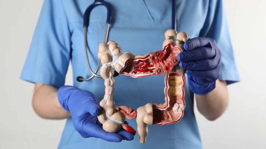
    Colon cancer, also known as colorectal cancer, is one of the most common forms of cancer. It originates in the colon or rectum, often as a growth called a polyp. Over time, these polyps can become cancerous, making it crucial to detect and treat them early. This cancer can be highly aggressive, spreading to other parts of the body if not diagnosed and treated in its early stages. Colorectal cancer (CRC) is the third most common cause of death for both men and women. It is most frequently diagnosed among persons aged 65 to 74, and approximately 10.5% of new colorectal cancer cases occur in people under 50.

    Colon Cancer Risk Factors

    The exact cause of colon cancer is not fully understood, but certain risk factors can increase your likelihood of developing the disease. They are discussed below:

    Non-modifiable risk factors of colon cancer:

    These are the risk factors you do not have control over. They are factors that you are born with, and you cannot do anything to modify them to decrease the risk.

    • Age: The rate of CRC increases with age, and nearly 94% of new cases of CRC occur in adults 45 years or older.
    • Ethnicity: Studies have shown that the rates of colorectal cancer incidence are higher in certain ethnic groups such as black, American Indian, and Alaskan natives. The reason for this higher incidence is unclear; these adults are at increased risk of developing CRC.
    • Family History: A person with a family history of colorectal cancer is at higher risk. Sadly, you do not need to have known inherited syndromes like lynch or familial adenomatous polyposis to have the heightened risk.
    • Inherited gene mutations: Rare genetic mutations, such as APC, MUTYH, or DNA mismatch repair gene mutations, can increase the risk of colon cancer.

    Modifiable risk factors of colon cancer:

    These are risk factors that you can modify through behavioral, medical, or environmental interventions. Some of the modifiable risk factors of colorectal carcinoma are:

    • Diet: A diet high in red and processed meats, as well as low in fiber, fruits, and vegetables, is associated with an increased risk of colon cancer.
    • Obesity: It has been found that excess body weight, particularly around the waist, is linked to an increased risk of colon cancer. Regular exercise and weight management can help lower this risk.
    • Physical Inactivity: A sedentary lifestyle has been shown to contribute to a higher risk of colon cancer.
    • Alcohol Consumption: Heavy alcohol consumption is associated with an elevated risk of colon cancer. It’s advisable to consume alcohol in moderation or abstain from it.
    • Smoking: Smoking is a common risk factor for numerous cancers, including colon cancer.
    • Inflammatory Bowel Disease (IBD): Conditions like Crohn’s disease and ulcerative colitis have increased the risk of colon cancer.

    Colon Cancer Symptoms

    Colorectal cancer may not present with symptoms in its early stage and can reach a point where it might be difficult to cure. It is essential to keep note of the symptoms that can help to detect these cancers in their early stage.

    1. Changes in Bowel Habits:

    • Unexplained Constipation: If you’ve noticed persistent and unexplained changes in your bowel movements, such as constipation, that won’t resolve with dietary changes or laxatives, it could be a sign of colorectal cancer.
    • Chronic Diarrhea: On the flip side, persistent diarrhea can also be a red flag, especially if it lasts several weeks or occurs with other symptoms.
    • Alterations in Stool Consistency: Keep an eye on your stool consistency. Thin or pencil-like stools, sometimes compared to “pencil-thin” in appearance, may indicate a blockage or tumor in the colon.

    2. Blood in the Stool:

    • Visible Blood: Perhaps one of the most unmistakable signs, seeing bright red blood in your stool or on toilet paper can be alarming and warrants immediate attention.
    • Dark or Tarry Stools: Blood that’s been digested may turn your stool dark or black. This can be a sign of bleeding higher up in the digestive tract.

    3. Abdominal Discomfort: Colorectal cancer can cause cramps or aches in your lower abdomen that don’t seem to go away, even with over-the-counter remedies.

    4. Unexplained Weight Loss: If you’re shedding pounds without trying, it could be due to changes in your digestive system caused by cancer.

    5. Iron-Deficiency Anemia: This occurs when your body doesn’t have enough iron, often due to chronic bleeding in the colon. Symptoms may include weakness, paleness, and shortness of breath.

    6. Feeling of Incomplete Evacuation: You may feel the need to have a bowel movement but can’t empty your bowels. This can be one of the signs of colon cancer.

    CRC( colorectal Cancer) Screening

    Colorectal cancer, when caught early, is highly treatable and often curable. Screening for colorectal cancer is a proactive step that can make all the difference in the world.

    Q. Who should be screened?

    Screening guidelines typically recommend that individuals at average risk for colorectal cancer begin screening at age 45. However, the age at which screening should begin may vary based on individual risk factors, family history, and genetics. Those with a family history of colorectal cancer or certain genetic syndromes may need to start screening earlier.

    Colon Cancer Screening Guidelines

    Screening methods

    • Fecal Occult Blood Test (FOBT): This non-invasive test checks for hidden blood in the stool, possibly indicating colorectal cancer.
    • Fecal Immunochemical Test (FIT): It is a similar test to FOBT, and FIT specifically detects blood from the lower digestive tract.
    • Colonoscopy: This is considered the gold standard for colorectal cancer screening. It involves thoroughly examining the entire colon and rectum using a long, flexible tube with a camera. Polyps can be removed during the procedure.
    • Sigmoidoscopy: This procedure examines the lower part of the colon. It is similar to colonoscopy; however, the part it examines is limited to the sigmoid colon.

    Screening frequency

    • The frequency of screening depends on the method used and individual risk factors.
    • For most people at average risk, colonoscopy is typically recommended every 10 years.
    • Stool-based tests like FOBT and FIT may be done annually.

    Colon Cancer Diagnosis

    Your doctor might employ various ways to detect and diagnose the presence of the carcinoma. Here is the list of tests that your doctor might suggest:

    1. Blood tests: Blood tests play a crucial role in the detection and monitoring of various medical conditions that you have, including colorectal cancer. It is important to remember that blood markers are not diagnostic on their own and are used as a part of a comprehensive assessment. The test includes:

    • Tumor marker: Carcinoembryonic antigen (CEA) is the most widely recognized blood marker for colorectal cancer. CEA is primarily used for monitoring disease progression, treatment response, and detecting disease recurrence. However, elevated CEA levels may indicate the presence of other cancers, including lung, breast, and pancreatic cancer, and it can also be elevated in non-cancerous conditions, such as inflammation or smoking.
    • Genetic and Molecular Markers: Advances in molecular biology have led to the identification of genetic and molecular markers, such as mutations in genes like KRAS, NRAS, and BRAF. These markers help predict treatment responses to targeted therapies and immunotherapies in colorectal cancer patients.
    • Complete Blood Count (CBC): These routine blood tests can provide information about overall health, including detecting anemia and deranged liver and kidney function, which can be affected by colorectal cancer or its treatment.

    2. Colonoscopy and biopsy: colonoscopy is a procedure that utilizes a flexible tube with a camera that offers real-time visualization of the colon and rectum, enabling the detection of precancerous polyps and tumors. If your doctor detects abnormalities, biopsy samples can be obtained during the procedure, allowing for a definitive diagnosis by examining the tissue under a microscope. These crucial steps confirm the presence of colorectal cancer, determine its type stage, and guide treatment decisions.

    3. Imaging: Various imaging techniques are employed in the context of colorectal cancer. Computed Tomography (CT) scans provide detailed cross-sectional images of the abdomen and pelvis, making them useful for staging and monitoring response to treatment. Magnetic Resonance Imaging (MRI) offers high-resolution images of the pelvis, aiding in assessing the local extent of the tumor and lymph node status. Positron Emission Tomography (PET) scans detect distant metastases and assess the metabolic activity of lesions, enhancing staging accuracy when combined with CT or MRI. Ultrasound is utilized for assessing rectal tumors, providing real-time imaging during certain procedures. Not everyone requires all the tests; your doctor will suggest the right test for you.

    Colon Cancer Treatment

    There are various treatment options available for colorectal cancer, and they depend on numerous factors, including the location of the tumor, tumor stage, overall health, and many more.

    Here is the list of treatment options available:

    1. Surgery: Surgery is the most common treatment for colon cancer at all stages, which involves removing the tumor during an operation. The nature of the cancer determines the type of surgery. It can be local excision for a limited tumor or extensive resection for a cancer that has spread.

    • Local excision: If the cancer is discovered relatively early, your doctor can remove it without penetrating the abdominal wall. Instead, the surgeon could insert a tube with a cutting instrument into the rectum and colon to remove the malignancy.
    • Resection of the colon with anastomosis: If the malignancy is more advanced, your doctor will perform a partial colectomy. The physician could then carry out anastomosis which is sewing the healthy parts of the colon together.
    • Resection of the colon with colostomy: A hole is created outside the body to allow waste to flow through if the doctor cannot stitch the two ends of the colon back together.

    2. Radiofrequency ablation: Radiofrequency ablation (RFA) is a minimally invasive medical procedure gaining prominence in treating colorectal carcinoma, particularly for certain cases of liver metastases originating from colorectal cancer. It employs high-frequency electrical currents to generate heat, effectively “cooking” and destroying cancerous tissue. RFA can be used alone or with other treatment modalities, such as surgical resection, chemotherapy, or targeted therapies.

    3. Chemotherapy: Chemotherapy is a systemic treatment strategy commonly employed in colorectal carcinoma. It utilizes drugs to target and destroy cancer cells throughout the body, making it particularly useful for advanced-stage colorectal cancer or when cancer has spread to other organs. Chemotherapy may be administered before surgery to reduce the size of tumors, post-surgery to eliminate any remaining cancer cells, or in combination with other therapies such as radiation or targeted therapies. Common chemotherapy drugs for colorectal carcinoma include 5-fluorouracil (5-FU), oxaliplatin, and irinotecan. Although chemotherapy can effectively slow cancer growth and extend survival, it may also produce side effects, such as fatigue, nausea, and lowered blood cell counts, which medical professionals closely monitor and manage.

    4. Radiation therapy: High-energy X-rays or other forms of radiation are used in radiation therapy, which helps to kill or stop cancer cells’ growth. External radiation therapy uses a device outside the body to direct radiation at the cancerous region. Internal radiation therapy uses catheters, seeds, wires, or needles with radioactive materials sealed inside that are inserted into or close to the malignancy.

    5. Targeted therapy: Targeted therapy is a form of medical care that isolates and destroys particular cancer cells using medicines or other substances. Compared to chemotherapy and radiation therapy, targeted therapies often don’t kill healthy cells as much.

    6. Immunotherapy: Immunotherapy is a cancer treatment that activates the patient’s immune system. The body’s natural defenses against cancer are boosted, directed, or restored using substances produced by the body or in a lab. Biologic therapy is a kind of treatment for cancer.

    Tips To Prevent Colon Cancer

    • Consume a fiber-rich diet
    • Limit consumption of alcohol
    • Quit smoking
    • Maintain a healthy body weight
    • Get regular colorectal screening

    Questions For Your Doctor

    1. What stage is my colon cancer, and what does that mean for my treatment options?
    2. What are the potential side effects of colon cancer treatment?
    3. How do you monitor for post-treatment colon cancer recurrence?
    4. How will my treatment impact my long-term health and quality of life?
    5. How often will I need to come in for check-ups and testing during and after treatment?
    6. Will I be able to work during my treatment and recovery?
    7. Can the treatment affect my daily life?
    8. Does the cancer get inherited by my children?

    References

    1. https://www.cdc.gov/cancer/colorectal/basic_info/prevention.
    2. https://www.cancer.org/cancer/colon-rectal-cancer/causes-risks-prevention/risk-factors.html

  • What is Multiple Myeloma? – Symptoms, Diagnosis, and Treatment

    What is Multiple Myeloma? – Symptoms, Diagnosis, and Treatment

    What Is Multiple Myeloma?

    Multiple myeloma is a blood cancer that begins in the bone marrow’s plasma cells. Normal plasma cells that produce antibodies to defend your body against infections can be found in the healthy bone marrow. In multiple myeloma, plasma cells are converted into malignant multiple myeloma cells, which grow uncontrollably and generate excessive amounts of an abnormal antibody called M protein.

    The stages of Multiple myeloma are based on four factors:

    • The body’s albumin (a type of blood protein) concentration
    • Blood beta-2-microglobulin (another blood protein) concentration
    • LDH (a cell enzyme) concentration in the blood, and
    • Cancer’s specific gene abnormalities (cytogenetics)

    Symptoms of Multiple Myeloma

    Myeloma sometimes has no symptoms. When myeloma is further advanced, symptoms might include:

    1. Spine or rib discomfort
    2. Fragile bones
    3. Fever for no apparent cause
    4. Recurrent infections
    5. Bruising or bleeding readily
    6. Breathing difficulties
    7. Arm or leg weakness and
    8. Extreme exhaustion.

    Risk factors and causes of Multiple Myeloma

    This condition primarily affects older adults, with the average age of diagnosis being 66. Other risk factors include:

    • The most significant risk for myeloma onset is age. Most patients who receive this cancer diagnosis are at least 65 years old. Rarely does this illness affect anyone under the age of 45. Those under 35 are diagnosed less than 1% of the time.
    • Men are more likely to acquire multiple myeloma than women.
    • A person is more likely to get myeloma if they have a sibling or parent with the disease.
    • Obesity and overweight are also associated with an increased risk of multiple myeloma.
    • Individuals with solitary plasmacytoma (an early-stage plasma cell malignancy) are more likely to develop multiple myeloma than people who do not have these conditions.
    • In rare instances, exposure to X-rays or other ionizing radiation may increase the chance of developing myeloma.

    Complications of Multiple Myeloma

    The most common complications brought about by multiple myeloma are mentioned below:

    1. Bone problems: Myeloma cells interfere with the bone-strengthening cells. Bones are continuously rebuilt to maintain their strength. Two types of bone cells cooperate to maintain strong and healthy bones: Osteoclasts break down old bone, while osteoblasts form new bone. A chemical produced by myeloma cells directs the osteoclasts to accelerate the breakdown of bone. As a result, old bone is degraded without being replaced by new bone, leaving the bones prone to breakage. Bone fractures are a severe issue for myeloma patients.
    2. Infections: The production of antibodies to fight against diseases is prevented because the myeloma cells crowd out the healthy plasma cells. The myeloma cells’ abnormal antibody is ineffective in preventing infections.
    3. Kidney problem: Myeloma cells produce an antibody that can deposit in and damage the kidneys, causing renal failure.

    Diagnosis of Multiple Myeloma

    Multiple myeloma can be challenging to diagnose because it often presents vague or non-specific symptoms in its early stages. Here are the key steps involved in the diagnosis of multiple myeloma.

    1. Blood test: It is one of the most important tests in diagnosing multiple myeloma, which includes various parameters that must be assessed.

    • Complete blood count (CBC): A CBC measures the number of red blood cells, white blood cells, and platelets in the blood. Multiple myeloma can cause anemia and other abnormalities in blood cell counts.
    • Blood chemistry panel: This panel includes an assessment of kidney function, calcium levels, and other metabolic markers, as multiple myeloma can affect these parameters. It can cause you to have a higher calcium level and deranged kidney function.
    • Serum immunofixation (IFE): Serum immunofixation is used further to characterize the type of M protein in the blood.

    2. Electrophoresis: Electrophoresis is a technique commonly used to diagnose multiple myeloma where an electric field is applied to a gel matrix containing the patient’s serum or urine.  Proteins migrate through the gel at different rates based on their charge and size, resulting in distinct bands or peaks on the electrophoresis graph. Specifically, two types of electrophoresis tests are often performed to assess the presence and characteristics of abnormal proteins, including monoclonal or M proteins, which are a hallmark of multiple myeloma. There are two types of electrophoresis:

    • Serum Protein Electrophoresis (SPEP): SPEP is a blood test that separates and quantifies the various proteins in the serum (the liquid portion of blood). The electrophoretic pattern obtained from SPEP can help identify the presence of a monoclonal spike or band, which is indicative of M proteins (M proteins are proteins that are produced by the cancerous plasma cells). The test provides information about the type and amount of M protein in the blood, aiding in diagnosing and monitoring multiple myeloma.

      • Urine Protein Electrophoresis (UPEP): UPEP is a urine test that separates and quantifies proteins found in the urine. It detects and quantifies M proteins that may be excreted in the urine, a condition known as Bence Jones proteinuria. UPEP is particularly useful when the M protein level in the blood is low, as it may be more readily detected in the urine.

      3. Bone marrow Aspiration and Biopsy: A bone marrow aspiration and biopsy are crucial for confirming the diagnosis of multiple myeloma. A sample of bone marrow is usually taken from the hipbone (pelvic bone) and examined under a microscope. This helps identify abnormal plasma cells and assess the extent of bone marrow involvement. A diagnosis of multiple myeloma requires a plasma cell tumor diagnosed by biopsy or at least 10% of the bone marrow to contain plasma cells.

      4. Imaging studies: X-rays, CT scans, MRI scans, or PET scans may be performed to evaluate the bones and detect any bone lesions or fractures caused by multiple myeloma. These imaging studies can also help determine the stage of the disease.

      Treatment of Multiple Myeloma

      Myeloma is difficult to cure, requiring different therapy when the disease reappears. Not everyone with myeloma needs immediate treatment; for instance, the illness may not bother anyone. If you do not require treatment, you will be monitored for any warning signals of progressive cancer. The most often-used treatment choices are listed below:

      1. Local treatment: Certain medical procedures are known as local therapies. This indicates that the tumor is treated without having an impact on the body as a whole. While they could be used in other circumstances, these therapies are more likely beneficial for tumors in an earlier stage (less advanced).

      • Surgery: While surgery is rarely used to treat multiple myeloma, it is occasionally performed to remove single plasmacytomas (tumors of plasma cells in soft tissues).
      • Radiation therapy: Radiation may be used to treat myeloma-damaged bone that is causing pain or may soon break but has not responded to chemotherapy or other medications.

      2. Systematic Treatment

      • Drug therapy: Multiple myeloma can be treated with a wide variety of medications such as Chemotherapy Corticosteroids (steroids), Immunomodulating agents, Proteasome inhibitors, Monoclonal antibodies, Nuclear export inhibitors, Bispecific T cell engager (BiTE), Intravenous immunoglobulin (IVIG), and treatment for anemia.
      • CAR T-cell Therapy: Cancer immunotherapy includes a novel therapy called chimeric antigen receptor (CAR) T-cell treatment. It helps the body’s immune system in locating and eliminating cancer cells.
      • Stem Cell Transplant: In order to destroy the bone marrow cells before a stem cell transplant, the patient receives high-dose chemotherapy. The patient is then given fresh, healthy stem cells that can produce blood. The new stem cells were derived from bone marrow in the early days of stem cell transplants. Hence, it is known as a “bone marrow transplant.”

      3. Supportive Treatments: Supportive care aims to prevent or relieve symptoms. However, it doesn’t cure cancer. Regardless of the cancer stage or the therapeutic aim, the primary objective of this type of therapy is to increase the patient’s comfort and quality of life.

      Multidisciplinary Approach to Treatment

      You must visit your doctor if you have any of the above-mentioned symptoms or concerns about your health. You might have to consult various medical professionals depending on your treatment options.

      ⇒ An orthopedic surgeon is a medical professional who performs surgery to cure bone problems.

      ⇒ A radiation oncologist is a physician who uses radiation treatment to treat cancer.

      ⇒ A medical oncologist is a physician who uses targeted therapies or chemotherapy to treat cancer.

      ⇒ A bone marrow transplant specialist is a cancer doctor performing bone marrow transplants.

      Questions For Your Doctor

      1. Has my cancer spread beyond where it started?
      2. Will I need other tests before we can decide on treatment?
      3. What is the stage of my multiple myeloma, and what does this mean for my prognosis and treatment options?
      4. What are the treatment options for multiple myeloma, and what are the benefits and potential side effects?
      5. What lifestyle changes can I make to improve my overall health?
      6. How often will I need to undergo tests and follow-up appointments to monitor my condition, and what should I expect during these visits?
      7. Are any support groups or resources available for people with multiple myeloma and their caregivers?

      References

      1. Myeloma (2022) Centers for Disease Control and Prevention. Centers for Disease Control and Prevention. Available at: https://www.cdc.gov/cancer/myeloma/index.htm (Accessed: April 1, 2023).
      2. Multiple myeloma (no date) American Cancer Society. Available at: https://www.cancer.org/cancer/multiple-myeloma.html (Accessed: April 1, 2023).
      3. Home: Multiple myeloma research foundation (2023) themmrf.org. Available at: https://themmrf.org/ (Accessed: April 2, 2023).

    1. UC Uncovered – Symptoms, Causes, and Top Treatment Solutions for Ulcerative Colitis

      UC Uncovered – Symptoms, Causes, and Top Treatment Solutions for Ulcerative Colitis

      In this comprehensive article, we will delve into the intricate world of Ulcerative Colitis (UC), a chronic inflammatory bowel disease that affects millions of people worldwide. We will explore the causes, symptoms, and effective management strategies for individuals dealing with this condition.

      What is Ulcerative Colitis?

      Ulcerative colitis is a chronic inflammatory bowel disease (IBD) characterized by ongoing inflammation and ulcers (sores) in the digestive tract. Ulcerative colitis is a common condition with a prevalence of more than 5 million cases worldwide.

      This condition falls under the umbrella of IBD alongside Crohn’s disease, but it has unique features and primarily affects the colon and rectum. The inflammation usually starts from the rectum and spreads to the entire colon.

      It has different names depending on the location it affects most.

      • Ulcerative colitis: Inflammation affecting the lower part of the colon and rectum.
      • Limited colitis: Inflammation only in a limited area of the colon
      • Pancolitis: Inflammation in the entire colon.

      What Are The Symptoms of Ulcerative Colitis?

      The symptoms of ulcerative colitis can vary widely depending on the severity of inflammation and the location. Some may experience mild, intermittent symptoms, while others may have more severe and persistent issues. Additionally, periods of remission, during which symptoms improve or disappear, can occur between flare-ups. Some of the common symptoms are:

      1. Diarrhea: Persistent and often urgent bowel movements with loose, watery stools are a hallmark symptom of ulcerative colitis. You can have a strong and sudden urge to have a bowel movement, often accompanied by an inability to control in some cases.
      2. Abdominal Pain: Crampy abdominal pain, usually in the lower left side of the abdomen, is a frequent symptom.
      3. Rectal Bleeding: If the location of inflammation is the rectum, blood in the stool or toilet paper is a common sign. The amount of bleeding can vary depending on the degree of inflammation. With ongoing bleeding for a long time, you can also have anemia, leading to fatigue and weakness.
      4. Tenesmus: It is a feeling of incomplete bowel evacuation, where you still feel the need to pass stool.
      5. Weight Loss: Unintended weight loss is another common symptom and may occur due to decreased appetite, malabsorption, and inflammation-induced metabolic changes.
      6. Joint Pain: Joint pain or swelling, known as arthralgia or arthritis, can be associated with ulcerative colitis in some cases and might be the most problematic symptom of all.
      7. Skin and Eye Problems: Inflammation from ulcerative colitis can affect the skin (rash) and eyes, causing uveitis or episcleritis in some individuals.

      What Are The Causes of Ulcerative Colitis?

      The exact cause of ulcerative colitis is unknown, and researchers believe that an overactive or dysregulated immune system is thought to play a significant role in ulcerative colitis. In a healthy immune response, the immune system defends the body against harmful invaders, such as bacteria and viruses. However, in people with ulcerative colitis, the immune system may mistakenly attack the cells lining the colon, leading to chronic inflammation.

      What Are The Complications of Ulcerative Colitis?

      Ulcerative colitis is a chronic disorder and can lead to various complications, some of which can be life-threatening. Some of the complications associated with ulcerative colitis are:

      1. Colon Cancer: Individuals with long-standing, extensive ulcerative colitis have an increased risk of developing colorectal cancer. Regular colonoscopies with biopsies are recommended for early detection and prevention.
      2. Toxic Megacolon: Inflammation in the colon can cause it to become severely dilated and lead to a life-threatening condition known as toxic megacolon. Symptoms include abdominal pain, distension, fever, and rapid heart rate.
      3. Perforation: Severe inflammation can weaken the walls of the colon, increasing the risk of perforation. This can lead to a serious abdominal infection called peritonitis.
      4. Strictures: It is the narrowing of the colon. Chronic inflammation can cause scar tissue formation and strictures in the colon, leading to bowel obstructions.
      5. Fistulas: Inflammation may create abnormal connections called fistulas between the colon and nearby organs, such as the bladder, skin, or other parts of the gastrointestinal tract.
      6. Osteoporosis: Malabsorption of nutrients and long-term use of corticosteroids can lead to reduced bone density and an increased risk of fractures.

      What Are The Risk Factors for Ulcerative Colitis?

      There are various risk factors associated with ulcerative colitis. It can affect people of any age or gender, or race, but the risk of getting it increases with the following factors:

      1. Genetic Predisposition: A family history of IBD, including ulcerative colitis, significantly increases the risk. People with close relatives with IBD are at a higher risk of developing the condition.
      2. Age: Although ulcerative colitis can develop at any age, it is most commonly diagnosed in individuals between the ages of 15 and 30 and those over 60.
      3. Ethnicity: Certain ethnic groups, such as Ashkenazi Jews, are at a higher risk of developing ulcerative colitis than others.
      4. Prior Gastrointestinal Infections: Some individuals who have experienced severe gastrointestinal infections may have a slightly increased risk of developing ulcerative colitis. This is an area of ongoing research.
      5. Non-steroidal anti-inflammatory Drugs (NSAIDs): Long-term use of NSAIDs for other conditions can also increase some individuals’ risk of developing ulcerative colitis.

      What Are The Measures to Diagnose Ulcerative Colitis?

      To diagnose ulcerative colitis, your doctors may order medical tests that include blood tests, stool tests, and imaging studies.

      1. Blood Tests: Blood tests can help assess the patient’s overall health and detect any signs of inflammation or anemia that can be associated with ulcerative colitis. Blood testing might also reveal signs of an infection or other gastrointestinal conditions that could have triggered the attack.
      2. Stool Sample Analysis: A stool sample may be collected and analyzed to rule out infections and check for blood in the stool.
      3. Imaging Studies: Imaging tests such as X-rays, computed tomography (CT) scans, or magnetic resonance imaging (MRI) may be performed to visualize the gastrointestinal tract and assess the extent of inflammation.
      4. Colonoscopy or sigmoidoscopy: These are crucial procedures for diagnosing ulcerative colitis. An endoscope is a tube with a camera at one end called an endoscope is inserted through the mouth, nose, or anus to examine the different parts of the digestive tract. A Colonoscopy is done to visualize the entire colon and rectum, whereas a sigmoidoscopy is used to view the lower part of the colon and rectum.
      5. Biopsy: Small tissue samples are often taken during colonoscopy to confirm the diagnosis by examining the tissue under a microscope. It helps to identify the area of changes in the colon lining, such as inflammation, ulcers, and abnormal cell patterns characteristic of ulcerative colitis.

      How is Ulcerative Colitis Treated?

      The management of ulcerative colitis is highly individualized, and treatment plans are tailored to each patient’s specific symptoms, disease severity, and response to therapy. It may involve a combination of approaches, and adjustments may be made over time to achieve and maintain remission.

      Medications

      Medications for ulcerative colitis can reduce colonic inflammation and promote tissue healing. It can reduce the frequency of symptom flare-ups and manage and suppress symptoms, maintaining remission.

      1. Anti-Inflammatory Medications: These are often the first line of treatment and may include aminosalicylates (e.g., sulfasalazine, mesalamine) to reduce inflammation in the colon.
      2. Corticosteroids: In cases of moderate to severe symptoms or during flare-ups, corticosteroids like prednisone may be prescribed to reduce inflammation quickly. These are typically not used for long-term treatment due to side effects.
      3. Immunomodulators: Drugs like azathioprine and 6-mercaptopurine can suppress the immune system and help maintain remission.
      4. Biologics: Biologic medications, such as anti-tumor necrosis factor (TNF) drugs like infliximab or adalimumab, target specific immune system proteins associated with ulcerative colitis and can be effective for moderate to severe cases.
      5. Janus Kinase (JAK) Inhibitors: Medications like tofacitinib may be used in certain cases to reduce inflammation by targeting specific enzymes in the immune system.
      6. Topical Medications: Suppositories or enemas containing corticosteroids or aminosalicylates may be prescribed for localized inflammation in the rectum.

      Dietary and Nutritional Management

      Maintaining a healthy diet is essential for ulcerative colitis patients. Common symptoms like diarrhea can also decrease the body’s capacity to absorb protein, fat, carbohydrates, water, vitamins, and minerals. In cases of malnutrition or nutrient deficiencies, nutritional supplementation may be necessary. While no specific diet universally treats ulcerative colitis, some individuals find relief by identifying and avoiding trigger foods.

      Surgery

      Surgery is an option if medications are not helping or you have life-threatening complications such as bleeding or to prevent/treat growths that are likely cancers. Studies have shown about 30% of people need surgery during their lifetime. The surgical procedure is:

      1. Proctocolectomy and ileoanal pouch: It is a common procedure for ulcerative colitis. However, it can require more than one surgery. In this procedure, the entire colon and rectum are removed. The surgeon then creates an internal J-pouch using a small intestine (ileum) section. The pouch is connected to the anal canal allowing for a normal bowel movement. This procedure is typically performed in two stages. In the first stage, the pouch is created, and a temporary ileostomy is constructed. In the second stage, the ileostomy is closed, allowing bowel movements to pass through the pouch and out through the anus.
      2. Ileostomy: In some cases, a temporary or permanent ileostomy may be necessary if a J-pouch or other procedures are unsuitable after proctocolectomy. An ileostomy diverts waste material directly from the small intestine to an ostomy pouch outside the body.
      3. Subtotal colectomy: This surgery involves the removal of a significant portion of the colon, leaving part of the colon intact. It may be considered when the disease is limited to a specific portion of the colon or when a complete colectomy is not necessary.

      When Do You Have To See The Doctors?

      You need to call your doctor if you experience:

      1. Blood in your stool
      2.  Ongoing diarrhea that doesn’t respond to medications
      3. Severe stomach discomfort or cramping
      4. High-grade fever

      Questions For Your Doctor

      1. What are the potential side effects of the medications used to treat ulcerative colitis?
      2. How often will I need to see you for follow-up appointments, and what should I expect during these appointments?
      3. Can I make any dietary changes to manage my symptoms?
      4. What are the potential long-term complications of ulcerative colitis?
      5. How will ulcerative colitis affect my ability to work, travel, and exercise?
      6. What can I do at home to ease my symptoms?
      7. What should I do if my symptoms worsen or if I experience new symptoms?

      References

      1. https://www.niddk.nih.gov/health-information/digestive-diseases/ulcerative-colitis/definition-facts
      2. https://my.clevelandclinic.org/health/diseases/10351-ulcerative-colitis

    2. Understanding Psoriasis – Causes, Symptoms, and Effective Treatments

      Understanding Psoriasis – Causes, Symptoms, and Effective Treatments

      Psoriasis is a common yet often misunderstood skin condition that affects millions of people worldwide. In this comprehensive article, we will delve into the intricacies of psoriasis, covering its causes, symptoms, and effective management strategies. Let’s embark on a journey to demystify this chronic skin disorder.

      What is Psoriasis?

      Psoriasis is a chronic skin condition that has affected about 3% of the US population and approximately 125 million people worldwide. It causes rashes that are itchy with scaly areas of skin, most commonly found on knees, elbows, and trunks. It is a condition that can affect any age group with a predilection for adults and has a waxing and waning nature with triggers that can be different for each person.

      What Are The Symptoms of Psoriasis?

      Psoriasis manifests with various symptoms that can vary in severity from person to person. The most common symptoms include:

      1. Appearance of thick, red, and scaly patches of skin, often covered with silvery-white scales. These patches, known as plaques, are typically itchy and may be painful.
      2. Psoriasis commonly affects the elbows, knees, scalp, lower back, and nails, but it can occur anywhere on the body.
      3. Nail changes, such as pitting or discoloration, are also common.
      4. You might also experience joint pain in your larger joints, like knees, known as psoriatic arthritis.

      What Are The Causes of Psoriasis?

      Psoriasis is considered an autoimmune disorder, where the immune system mistakenly attacks healthy skin cells. T-cells, a type of white blood cell, become overactive, leading to inflammation and causing the skin to grow and mature at an accelerated rate. It usually takes about a month for new skin cells to grow and replace the older ones. But this process happens very fast in psoriasis, and new skin cells mature within 3 to 4 days, forming thick and scaly skin.

      What Are the Types of Psoriasis?

      There are different types of psoriasis depending on their morphological pattern and location.

      1. Plaque psoriasis: It is the most common type of Psoriasis, representing approximately 80% to 90% of cases. This type manifests as elevated, reddish areas of skin that are coated in scales that are silvery white. The patches often emerge on the scalp, trunk, and limbs, notably the elbows and knees. They normally form symmetrically over the body and may cause itching and burning sensation.
      2. Guttate Psoriasis: This type is marked by small, red dots, usually on the torso or limbs, and typically affects children or young adults. This type usually appears after an upper respiratory tract illness caused by Streptococcus.
      3. Inverse Psoriasis: This type of psoriasis appears in skin folds, such as under the breasts, armpits, or the groin area. It is characterized by smooth, red, and inflamed patches of skin and is particularly prone to irritation and discomfort due to friction and sweating.
      4. Erythrodermic Psoriasis: This is a rare but severe form of psoriasis that can affect the entire body. It causes widespread redness, inflammation, and scaling of the skin. Erythrodermic psoriasis can be life-threatening and requires immediate medical attention.
      5. Pustular Psoriasis: Pustular psoriasis is marked by the formation of pus-filled blisters (pustules) surrounded by red skin. These pustules are not infectious but can be painful and may appear on localized areas or across the entire body. There are two main forms of pustular psoriasis: localized and generalized pustular psoriasis.

      Complications of Psoriasis

      • Psoriasis is commonly associated with significant medical and mental health problems such as anxiety, depression, and low self-esteem.
      • Psoriasis-related inflammation can affect the body’s other organs and tissues.
      • Psoriatic arthritis can occur in one out of every three patients with Psoriasis.
      • Psoriasis-Arthritis symptoms include swelling, stiffness, and discomfort in the joints and the tissues around them. Along with these conditions, people living with Psoriasis may also be at an increased risk for Crohn’s disease, diabetes, metabolic syndrome, obesity, osteoporosis, uveitis (inflammation of the middle of the eye), liver disease, and kidney disease.

      Triggers for Psoriasis- Explained

      Psoriasis is a complex condition, and its triggers can vary from person to person. While the exact cause of psoriasis remains unclear, several common triggers and exacerbating factors have been identified:

      1. Stress: Emotional stress is a well-recognized trigger for psoriasis flare-ups. High-stress levels can lead to an overactive immune response, exacerbating the condition.
      2. Infections: Certain infections, especially streptococcal throat infections, can trigger or worsen guttate psoriasis in some individuals.
      3. Skin Trauma: Injuries to the skin, including cuts, burns, insect bites, or friction, can trigger the development of psoriasis lesions, a phenomenon known as the Koebner response.
      4. Medications: Some medications, such as certain antimalarial drugs, beta-blockers, and lithium, can exacerbate psoriasis or induce the condition in susceptible individuals. If you have psoriasis or a family history, you must inform your healthcare provider about your condition to explore alternative medications when necessary.
      5. Alcohol and Smoking: Excessive alcohol consumption and smoking have been linked to a higher risk of psoriasis and more severe symptoms. Reducing or quitting these habits has been shown to decrease the symptoms, thus making them important triggers to avoid.
      6. Hormonal Changes: Hormonal fluctuations during puberty, pregnancy, and menopause can affect psoriasis symptoms. Interestingly, some women experience relief during pregnancy but may notice flare-ups after giving birth.
      7. Weather and Climate: Cold, dry weather conditions can worsen psoriasis symptoms, while warm, humid climates may provide relief.

      How Can a Diagnosis of Psoriasis Be Made?

      A dermatologist or healthcare provider typically diagnoses psoriasis through clinical examination and medical history assessment. Since psoriasis has a characteristic appearance that your doctor can recognize, no test is usually required to diagnose it. However, the diagnosis can sometimes be challenging and mistaken for other disorders like eczema. That is when a skin biopsy may be performed. During a skin biopsy, your doctor will remove a small sample of skin tissue and send it for examination under a microscope.

      Treatment of Psoriasis – Explained

      The treatment of psoriasis aims to alleviate symptoms, reduce inflammation, and slow down the skin cell turnover that characterizes the condition. Treatment options for psoriasis can vary depending on the type, severity, and individual patient preferences. Here are some common treatment approaches:

      Topical Treatments

      1. Corticosteroids: These anti-inflammatory creams or ointments can reduce redness, itching, and scaling. They are often used for mild to moderate psoriasis.
      2. Topical Retinoids: Retinoids are substances derived from vitamin A, and these creams can help slow skin cell growth and reduce inflammation.
      3. Calcineurin Inhibitors: If you have psoriasis on sensitive areas of the skin, such as the face and genitals, then your doctor may prescribe you topical calcineurin inhibitors like tacrolimus and pimecrolimus. These drugs help to reduce inflammation.

      Phototherapy (Light Therapy)

      1. UVB Phototherapy: You would be exposed to controlled doses of ultraviolet B (UVB) light that slows down skin cell turnover and reduces inflammation. UVB treatment can be administered in a hospital or home using a prescribed UVB lamp.
      2. Psoralen plus Ultraviolet A (PUVA) Therapy: PUVA involves taking a light-sensitizing medication (psoralen) before exposure to UVA light. It’s effective for severe psoriasis but requires careful monitoring due to potential side effects.

      Systemic Medications

      1. Oral Medications: In cases of moderate to severe psoriasis, oral medications like methotrexate, acitretin, or cyclosporine may be prescribed. These medications work by suppressing the immune system or regulating skin cell production.
      2. Biologics: Biologic drugs, such as TNF-alpha inhibitors (e.g., adalimumab, etanercept) and IL-17 inhibitors (e.g., secukinumab, ixekizumab), target specific parts of the immune system responsible for psoriasis inflammation. They are often used for severe psoriasis and psoriatic arthritis.
      3. Apremilast: This oral medication is a phosphodiesterase-4 inhibitor that helps reduce inflammation and treats moderate to severe psoriasis.
      4. Combination Therapies: Some individuals may benefit from combining treatments, such as topical creams and phototherapy or biologics, along with other medications, to achieve better symptom control.

      Lifestyle and Home Remedies for Psoriasis

      Certain lifestyle changes can complement medical treatments and reduce psoriasis flare-ups:

      1. Moisturizers: You should keep your skin well-hydrated with moisturizers, which can help reduce dryness and itching.
      2. Stress Management: Stress reduction techniques, such as meditation and yoga, is a crucial part of the treatment as they can help prevent stress-related flare-ups.
      3. Avoid Triggers: You should identify and avoid specific triggers, such as certain foods or environmental factors, that may help manage psoriasis symptoms.

      How is Psoriasis Prevented?

      Preventing psoriasis is challenging because it is a complex condition with genetic and immune system factors at play. However, some strategies may help prevent flare-ups in individuals with an existing diagnosis. Here are some of the measures:

      1. Manage Stress: You should practice stress-reduction techniques, such as meditation, yoga, deep breathing, or mindfulness, to lower stress levels, which can trigger or worsen psoriasis.
      2. Maintain a Healthy Lifestyle: You are encouraged to consume a balanced diet of fruits, vegetables, whole grains, and lean proteins. Omega-3 fatty acids in fish and flaxseeds may have anti-inflammatory benefits.
      3. Infections: You should practice good hygiene to reduce the risk of streptococcal infections, which can trigger guttate psoriasis in some individuals. In such people, it is of utmost importance that any infection in the skin must be treated to minimize the risk of exacerbating psoriasis.
      4. Smoking and Alcohol: If you smoke, consider quitting, as smoking has been linked to an increased risk of psoriasis and more severe symptoms. You should also limit alcohol consumption, as excessive drinking can worsen psoriasis.
      5. Medications and Triggers: You should discuss your medications with your healthcare provider to ensure they do not exacerbate or trigger psoriasis. In addition to medications, you also should identify and avoid personal triggers, such as stress, injuries, or specific foods, that may worsen your symptoms.
      6. Sun Protection: While controlled sun exposure can help some individuals with psoriasis, excessive sunburn can trigger flare-ups. Use sunscreen and protective clothing when spending time outdoors.
      7. Regular Checkups: If you have a family history of psoriasis or other risk factors, consider regular checkups with a dermatologist to monitor your skin health.

      When to Consult a Dermatologist?

      A visit to a dermatologist should be taken into account if:

      1. You have symptoms of Psoriasis
      2. Your quality of life is significantly impacted, and
      3. You have a severe and widespread illness

      Questions For Your Doctor

      1. Are there any lifestyle changes that could help manage my symptoms?
      2. What triggers my psoriasis flare-ups, and how can I avoid them?
      3. Can you recommend any over-the-counter treatments that may help relieve my symptoms in case of flare-ups?
      4. Are there any prescription medications that can interact with my current medicines or health conditions?
      5. How often should I come in for follow-up appointments, and what should I expect during those appointments?
      6. What should I do if my symptoms worsen or I develop new symptoms?
      7. Is there a permanent cure for this disease?

      References

      1. https://www.cdc.gov/psoriasis/
      2. https://my.clevelandclinic.org/health/diseases/6866-psoriasis
      3. https://www.mayoclinic.org/diseases-conditions/psoriasis/diagnosis-treatment/drc-20355845

    3. Unlocking the Power of Dialysis: Types, Benefits, and Your Healthcare Journey

      Unlocking the Power of Dialysis: Types, Benefits, and Your Healthcare Journey

      Dialysis is a crucial medical procedure that has transformed the lives of millions of individuals suffering from kidney disease. In this comprehensive article, we will delve into the world of renal treatment, exploring its various types, the process involved, its significance, and the impact it has on the lives of patients. Whether you’re a patient seeking information or simply curious about this life-saving treatment, this article will provide you with a deep understanding of renal treatment.

      What Is Dialysis?

      Dialysis is like a superhero when your kidneys cannot work as they should. Think of your kidneys as the body’s natural filter, removing waste and excess fluids from your blood. When they’re not working well, harmful substances can build up in your body, making you feel unwell. Renal treatment is an artificial filter, taking over the job your kidneys can’t do. It’s like having a helping hand to keep your body balanced and healthy when your kidneys need a little extra support. There are mainly two types of renal treatment. One of them is hemodialysis, and the other one is peritoneal dialysis. But which renal treatment is better for you? Remember, the ideal form of renal treatment for you depends on your preferences and health. Your doctor will assist you in making the best decision possible for you.

      Hemodialysis Explained

      Hemodialysis is a treatment where blood is cleaned using a renal treatment machine and a specific filter called an artificial kidney or a dialyzer. The cleaned blood then travels back to your body through tubes from the machine connected to your vessels.

      FAQs (Frequently Asked Questions)

      1. What needs to be done before starting hemodialysis?

      Before starting hemodialysis, you need to create a port for your blood to flow in and out of your body to the renal treatment machine. Therefore, your doctor will order a minor surgery to get vascular access through your blood vessels. The port, in simple terms, is called a fistula. These fistula are usually made on your arms.

      2. How will your blood flow from your body to the renal treatment machine?

      Initially, when you reach the center, nurses will take your vitals to ensure they have your baseline information. Then, the nurse or the renal treatment technician will insert two needles into your arm through the fistula. Your blood will flow from one needle to the renal treatment machine, and then from the machine, the other needle will flow the cleaned blood back to your body. After setting up the flow rate, the renal treatment machine monitors your blood pressure (High Blood Pressure) and regulates how rapidly your blood passes through the filter, removing bodily fluids.

      3. Can you do hemodialysis at home?

      Hemodialysis was traditionally done at the renal treatment center, but these days, it can be done in the comfort of your home. You have to train yourself and someone else for emergency situations when you cannot do it. However, you must get your doctor’s appointment monthly to ensure your kidney numbers are good. That will, additionally, allow your doctor to decide the frequency of your renal treatment.

      4. How long will you have to stay there attached to the machine?

      Usually, each hemodialysis session lasts about three to four hours and is done three times a week. If you do it at home, you might need it four to six times weekly. It can be done at any time of the day or night. However, the time you need for renal treatment and the frequency depends on various factors:

      • Your current kidney function
      • The number of times you have symptoms like shortness of breath and edema between treatments.
      • Your body habitus.

      5. Do you need to change your diet?

      When you have reached a dialysis stage due to kidney failure, you must be strict with your diet, with limitations over certain foods rich in potassium and phosphorus, like bananas, beans, avocados, and whole wheat. You must also avoid taking high sodium-containing foods like canned and processed foods. But, if you are having home dialysis and your sessions are regular, you might be less strict with your diet. However, that may not apply to all.

      6. Can you travel when you are having hemodialysis?

      There are no restrictions to travel just because you are having hemodialysis. You can have your hemodialysis at any center, so you can feel free to travel, given that the city you are visiting has a renal treatment center. If you are on home hemodialysis, there are certain machines these days that are small enough to carry. You have to ensure someone is available to help you with each session.

      7. Can you work while you are on hemodialysis?

      One can always choose to work while having renal treatment, but you have to schedule your work around your renal treatment time. There is a little bit more flexibility when it comes to work and home renal treatment.

      Peritoneal Dialysis Explained

      Peritoneal dialysis is one type of renal treatment treatment similar to hemodialysis for kidney failure. It uses a fluid that is introduced into your belly, and that helps to remove the toxin from your blood.

      What Needs To Be Done Before Starting Peritoneal Dialysis? (FAQs)

      A few weeks before beginning peritoneal renal treatment, a surgeon will insert a soft tube into your abdomen known as a catheter. This is a minor surgery where only the area where your catheter gets inserted will be numbed. This catheter will transport fluid in and out of your belly. Additionally, you will get your initial peritoneal renal treatment sessions in the center to get used to the process. After the initial stage of training, you will get to do it on your own, and once you are confident, you can start doing it at home.

      How Does A Peritoneal Dialysis Work?

      → To begin with, you will use the catheter to fill your belly with the dialysate fluid. The fluid is rich in sugar, which will help to pull the fluid and waste from your blood. The tissue called the peritoneum, which covers most of your organs, will act as a filter

      → The dialysate will stay in your belly for a certain period, lasting four to six hours. During this time, the toxins will accumulate in the fluid.

      → After four to six hours, you will have to drain the fluid and refill it with fresh dialysate. This is called exchange, which will be determined by your doctor as to how many times you are supposed to do it in each session.

      Depending on who will do the exchange process, there are two types of peritoneal dialysis:

      1. Continuous Ambulatory Peritoneal Dialysis (CAPD)

      In this type, you will use the force of gravity to do the exchange process. During this process, you will attach the catheter in your belly to the dialysate fluid in a plastic bag. The bag will be hung in a stand and kept at the level of your shoulder. Gravity will pull the liquid into your belly. While that happens, you can walk around and do all your chores, ensuring you do not pull the catheter. Once the bag is empty, it is discarded. The dialysate will stay in your belly between the exchanges.

      Usually, this is done at the beginning of the day so that the fluid can be drained at the end of the day, and you refill it one last time before you go to bed. In the morning, you will have to start the same process. In 24 hours, this procedure is often carried out three to five times, and each session lasts between 30 and 40 minutes.

      2. Automated Peritoneal Dialysis

      A device known as a cycler fills and empties your abdomen three to five times throughout an automated peritoneal dialysis treatment. You start the day with a new solution that the machine will fill in your belly. Then, you let that fluid stay there all day while you do your work. At night, you attach your catheters to the machine and program it to do three to five cycles. For this purpose, one bag of solution for each exchange is connected to the three to five bags of dialysis solution that go into the cycler.

      Tips To Prevent Peritoneal Catheter Infection

      1. Ensure hand hygiene and wash the catheter with soap and water before touching any catheter parts.
      2. Clean the area with an antibiotic solution where your catheter enters your belly.
      3. Keep your catheter dry, as leaving it moist might attract microbes.
      4. Make sure to do the exchanges in a clean and dry place.
      5. Keep your storage area clean and dry for your dialysate supplies.

      Red Flags For Peritoneal Catheter Infection: When To Call Your Doctor?

      1. Your vascular access point is bleeding.
      2. If you see redness, pus, and swelling on your vascular access site.
      3. If you have a fever.
      4. Signs of infection, such as pus, warmth, discomfort, redness, and swelling around the vascular access site.
      5. When the catheter-placed arm swells, the cold sensation spreads to the hand.

      Questions For Your Doctor

      1. What type of renal treatment do you recommend and why?
      2. What changes will I make when starting peritoneal dialysis?
      3. How often will I need to undergo renal treatment, and for how long each session last?
      4. What are the potential complications of renal treatment, and how can they be managed or prevented?
      5. How will dialysis affect my overall health and quality of life?
      6. What should I do if I experience discomfort or side effects during or after dialysis?

      References

      1. National Kidney Foundation. Available at: https://www.kidney.org/atoz/content/hemodialysis#dialysis-covid-19 (Accessed: March 19, 2023).
      2. MedlinePlus. U.S. National Library of Medicine. Available at: https://medlineplus.gov/dialysis.html (Accessed: March 19, 2023).
      3. National Kidney Foundation. Available at: https://www.kidney.org/atoz/content/peritoneal#what-dialysis (Accessed: March 19, 2023).
      4. National Institute of Diabetes and Digestive and Kidney Diseases. U.S. Department of Health and Human Services. Available at: https://www.niddk.nih.gov/health-information/kidney-disease/kidney-failure/hemodialysis (Accessed: March 19, 2023).
      5. National Institute of Diabetes and Digestive and Kidney Diseases. U.S. Department of Health and Human Services. Available at: https://www.niddk.nih.gov/health-information/kidney-disease/kidney-failure/peritoneal-dialysis#types (Accessed: March 19, 2023).
      6. U.S. National Library of Medicine. Available at: https://medlineplus.gov/ency/patientinstructions/000707.htm (Accessed: March 19, 2023).
      7. https://www.kidneyfund.org/treatments/dialysis/peritoneal-dialysis

    4. Pulmonary Embolism – Risk Factors, Symptoms, Diagnosis and Treatment

      Pulmonary Embolism – Risk Factors, Symptoms, Diagnosis and Treatment

      Pulmonary embolism can be very dangerous as it hinders blood flow to the lungs. However, quick treatment can greatly reduce the risk of death. Preventing blood clots in your legs is essential to protect yourself from a pulmonary embolism. In this article, we will explore the key risk factors of PE, with it’s symptoms, diagnosis and treatment options.

      Pulmonary Embolism – What Is It?

      pulmonary embolism (PE) happens when a blood clot blocks an artery in the lung, stopping the blood flow. Usually, the blood clot starts in a leg vein and travels to the lung. In some cases, it forms in other parts of the body. This kind of blood clot in the deep veins is called deep vein thrombosis (DVT).

      What Are The Risk Factors Of Pulmonary Embolism?

      Pulmonary embolism can affect anyone, but certain factors can increase your chances of developing blood clots. Understanding these risk factors is essential for taking proactive measures to protect your health and prevent potential complications.

      1. Inherited blood disorder: If you or any of your blood relatives, such as parents or siblings, have experienced venous blood clots or pulmonary embolisms in the past, you may be at a higher risk. Conditions like factor V Leiden deficiency, protein c, and s are common inherited conditions that increase your risk of pulmonary embolism.

      2. Medical Conditions and Treatments: Certain medical conditions and treatments can elevate your risk of developing blood clots, including:

      • Surgery: Surgical procedures are one of the primary causes of problematic blood clots. Doctors may administer blood thinner medication before and after major surgeries, such as joint replacements, to prevent clots.
      • Cancer: Certain cancers involving the brain, ovary, pancreas, colon, stomach, lung, and kidney can increase your risk of blood clots. Chemotherapy further increases this risk. Individuals with a personal or family history of breast cancer taking medications like tamoxifen or raloxifene (Evista) also have a higher risk of blood clots.
      • Coronavirus disease 2019 (COVID-19): People with severe symptoms of COVID-19 are at an increased risk of developing pulmonary embolism due to the viral infection’s impact on the body.

      3. Extended Periods of Inactivity: Long periods of inactivity can lead to blood clot formation, especially during bed rest after surgery, a heart attack, leg fracture, trauma, or a serious illness. Blood flow in the legs slows down when lying flat for extended periods, increasing the risk of clot development. Similarly, sitting for extended periods during lengthy plane or car trips can also slow blood flow in the legs, raising the risk of clots.

      4. Other Risk Factors:

      Several other risk factors can contribute to the likelihood of developing blood clots.

      • For reasons not entirely understood, smoking can increase the risk of clots, especially when combined with other risk factors.
      • Being overweight has also been shown to play a role, particularly in individuals with other risk factors.
      • The use of estrogen-based birth control pills or hormone replacement therapy can increase clotting factors in the blood, especially in those who smoke or are overweight.
      • Pregnancy can lead to blood pooling in the legs, increasing the chances of clot formation.

      How DVT Leads To Pulmonary Embolism?

      DVT can lead to pulmonary Embolism through a process called embolization. Embolization occurs when a blood clot, known as an embolus, breaks loose from its original location and travels through the bloodstream to another part of the body, where it becomes lodged and obstructs blood flow.

      Here’s a simple way to understand how DVT can lead to PE:

      • Formation of DVT: DVT occurs when a blood clot forms within a deep vein, usually in the leg. The clot partially or completely obstructs the vein, causing blood flow to slow down or stop in that area.
      • Embolus Formation: If the DVT remains undetected or untreated, there is a risk that the blood clot can become dislodged from the vein wall.
      • Embolus Travel: Once dislodged, the blood clot, now called an embolus, can travel through the bloodstream. It moves towards the heart through the large veins in the body.
      • Lodging in the Lung: The embolus eventually reaches the right side of the heart and then enters the pulmonary artery, the blood vessel that carries blood from the heart to the lungs. If the embolus is large enough, it can get stuck in one of the smaller arteries within the lungs, blocking blood flow to that area.
      • Pulmonary Embolism: When the embolus blocks blood flow to a portion of the lung, that is what we call a pulmonary embolism.

      What Are The Symptoms Of Pulmonary Embolism?

      The symptoms of pulmonary embolism can vary depending on how much of the lung is affected, the size of the blood clots, and if there are any existing lung or heart conditions. Here are the symptoms one might experience if they have a pulmonary embolism:

      • Sudden onset Shortness of Breath: You may experience sudden and severe shortness of breath, even at rest or after minimal exertion.
      • Unexplained Chest Pain: You might feel sharp and intense pain in the chest is a common symptom of PE, and it may resemble the symptoms of a heart attack. The pain may worsen while breathing deeply, coughing, bending, or leaning over.
      • Coughing with or without Bloody Mucus: Some people with PE may experience a persistent cough that may produce blood-tinged or bloody mucus.
      • Pale or Bluish Skin: The skin may appear pale, clammy, or bluish due to reduced blood oxygen levels. This can be a frightening experience for many.
      • Rapid Heartbeat: When the artery in the lungs is blocked, your heart gets affected. It will have to work against that clot to pump blood to the lungs, thus leading to an increased heart rate.
      • Feeling Anxious, Lightheaded, or Fainting: In some cases, individuals with PE may experience anxiety, dizziness, or fainting due to reduced blood flow and oxygen supply to the brain.
      • Wheezing: Wheezing or abnormal breath sounds may be heard, in patients with PE, indicating potential airway obstruction.

      How Is A Diagnosis Of Pulmonary Embolism Made?

      Diagnosing a pulmonary embolism can be challenging, especially if underlying heart or lung issues exist. Here is the list of tests your doctor will use to diagnose pulmonary embolism:

      • CT Pulmonary Angiography (CTPA): CTPA is a highly effective diagnostic imaging test to detect pulmonary embolism. During a CTPA, X-rays create detailed cross-sectional images of the blood vessels in the lungs, providing valuable information about blood clots’ presence, location, and size. The procedure involves injecting a contrast dye into a vein to enhance the visualization of the pulmonary arteries. As the dye circulates through the lungs, it highlights any areas of blood flow obstruction caused by clots. CTPA is considered the gold standard for diagnosing PE because it produces high-resolution 3D images, allowing healthcare professionals to identify and assess the condition’s extent accurately.
      • Blood Tests: The D dimer test is one of the most commonly used blood tests to check for the presence of a blood clot. It measures the clot-dissolving substance in the blood. Elevated d dimer levels may indicate an increased likelihood of blood clots, but other factors can also affect them, especially infections.
      • Chest X-ray: While a chest X-ray cannot directly diagnose PE, it is useful for ruling out other conditions with similar symptoms. It provides images of the heart and lungs, which may appear normal or show signs of other lung or heart conditions.
      • Ultrasound (Duplex Ultrasonography): This is a noninvasive test that uses sound waves to scan veins in the thigh, knee, calf, and arms for deep vein blood clots. The absence of clots reduces the likelihood of DVT, a common cause of PE.
      • Ventilation-Perfusion (V/Q) Scan: In conditions when avoiding radiation or contrast material is necessary due to a medical condition, a V/Q scan can be used. During this test, a small quantity of a radioactive substance known as a tracer is injected into a vein in the arm. The tracer maps the blood flow (perfusion) and compares it with the airflow (ventilation) in the lungs. This test is helpful in identifying whether blood clots are responsible for symptoms related to pulmonary hypertension.
      • Pulmonary Angiogram: This test provides a clear picture of blood flow in the lungs’ arteries and is the most accurate method for diagnosing PE. It involves inserting a catheter into a large vein, guiding it to the pulmonary arteries directly, and injecting a special dye to visualize the blood vessels through X-rays. Pulmonary angiograms are reserved for cases where other tests are inconclusive due to the procedure’s complexity and potential risks.
      • MRI (Magnetic Resonance Imaging) of the lungs: MRI is used in specific cases, such as pregnant individuals, to avoid exposing the baby to radiation or in people with kidney concerns about contrast dye used in other tests. It creates detailed images of organs and tissues using a magnetic field and radio waves, helping diagnose underlying conditions or complications.

      How Is Pulmonary Embolism Treated?

      If you have a pulmonary embolism, you require immediate medical attention. Breaking up clots and preventing the formation of new ones are the two main objectives of treatment. Both medications and surgeries are available as treatments.

      Medications

      • Anticoagulants: These are the drugs often known as blood thinners, preventing blood clots from becoming bigger and creating new clots. You might know them as heparin, Lovenox, coumadin, Eliquis, or Xarelto. They may be administered intravenously, as a tablet, or as an injection. The most important thing to remember is these drugs can result in bleeding, mainly if you take aspirin or other medications that thin the blood. Your doctor will discuss with you the risks vs. benefits of being anticoagulation and the risks of bleeding
      • Thrombolytics: These medications are clot busters used to dissolve blood clots. They can be prescribed if you have big clots that result in painful symptoms or other critical consequences. Thrombolytics are given when your pulmonary embolism is severe and possibly life-threatening since they can result in life-threatening bleeding. They help to reduce the embolic load quickly and enhance cardiorespiratory blood circulation. However, bleeding is a side effect of thrombolytic therapy that can be fatal, so these drugs are prescribed only after assessing your comorbidities.

      Surgery

      Surgical management of pulmonary embolism is typically reserved for severe cases or situations where other treatment options are ineffective or contraindicated.

      • Catheter-assisted thrombus removal: This minimally invasive procedure involves inserting a catheter through a blood vessel, usually in the groin, and guiding it to the site of the blood clot in the lungs. Using specialized tools attached to the catheter, the clot is carefully broken down or suctioned out, restoring blood flow to the affected lung tissue.
      • Vena cava filter: A small, cone-shaped medical device inserted into the vena cava, the large vein that carries deoxygenated blood from the lower body to the heart. The primary purpose of this filter is to prevent blood clots from traveling to the lungs and causing a potentially life-threatening condition known as pulmonary embolism. Vena cava filters are typically recommended for individuals at high risk of developing blood clots but cannot tolerate or respond to standard blood-thinning medications. The filter works by trapping large blood clots that may form in the lower extremities, preventing them from reaching the lungs. A vena cava filter is inserted through a minimally invasive procedure, usually using a catheter to place the filter into the vena cava. Over time, the filter may be removed if the risk of blood clots subsides or if there are concerns about potential long-term complications. While vena cava filters can be life-saving for some patients, their use is carefully considered and individualized based on each patient’s medical condition and risk factors.

      What Are The Complications Of Pulmonary Embolism?

      Pulmonary embolism poses a significant risk to life. Approximately one-third of individuals with an undetected and untreated pulmonary embolism do not survive. Yet, the mortality rate decreases significantly when the condition is identified and managed promptly. The most common complications of pulmonary embolism are:

      • Pulmonary Hypertension: When blood clots obstruct the pulmonary arteries for an extended period, it can lead to increased lung pressure, causing pulmonary hypertension. This condition increases the work that your heart has to do and can lead to heart failure if left untreated.
      • Right Ventricular Dysfunction: Severe pulmonary embolism can impair the function of the right ventricle, the heart chamber responsible for pumping blood to the lungs. This dysfunction can cause a decrease in the output of your heart and potentially lead to shock.
      • Pulmonary Infarction: Large blood clots may completely block blood flow to a portion of the lung, causing lung tissue to become deprived of oxygen. This can result in pulmonary infarction, leading to localized lung tissue damage.
      • Pleural Effusion: Pulmonary embolism may cause inflammation in the lining of the lungs (pleura), accumulating fluid in the pleural space, known as pleural effusion. This can, in turn, cause chest pain and difficulty breathing.
      • Recurrent PE: There is always a chance that you have it again, especially if the underlying risk factors are not adequately managed. Most commonly, recurrent PE is seen in people with inherited conditions that went undiagnosed.
      • Hemoptysis: In some instances, pulmonary embolism can cause lung bleeding, leading to coughing up blood, a condition known as hemoptysis.

      What Are The Measures To Prevent Pulmonary Embolism?

      Preventing blood clots in the deep veins of the legs is essential for avoiding pulmonary embolism. You can take the following measures to prevent pulmonary embolism:

      • Blood Thinners: These medicines are administered before and after surgery, as well as to hospitalized individuals with certain medical conditions like heart attack, stroke, or cancer complications. Those who cannot mobilize immediately must be careful and take their blood thinners on time.
      • Early mobilization: Early movement after surgery can prevent pulmonary embolism and expedite overall recovery. Thus, motivate yourself to move after your surgery.
      • Leg Elevation: Elevating the legs, particularly during rest and at night, can effectively reduce clot risk. Raising the bottom of the bed four to six inches with blocks or books can also be helpful.
      • Pneumatic Compression Device: These are the devices that are placed on the thighs or calf, They have a mechanism to automatically inflate and deflate that massages and squeezes leg veins, improving blood flow from the leg to the heart and reducing the risk of formation of DVT.

      How To Prevent Blood Clots During Travel?

      It is essential to make sure that you follow this advice for your next travel to ensure that you do not have a pulmonary embolism:

      • Staying Hydrated: Drink plenty of water to prevent dehydration, a risk factor for blood clots. Avoid alcoholic beverages, which contribute to fluid loss and the formation of blood clots in a predisposed individual.
      • Frequent Movements: Take breaks during long-haul travel, walk around the airplane cabin, or perform knee bends if driving.
      • Seat Exercises: Make ankle movements and raise toes up and down every 15 to 30 minutes while seated.
      • Support Stockings: Compression stockings can be worn to promote leg circulation and fluid movement, reducing clot risk during travel.

      Questions For Your Doctor

      Here are some questions that you may consider asking your doctors:

      1. What could have caused my pulmonary embolism?
      2. Is the clot large or small, and how severe is my condition?
      3. How long will I need to take blood thinners or other medications?
      4. Are there any lifestyle changes I should make to reduce the risk of future blood clots?
      5. Can I continue my regular activities and exercise routines while undergoing treatment?
      6. What should I do if I experience new or concerning symptoms during treatment?
      7. Are there any dietary considerations I should consider while on blood thinners?
      8. Is it safe for me to travel or fly, considering my condition?
      9. Are there any alternative treatment options or complementary therapies that may help support my recovery?

      References

      1. https://my.clevelandclinic.org/health/diseases/17400-pulmonary-embolism#symptoms-and-causes
      2. https://www.mayoclinic.org/diseases-conditions/pulmonary-embolism/symptoms-causes/syc-20354647
      3. https://medlineplus.gov/pulmonaryembolism.html

    5. Aortic Stenosis – Symptoms, Treatment, Diagnosis and Prevention

      Aortic Stenosis – Symptoms, Treatment, Diagnosis and Prevention

      What is Aortic Stenosis?

      Aortic stenosis is a heart condition that affects the aortic valve, one of the four valves in your heart. Aortic stenosis occurs when the aortic valve narrows, reducing blood flow from the heart into the aorta, the main blood vessel that carries oxygenated blood to the rest of the body.

      Symptoms Of Aortic Stenosis

      Symptoms of aortic valve stenosis range from mild to severe. Most people with aortic valve stenosis would not experience any major symptoms for many years. Symptoms generally occur when the narrowing of the valve is severe:

      • Chest pain

      This is often felt during physical activity or when the heart has to work harder. This pain is often termed as angina.

      • Shortness of breath

      You can have shortness of breath, especially during exercise or when lying flat.

      • Dizziness

      You might feel lightheaded or dizzy due to reduced blood flow to the brain through the narrowed lumen of the valve. You might even have a syncope if it is too narrow.

      • Fatigue

      As the condition progresses, you can tire easily, usually much faster. You might not be able to conduct the regular activities you used to do.

      • Poor growth

      Children with congenital aortic stenosis can have poor growth. They can be low in weight and height about their age.

      • Heart failure

      When the disease progresses to very advanced stages, patients can present with heart failure and have worsening symptoms.

      Risk Factors of Aortic Stenosis

      Several factors can increase the risk of developing aortic stenosis, which often develops gradually over time. Some of them are discussed below:

      • Age

      As we age, the risk of aortic stenosis increases. The valve may naturally thicken and become less flexible over time.

      • Congenital heart conditions

      Some people are born with abnormal aortic valves that may eventually lead to stenosis. The bicuspid aortic valve is the most common congenital heart defect affecting 1% of the general population and is one of the leading causes of stenosis in the young population.

      • Aortic valve calcification

      Calcium is a mineral present in the blood. As blood flows through the valves, calcium can deposit on the heart valves, and the process gets hastened if the valve is highly pressured, like the aortic, and has microtrauma.

      • High cholesterol

      High cholesterol levels can lead to the buildup of fatty deposits on the aortic valve, narrowing it.

      Uncontrolled hypertension can strain the aortic valve and contribute to its deterioration.

      • Rheumatic fever

      Rheumatic fever is one of the most common conditions seen among children. A previous bout of rheumatic fever can damage the aortic valve, increasing the risk of stenosis.

      Treatment Of Aortic Stenosis

      Treatment options for aortic stenosis depend on the symptoms and the severity of the condition. While you might need a regular follow-up with your doctor if there is mild aortic valve disease or no symptoms, you may need surgery to repair or replace the valve for severe disease. There are less invasive options available, too, but that depends on your overall health and your choice.

      The treatment options for aortic valve stenosis are:

      • Balloon valvuloplasty

      This minimally invasive procedure is used in infants and children. The procedure is limited in adults, as the valve gets narrower afterward. So, it is usually only done if you are young or waiting for a valve replacement. In this procedure, a tube with a balloon tip is inserted into an artery through the arm or the groin. It is then taken to the aortic valve. Once it reaches the valve, the balloon is inflated, which widens the valve opening. The balloon is then deflated, and the catheter is withdrawn.

      • Aortic valve replacement

      It is the ultimate treatment for aortic stenosis. The damaged valve is replaced with a mechanical valve made from cows, pigs, or human tissue. While biological valves break down over time and need to be replaced again, mechanical valves remain longer than the biological valve, but the patient will have to take a blood thinning agent.

      The valve can be replaced by either of the following two procedures:

      • Surgical aortic valve replacement (SAVR):

      Aortic valve replacement is done via an open-heart surgical procedure. During an SAVR, you would be put under general anesthesia and placed on a ventilator while on a heart-lung bypass machine. A surgeon would then cut open your breastbone to get to the heart. In some patients, less invasive approaches can be used. The aortic stenosis valve is cut out and replaced by the new valve. Recovery in the hospital usually takes about five days. This procedure can use both bioprosthetic as well as mechanical valves.

      • Transcatheter aortic valve replacement (TAVR):

      TAVR is an alternative to SAVR and is done for those not candidates for open-heart surgery due to other comorbid conditions. This minimally invasive procedure requires no sternotomy or a heart-lung bypass machine. The replacement valve is usually inserted using a catheter into an artery in the leg, then threaded through arteries to the heart. Once within the diseased aortic valve, the folded TAVR valve is opened with a balloon and placed in the right place. Some valves can, however, self-expand and do not need re-positioning. The surgeon then removes the catheter once the new valve is securely placed. It is to be noted that this procedure uses only bioprosthetic valves only.

      Diagnostic Evaluation Of Aortic Stenosis

      Several diagnostic tests are performed to confirm and determine the cause and its severity. Tests for aortic valve stenosis may include:

      • Electrocardiogram (EKG)

      It is one of the routine tests performed that measures the electrical activity of the heart. It can show how fast or slow your heart is beating. Your doctor will look for the strain pattern in the EKG if you have aortic stenosis.

      • Echocardiogram (ECHO)

      In simple terms, it is an ultrasound of your heart, and it creates images of the heart. It is a fast and painless investigation that helps to evaluate the structure and function of the heart. It can recognize a weakened heart muscle, changes in the valves of the heart, and look at how the blood flows through the valves. With an ECHO, your doctor can determine the severity of aortic valve stenosis.

      • Chest X-ray

      Chest X-ray gives you a view of the heart and lungs together. It helps to determine if there is an enlarged heart, a common finding of aortic stenosis. It can also show an enlarged aorta and calcium build-up.

      • Cardiac computed tomography(CT)

      This test utilizes multiple X-rays to obtain detailed heart pictures. It is also utilized to evaluate the size of the aorta and calcium deposits on the aortic valves and to guide surgical management of the condition.

      • Cardiac catheterization

      This is a test where a catheter is guided from a vessel in the wrist or groin leading to the heart, and a contrast dye is injected through it. The dye goes to the arteries of the heart and helps to assess their patency. The test is not used to diagnose aortic valve disease but can be used if other tests are not able to diagnose the condition. A coronary angiogram can be used to identify patients who also might benefit from surgery on the blood vessels around the heart along with an aortic valve replacement procedure. Therefore, every patient with aortic stenosis should undergo a coronary angiogram to detect and treat any coexisting coronary vessel disease.

      Complications Of Aortic Stenosis

      While aortic stenosis can initially be asymptomatic, it can lead to several complications as the condition progresses. Watch out for these complications:

      • Reduced exercise tolerance

      As the condition progresses, reduced blood flow can lead to decreased exercise tolerance, fatigue, and a decline in overall physical capacity.

      • Heart failure

      As the aortic valve narrows further, the heart must work harder to pump blood through the narrowed valve. Over time, this extra strain on the heart can lead to left ventricular hypertrophy and, eventually, heart failure.

      • Arrhythmias

      Aortic stenosis can increase the risk of abnormal heart rhythms or arrhythmias. The irregular heartbeats can further compromise the heart’s ability to pump blood effectively.

      • Infection (Infective Endocarditis)

      Aortic stenosis may make the heart valve more susceptible to infection. Bacterial or microbial infections of the heart valve can lead to a condition called infective endocarditis, which is a serious and potentially life-threatening complication.

      Prevention of Aortic Stenosis

      You can take steps to strengthen your heart and guard against developing aortic stenosis. These include:

      • Make heart-healthy choices 

      Healthy choices such as daily physical exercise and quitting smoking can prevent calcium buildup in arteries and aortic valve. If you have high blood pressure, high cholesterol, diabetes, or kidney problems, you should consult your doctor regarding adequate management of these chronic health conditions.

      • Keep up with dental checkups

      Gum infections can lead to infection and development of aortic valve stenosis. A regular dental checkup can avoid aortic valve disease secondary to gum infections.

      • Treat infections

      The bacteria that cause strep throat can sometimes cause rheumatic fever if left untreated. Rheumatic fever often happens in children and young adults. This is a condition that can be easily treated with antibiotics. So, getting treated for strep throat is important to prevent valvular heart disease.

      Question For Your Doctor

      1. What symptoms of Aortic Stenosis should prompt me to seek immediate medical attention?
      2. Should I limit my physical activity because of Aortic Stenosis?
      3. How often should I have my heart checked yearly?
      4. What are the treatment options?
      5. Are there any medications that I can take for this condition?
      6. How do I know if surgical or Transcatheter Aortic Valve Replacement is a better treatment option for me?
      7. What changes happen to the heart after having aortic stenosis over time?

      References

    6. Sickle Cell Disease – Symptoms and Treatments Demystified

      Sickle Cell Disease – Symptoms and Treatments Demystified

      What is Sickle Cell?- Understanding The Disease

      Sickle cell disease (SCD) comprises a collection of hereditary blood cell disorders impacting hemoglobin, the protein responsible for transporting oxygen throughout the body. This condition affects over 100,000 individuals in the United States and around 20 million worldwide. In healthy individuals, red blood cells possess a disc-like shape and flexibility, enabling smooth movement through blood vessels. However, SCD causes the red blood cells to adopt a crescent or “sickle” shape. These abnormal cells lose their ability to bend and move easily, leading to potential blockages in blood flow to various parts of the body.

      Sickle Cell Disease and Its Risk Factors

      SCD predominantly affects individuals of African ancestry or those identifying as Black in the United States. Around 1 in 13 Black or African American babies are born with sickle cell trait, while approximately 1 in every 365 African American babies are born with SCD. Moreover, the condition also affects individuals from Hispanic, Southern European, Middle Eastern, or Asian Indian backgrounds.

      Sickle Cell Disease Symptoms

      SCD is a genetic condition that you’re born with, but its effects might not appear until around 5 or 6 months. SCD symptoms can vary from person to person and change over time. Some of the most common signs that are seen in SCD are:

      1. Pain Crisis (Sickle Cell Crisis): This is one of the hallmark symptoms of SCD. Sickle cells can block blood flow through small blood vessels, causing episodes of severe pain. These episodes are also called the pain crises. These crises can occur anywhere in the body but often affect the bones, joints, chest, and abdomen.
      2. Anemia: The abnormal shape of sickle cells makes them fragile and prone to breaking apart, leading to a shortage of red blood cells. This can result in anemia, characterized by fatigue, weakness, and paleness.
      3. Jaundice: Sickle cells have a shorter lifespan than normal red blood cells, leading to a buildup of bilirubin in the blood. This can cause a yellowish tint to the skin and eyes, a condition known as jaundice.
      4. Infections: Sickle cells can impair the immune system’s ability to fight off infections, making individuals with SCD more susceptible to bacterial infections, particularly those caused by certain types of bacteria, such as Streptococcus pneumoniae.
      5. Organ Damage: The repeated episodes of blocked blood flow can damage various organs, including the spleen, kidneys, liver, and lungs. This can lead to organ dysfunction and long-term complications.
      6. Stroke: Sickle cells can also block blood vessels leading to the brain, causing a stroke. Symptoms may include sudden weakness, numbness, confusion, and difficulty speaking or walking.
      7. Acute Chest Syndrome: This is a serious condition that resembles pneumonia. It occurs when sickle cells block the blood vessels in the lungs, causing chest pain, cough, fever, and difficulty breathing.
      8. Priapism: It is a condition where individuals experience a prolonged and painful erection that can last for more than 4 hours. It happens due to blocked blood vessels in the penis and requires immediate management.
      9. Eye Problems: SCD can affect the blood vessels in the eyes, potentially leading to vision problems and even blindness.

      Diagnosis of sickle cell disease

      1. Blood Tests: Blood tests play a crucial role in diagnosing SCD. These tests include:

      • Complete Blood Count (CBC): This measures the number of red blood cells, white blood cells, and platelets in your blood. People with SCD often have fewer red blood cells and higher levels of reticulocytes (young red blood cells).Hemoglobin
      • Electrophoresis: This test identifies the types of hemoglobin present in your blood. In SCD, there is a higher hemoglobin S (HbS) level, responsible for the characteristic sickling of red blood cells, which can be seen with the help of electrophoresis.

      2. Sickling Test: In this test, a sample of your blood is exposed to low oxygen levels to observe how your red blood cells behave. In SCD, the cells will take on a characteristic sickle shape under these conditions.

      3. Genetic Testing: Genetic tests can confirm the presence of abnormal hemoglobin genes that cause SCD. This is particularly important for carrier screening and prenatal testing. Hemoglobin gene mutations are identified through DNA analysis.

      4. Newborn Screening: In many countries, including the United States, newborns are screened for SCD in routine newborn screening programs. This helps identify babies with SCD early on so that appropriate care can be provided. During a newborn screening, drops of blood from a hill prick are collected on a special type of paper. The screening helps to determine whether your baby has a sickle cell trait or is a carrier.

      Treatments of Sickle Cell Disease

      The treatments of SCD aim to manage symptoms, prevent complications, and improve the quality of life for individuals with the condition. Treatment approaches can vary based on the severity of the disease and the specific symptoms experienced. Here’s an overview of treatment strategies for SCD:

      1. Pain Management

      • Pain is a common symptom of SCD, often resulting from blocked blood vessels and tissue damage. Pain crises can be managed with pain medications, both over-the-counter and prescription medications.
      • Hydration and warm baths can also help alleviate pain during a crisis.
      • For severe pain, hospitalization and stronger pain medications may be necessary.

      2. Blood Transfusions

      • Regular blood transfusions may be recommended for individuals with severe anemia, acute chest syndrome, or other complications.
      • Transfusions help increase the number of healthy red blood cells in circulation.

      3. Hydration

      You need to stay well-hydrated to prevent the formation of sickle cells and reduce the risk of pain crises.

      FDA-Approved Treatment

      • Hydroxyurea: Hydroxyurea is a well-established medication for SCD. By increasing the production of fetal hemoglobin (HbF), it helps inhibit the sickling of red blood cells and prolong their lifespan. This can reduce pain crises, episodes of acute chest syndrome, and the need for blood transfusions. While hydroxyurea offers substantial benefits for many individuals with SCD, its usage requires careful monitoring due to potential side effects such as changes in blood counts and liver function.
      • L-Glutamine (Endari): L-Glutamine is an oral medication approved for treating SCD. This amino acid enhances antioxidant levels in red blood cells, decreasing oxidative stress and improving their function. L-glutamine can help mitigate the frequency of pain crises and hospitalizations, particularly in children and adults with SCD. Its unique formulation as a powder mixed with a liquid makes it convenient for daily use.
      • Voxelotor (Oxbryta): Voxelotor is an oral medication that offers a promising approach to managing SCD. By increasing the oxygen affinity of hemoglobin, it prevents the sickling of red blood cells and reduces hemolysis. This results in improved anemia and a decrease in pain crises. Voxelotor is approved for individuals aged 12 and older with SCD.

      4. Crizanlizumab (Adakveo)

      Crizanlizumab is a monoclonal antibody infusion designed to prevent vaso-occlusive crises in SCD. By blocking the interaction of sickle cells with blood vessel walls, it reduces the frequency and severity of painful episodes. Administered through intravenous infusions, crizanlizumab has shown promise in improving the quality of life for people with SCD.

      5. Bone marrow or stem cell transplantation

      Bone marrow also known as hematopoietic stem cell transplantation (HSCT), presents a potential curative avenue for SCD. This procedure involves substituting the patient’s malfunctioning bone marrow with healthy donor marrow or stem cells to produce normal red blood cells. Though promising, HSCT is complex and involves risks, including graft-versus-host disease, infection, and organ damage. It is generally reserved for individuals with severe SCD and high-risk complications. Many people who have the disease do not have a relative who is a close match to be a donor. HSCT necessitates meticulous patient selection, close monitoring, and specialized post-transplant care.

      Prevention of Sickle Cell Disease

      Preventive measures for SCD aim to minimize complications, improve quality of life, and enhance overall health. These strategies focus on managing symptoms, reducing the frequency of pain crises, and preventing infections. Key preventive measures include:

      Lifestyle behavior

      • Drink a lot of water; it helps prevent sickle cell formation.
      • Avoid extremes of temperature, as very cold and hot temperatures can trigger pain crises.

      Interventions & Medical Screenings to Reduce SCD Complications

      • Vaccines: You or your child with SCD should get all the vaccinations as scheduled, particularly against infections like influenza, pneumonia, and meningococcal disease. Vaccination will help to prevent the disease that can trigger SCD complications.
      • Penicillin prophylaxis: Children with SCD need to be given daily doses of penicillin or other antibiotics that help to reduce the risk of bacterial infection in the early days of their growth and development.
      • Eye examination: It is recommended that individuals with SCD should receive annual eye examinations to check for possible retinal damage, which is a common complication of SCD.
      • Transcranial Doppler ultrasonography (TCD), a specialized type of examination, can be used to detect children at risk for stroke.
      • To prevent severe anemia, the patient may be treated with blood transfusions.

      When To See Your Doctor?

      It is crucial to recognize when immediate medical attention is needed, as SCD can sometimes lead to severe and life-threatening health issues. If you notice the following symptoms or complications, it’s essential to seek medical help or call 9-1-1 without delay:

      • Severe Anemia Symptoms: These include extreme tiredness, shortness of breath, dizziness, or irregular heartbeat. Conditions like splenic sequestration or aplastic crises can lead to severe anemia, posing serious risks.
      • Fever: If someone with SCD has a fever above 101.3°F (38.5°C), seeing a healthcare provider promptly is critical. Antibiotics may be necessary, and hospitalization might be required in some cases.
      • Acute Chest Syndrome Symptoms: Symptoms encompass chest pain, coughing, fever, and difficulty breathing. Admission to a hospital becomes necessary, where treatments like antibiotics, oxygen therapy, or blood transfusions may be administered.
      • Stroke Warning Signs: These include sudden weakness, numbness on one side of the body, confusion, and trouble with speaking, seeing, or walking. These signs would indicate a stroke requiring immediate medical intervention.
      • Priapism: In cases where an erection lasts for more than 4 hours, it’s advised to visit a hospital. Consultation with a hematologist and a urologist is crucial, as they specialize in blood conditions and male reproductive/urinary system issues, respectively

      Questions For Your Doctor

      1. What type of sickle cell disease do I have?
      2. What are the common symptoms and complications associated with my type of SCD?
      3. Is there anything I can do at home to improve my health?
      4. Are there specific triggers I should be aware of?
      5. When should I go to the emergency room?
      6. Can my children get this from me?
      7. What are my family planning options?

      References

      1. Sickle cell disease | sickle cell anemia (no date) MedlinePlus. Available at: https://medlineplus.gov/sicklecelldisease.html
      2. (What is sickle cell disease? (2023) Centers for Disease Control and Prevention. Available at: https://www.cdc.gov/ncbddd/sicklecell/facts.html
      3. https://www.nhlbi.nih.gov/health/sickle-cell-disease/treatment

    7. Heart Health – Understanding and Managing Coronary Artery Disease

      Heart Health – Understanding and Managing Coronary Artery Disease

      What is coronary artery disease (CAD)?

      Coronary Artery Disease (CAD) is a prevalent and potentially life-threatening cardiovascular condition affecting millions worldwide. It is the leading cause of heart-related morbidity and mortality in the US, imposing a significant burden on public health systems and individuals alike. CAD develops when the coronary arteries, the blood vessels responsible for supplying oxygen and nutrients to the heart muscle, become narrowed or blocked due to atherosclerosis, characterized by the accumulation of fatty deposits and plaque on the arterial walls.

      As the disease progresses, it hinders blood flow to the heart, depriving the cardiac muscle of the necessary oxygen and nutrients required for proper functioning. This can lead to various symptoms, such as chest pain (angina), shortness of breath, and fatigue. In severe cases, CAD can result in a heart attack, where a sudden and complete coronary artery blockage leads to the death of a portion of the heart muscle.

      Forms of coronary artery disease

      Coronary artery disease can present in various forms, each with its distinct characteristics and implications for heart health. It is essential to recognize and diagnose the specific form of CAD in each individual, as treatment strategies and management approaches may vary.

      The different forms of coronary artery disease include:

      • Stable Angina: Stable angina is the most common form of CAD. It occurs when there is a partial blockage or narrowing of the coronary arteries, leading to chest pain or discomfort during physical exertion or emotional stress. The pain is predictable and tends to subside with rest or medication.
      • Unstable Angina: Unstable angina is a more severe and unpredictable form of CAD. It happens when a plaque in the coronary artery ruptures, causing a sudden and severe reduction in blood flow. Unstable angina can occur at rest or with minimal exertion and requires immediate medical attention, as it may lead to a heart attack.
      • Acute Myocardial Infarction (Heart Attack): A heart attack occurs when a coronary artery becomes completely blocked by a blood clot, cutting off blood flow to a portion of the heart muscle. This leads to the death of heart tissue. Heart attacks are medical emergencies that require urgent treatment to restore blood flow and prevent further damage.
      • Chronic Total Occlusion (CTO): CTO refers to a complete blockage of a coronary artery that persists for an extended period, often over three months. People with CTO may experience chronic chest pain, reduced exercise tolerance, and, in severe cases, heart failure.
      • Silent Ischemia: Some individuals with coronary artery disease may not experience typical symptoms like chest pain or discomfort. Instead, they may have “silent ischemia,” where the heart muscle does not receive enough blood and oxygen without noticeable symptoms. This type of CAD is concerning as it can lead to heart damage without the person knowing it.

      Risk factors of coronary artery disease

      Coronary artery disease are often associated with modifiable risk factors, such as an unhealthy diet, sedentary lifestyle, smoking, high blood pressure, high cholesterol levels, and diabetes. However, genetic predisposition and older age are the 2 major non-modifiable risk factors that significantly affect the development of coronary artery disease.

      Symptoms of coronary artery disease

      The symptoms of CAD may not be immediately apparent, as it is a chronic condition that progresses slowly over many years, possibly even decades. However, as the arteries narrow due to plaque accumulation, mild symptoms may emerge, indicating that the heart is working harder to supply oxygen-rich blood to the body.

      Common symptoms of chronic CAD include:

      1. Chest Discomfort: One of the most common and characteristic symptoms of coronary artery disease is chest discomfort, often described as a feeling of pressure, tightness, squeezing, or heaviness in the chest. This discomfort may be mistaken for indigestion or heartburn, but it typically occurs during physical exertion or emotional stress and eases with rest. Such kind of pain is seen with stable angina. It is essential not to ignore persistent or recurrent chest discomfort that does not go away with rest or nitroglycerine then, it might be more severe conditions like unstable angina or myocardial infarction.
      2. Shortness of Breath: People with CAD may experience shortness of breath, especially during activities that once seemed effortless. As the coronary arteries become narrowed, the heart may struggle to pump blood efficiently, leading to reduced oxygen supply to the body. Shortness of breath can occur even during mild physical exertion or at rest in more advanced stages of the disease.
      3. Fatigue: Persistent fatigue is another common symptom of CAD. As the heart’s blood supply diminishes, the heart muscle may not receive enough oxygen, leading to a feeling of constant tiredness and reduced stamina. Fatigue may be particularly pronounced after physical activity or during stressful situations.
      4. Unexplained Pain in Other Areas: In some cases, the pain or discomfort associated with coronary artery disease may radiate to other body parts. Pain may be felt in the arms (usually the left arm), shoulders, neck, jaw, or back. This referred pain can vary in intensity and may come and go, making it important to recognize these unusual symptoms.
      5. Dizziness and Lightheadedness: Insufficient blood flow to the brain due to narrowed coronary arteries can cause dizziness or lightheadedness. People with CAD may experience fainting spells or suddenly feel dizzy when standing up.
      6. Palpitations: CAD can disrupt the heart’s rhythm, leading to palpitations or a sensation of irregular heartbeats.

      Tests to detect coronary artery disease

      Tests play a crucial role in diagnosing CAD and evaluating heart function. Besides a thorough medical assessment, your healthcare provider may recommend the following tests to aid the diagnosis:

      1. Blood Tests: These tests analyze specific substances in your blood that may indicate artery damage or an increased risk of developing CAD. Elevated levels of certain biomarkers like Troponin, CK-MB, and BNP can provide valuable insights into your heart health.
      2. Electrocardiogram (EKG/ECG): By recording your heart’s electrical activity, an EKG/ECG can detect previous or current heart attacks, ischemia (reduced blood flow to the heart), and irregular heart rhythms that might be associated with CAD.
      3. Echocardiogram: Utilizing sound waves, an echocardiogram evaluates the structure and function of your heart. It provides valuable information about heart chamber sizes, pumping capacity, and any abnormalities in heart valves, contributing to diagnosing CAD.
      4. Stress Test: This test assesses how your heart responds to physical exertion. It helps detect angina (chest pain) and can identify potential blockages in the coronary arteries by monitoring the heart’s performance during exercise.
      5. Cardiac Catheterization: Considered the gold standard for diagnosing coronary artery disease, this procedure involves inserting thin tubes (catheters) into the coronary arteries to evaluate their condition. It can confirm the presence and severity of blockages, guiding further treatment decisions.
      6. Computed Tomography Coronary Angiogram: Using advanced imaging technology and contrast dye, a CT coronary angiogram generates detailed 3D images of your heart’s movement. This non-invasive test allows the detection of blockages in the coronary arteries, aiding in CAD diagnosis.
      7. Coronary Calcium Scan: This test measures the amount of calcium in the walls of your coronary arteries, which indicates atherosclerosis. Though it does not directly determine significant blockages, it helps assess your risk for developing coronary artery disease.

      What is Cardiac catheterization?

      Coronary catheterization, also known as cardiac catheterization or coronary angiography, is a widely used and minimally invasive medical procedure that plays a crucial role in assessing the condition of the coronary arteries—the blood vessels responsible for supplying oxygen-rich blood to the heart muscle.

      During this diagnostic test, a skilled medical team performs the procedure in a specialized area called the catheterization laboratory or “cath lab.” The patient is usually awake during the procedure, as it involves only local anesthesia to numb the area where the catheter will be inserted.

      To begin the process, a small incision is made in the wrist or groin area, where a thin, flexible tube called a catheter is carefully inserted into the blood vessels. The choice of insertion site depends on various factors, such as the patient’s medical history and the preference of the medical team.

      Once the catheter is in place, a special dye known as contrast is injected through the catheter and into the coronary arteries. This dye is visible on X-ray images, creating what is called an “angiogram.” As the contrast moves through the arteries, the medical team can visualize the blood flow and identify any potential blockages, narrowings, or abnormalities in the coronary arteries. The X-ray images obtained during the procedure provide valuable information about the condition of the heart’s blood vessels, helping doctors diagnose conditions such as CAD or other heart-related issues.

      What is coronary artery calcium scan?

      A coronary artery calcium scan, also known as a coronary calcium scan or cardiac CT for calcium scoring, is a specialized imaging test that helps assess the amount of calcium present in the walls of the coronary arteries. This test estimates a person’s risk of developing CAD.

      During the scan, the CT machine takes detailed images of the heart, focusing on the coronary arteries. The presence and quantity of calcium deposits in the arterial walls are measured and scored using a numerical scale called the Agatston score.

      A higher Agatston score indicates a greater amount of calcified plaque and suggests an increased risk of CAD. The results of the coronary artery calcium scan can help healthcare professionals identify individuals who may benefit from early intervention or lifestyle changes to reduce their risk of heart-related problems.

      What is the treatment of coronary artery disease?

      CAD is a chronic condition that requires comprehensive management to prevent progression and reduce the risk of complications. The treatment approach for CAD typically involves a combination of medications and, in some cases, surgical procedures.

      Medications

      • Antiplatelet Drugs: Medications like aspirin and clopidogrel help prevent blood clots from forming by inhibiting platelet aggregation. They are often prescribed to individuals with CAD or those who have undergone coronary stent placement.
      • Cholesterol-Lowering Drugs: Statins are the most commonly prescribed medications to lower LDL cholesterol levels, reducing the risk of plaque buildup in the arteries. Other lipid-lowering drugs, such as ezetimibe or PCSK9 inhibitors, may also be used with statins.
      • Beta-Blockers: These drugs help reduce the heart’s workload by slowing the heart rate and reducing blood pressure. Beta-blockers can alleviate angina symptoms and improve heart function too. These drugs help prevent the changes in the heart muscle called remodeling.
      • Angiotensin-Converting Enzyme (ACE) Inhibitors and Angiotensin II Receptor Blockers (ARBs): These medications are used to lower blood pressure and reduce strain on the heart. They are often prescribed to individuals with coronary artery disease and heart failure.
      • Nitroglycerin: Nitroglycerin is used to relieve angina symptoms by relaxing and dilating the coronary arteries, increasing blood flow to the heart.
      • Calcium Channel Blockers: These medications help widen blood vessels, improve blood flow, and reduce blood pressure. They are sometimes prescribed as an alternative to beta-blockers for angina management.
      • Antianginal Medications: Ranolazine is a newer antianginal drug that can manage chronic angina when other medications are insufficient.

      Surgical Procedures

      • Angioplasty and Stent Placement: Percutaneous Coronary Intervention (PCI) involves inserting a catheter with a balloon at the tip into the narrowed coronary artery. The balloon is inflated to widen the artery, and a stent (a mesh-like tube) is placed to keep the artery open and improve blood flow.
      • Coronary Artery Bypass Grafting (CABG): In this surgical procedure, a healthy blood vessel from another part of the body is grafted to the blocked coronary artery, bypassing the blocked segment and restoring blood flow to the heart muscle.
      • Enhanced External Counterpulsation (EECP): EECP is a non-invasive treatment option for individuals with refractory angina. It involves applying external pressure cuffs to the legs to improve blood flow to the heart.
      • Transmyocardial Laser Revascularization (TMR): This procedure involves using a laser to create tiny channels in the heart muscle to improve blood flow to areas with reduced blood supply.

      Importance of blood thinners after angioplasty

      The importance of blood thinners after angioplasty cannot be overstated, as these medications play a vital role in preventing complications and ensuring the success of the procedure.

      Angioplasty involves the insertion of a stent to open narrowed or blocked coronary arteries, improving blood flow to the heart. However, this procedure can cause the inner layer of the blood vessel to be damaged, leading to the formation of blood clots.

      Blood thinners, also known as antiplatelet medications, such as aspirin, Plavix or brilinta, are prescribed after angioplasty to prevent these clots from forming. Your cardiologist will recommend dual antiplatelet therapy like as aspirin and plavix or aspirin or brilinta after angioplasty for at least one year. By inhibiting platelet aggregation and reducing the risk of clot formation, blood thinners help maintain the newly opened artery’s patency and reduce the chance of restenosis (re-narrowing) at the stent site.

      Additionally, these medications lower the risk of heart attacks and other cardiovascular events, providing essential protection to individuals who have undergone angioplasty, and contributing to improved heart health and overall well-being. It is crucial for you to adhere to their prescribed blood thinner regimen even while you are on vacation with your friends.

      What is cardiac rehabilitation?

      Cardiac rehabilitation is a medically supervised program aimed at improving cardiovascular health for individuals who have undergone heart attack, heart failure, angioplasty, or heart surgery.

      The program encompasses three essential components: exercise counseling and training to promote heart health through physical activity, education on heart-healthy living to manage risk factors and make nutritious choices, and counseling to address and reduce stress, which impacts heart health. Cardiac rehab involves a collaborative effort, with doctors, nurses, pharmacists, family, and friends working together to manage lifestyle choices and habits affecting the heart.

      To get started, individuals need to consult their doctor for eligibility, register for a cardiac rehab program, set heart health goals with the medical team, and actively participate in their care to achieve these objectives. Consistent adherence to medication and promptly seeking medical attention for any new or worsening symptoms are also emphasized for a successful cardiac rehabilitation journey.

      What are the ways to prevent coronary artery disease?

      Preventing coronary artery disease is not always entirely within our control, as some risk factors are beyond our influence. However, we can take numerous proactive steps to lower our risk and manage the condition effectively if diagnosed. To reduce the risk of CAD and slow its progression, consider the following strategies:

      1. Quit Smoking and Tobacco Use: Connect with resources and support groups to aid in the challenging process of quitting. Seek recommendations from healthcare providers for assistance in your community.
      2. Adopt a Heart-Healthy Diet: Avoid foods high in saturated and trans fats, sodium, and sugar. Opt for whole grains over refined carbohydrates and learn about nutrition’s impact on cholesterol levels.
      3. Prioritize Quality Sleep: Strive for seven to nine hours of restful sleep per night. Discuss strategies with healthcare providers to ensure adequate sleep to support heart health.
      4. Maintain a Healthy Weight: Work with healthcare providers to determine your ideal weight and set achievable goals. Avoid extreme diets and focus on lifestyle changes for long-term success.
      5. Assess Heart Disease Risk: Undergo regular risk screenings with healthcare providers starting around age 20. These screenings include blood pressure and BMI measurements and may use risk calculators to predict future heart disease risk.
      6. Limit Alcohol Consumption: Be mindful of alcohol intake, aiming for no more than two drinks per day for men and people assigned male at birth (AMAB) and one drink per day for women and people assigned female at birth (AFAB).
      7. Engage in Regular Exercise: Aim for 150 minutes of planned and intentional weekly exercise, such as 30-minute walks on five days. Incorporate additional movement throughout the day, like parking farther away or walking around the house during daily activities.
      8. Adhere to Prescribed Medications: Take medications as healthcare providers recommend to reduce CAD risk and prevent heart attacks if diagnosed. Understand their benefits and how they contribute to prolonging life.

      Questions for your doctor

      1. What are my treatment options?
      2. How often should I have follow-up appointments?
      3. How can I make lifestyle changes to improve my condition and overall health?
      4. How does this condition affect my daily life or work?
      5. Are there any clinical trials or new treatment options I should know?

      References

    Sign Up to Our Newsletter

    Be the first to know the latest updates