Blog

  • Signs of Gender Dysphoria in Children – Diagnosis & Treatment by Age

    Signs of Gender Dysphoria in Children – Diagnosis & Treatment by Age

    | Highlights Of Gender Dysphoria

    • Gender Dysphoria is the distress experienced by individuals due to a mismatch between their assigned sex at birth and their gender identity.
    • Gender dysphoria is different for each person, causing distress, depression, anxiety, restlessness, or unhappiness alongside other mental health issues.
    • Not all transgender or gender non-confirming individuals experience gender dysphoria.
    • Signs of gender dysphoria may include a strong desire to be another gender, gender non-conforming behavior, and disliking their sexual anatomy.
    • Treatment options include hormone therapy and surgical reconstruction, along with supporting treatments.

    What Is Gender Dysphoria?

    Gender dysphoria is a profound sense of discomfort or distress that occurs when a person’s gender identity doesn’t align with the sex they were assigned at birth. It’s not just a matter of preference or choice but rather a deeply felt incongruence between one’s internal sense of gender and one’s physical body.

    This can manifest in various ways, such as feeling uncomfortable with one’s body, voice, or how others perceive them — and the signs of gender dysphoria in children can look different from those in teens and adults.. However, it is important to note that not all transgender or gender non-conforming individuals experience gender dysphoria.

    Sex vs Gender Identity

    While people have interchangeably used sex and gender, they are different. Sex refers to the sex assigned at birth based on external genitals and chromosomes, while gender identity is based on a deeper internal sense of who you are. For example, you might be labeled female at birth but feel deep down that you’re male.

    Terminologies To Know In Gender Dysphoria

    1. Cisgender

    Describes individuals who identify with the sex assigned to them at birth; “cis” means “on the same side”.

    2. Trans Woman

    Individuals are assigned male at birth but identify as women; some may identify as female-to-male (FTM or F2M).

    3. Trans Man

    Individuals are assigned females at birth but identify as men; some may identify as male-to-female (MTF or M2F).

    4. Genderqueer

    Individuals who don’t conform to the gender binary, neither identifying strictly as male or female.

    Signs of Gender Dysphoria In Children

    Gender dysphoria can start showing up when they are still kids.  That being said, it’s quite common for kids to act this way as they grow up. Just because a child shows these behaviors doesn’t always mean they have gender dysphoria or other gender identity concerns.

    Three important signs of gender dysphoria in children to watch out for include :

    1. Desire to be another gender

    The child strongly wants to be the other gender and insists on identifying as that gender.

    2. Gender non-conforming behavior

    • Boys might want to wear girls’ clothing, while girls might strongly prefer wearing boys’ clothing or resist typical feminine attire.
    • They like to pretend to be the opposite gender during make-believe play.
    • They enjoy toys, games, or activities usually linked with the opposite gender and avoid those associated with their own.
    • They want to have playmates who are of the opposite gender.
    • They want the physical and sexual characteristics to match the gender they identify with.

    3. Strong aversion to their sexual anatomy

    This could make them unhappy or disconnected from their own body. For example, a child born as a female but identifies as a boy might feel very upset about growing breasts or starting periods. Similarly, a child born as a male but identifies as a girl might feel bothered about growing facial hair or their voice getting deeper during puberty.

    When kids experience the mismatch between their internal sense of gender and societal expectations, they may feel distress, anxiety, and even depression. This can lead to social isolation, taking unnecessary risks, and neglecting themselves.

    Gender Dysphoria In Adolescents and Adults

    In adolescents and adults, the effects of gender dysphoria vary from those in children, presenting unique challenges and experiences. Individuals in this age group may strongly desire to be treated according to their identified gender, seeking respectful acknowledgment and recognition. Additionally, many may express a wish for gender-affirming treatments, such as altering physical characteristics to align with their gender identity. This longing often intensifies during puberty, heightening feelings of discomfort and distress. Such emotions can significantly impact various aspects of daily life, including work and relationships, leading to feelings of loneliness and isolation. Furthermore, individuals may encounter external pressures from peers, colleagues, or family members to conform to societal expectations, facing potential bullying and harassment for expressing their true selves.

    Diagnosis of Gender Dysphoria

    The diagnosis of gender dysphoria involves a thorough assessment by mental health professionals trained in working with gender-diverse individuals.

    Mental health professional assessing the signs of gender dysphoria in children during a behavioral health evaluation

    1. Behavioral Health Evaluation

    The process typically begins with an in-depth evaluation of the individual’s experiences, feelings, and thoughts about gender identity. This may include discussions about their childhood, family dynamics, social interactions, and previous gender-related experiences.

    2. Tools for assessment

    Doctors may use various assessment tools and techniques to gather information and diagnose accurately. These may include interviews, questionnaires, psychological testing, and observations of the individual’s behavior and interactions.

    3. Diagnostic criteria

    Diagnostic criteria for gender dysphoria are outlined in the Diagnostic and Statistical Manual of Mental Disorders (DSM-5), published by the American Psychiatric Association. According to the DSM-5, the diagnosis of gender dysphoria requires the presence of significant distress or impairment related to one’s assigned gender at birth and a strong desire to be of another gender. The criteria are different for different age groups.

    Things To Consider If You Have Gender Dysphoria

    Individuals with gender dysphoria often face significant challenges in expressing their true gender identity. However, some steps can be taken to help align their outward expression with their internal sense of self. These include changing one’s name and pronouns to match one’s preferred gender identity, updating official identifying documents like passports or driver’s licenses to reflect one true gender, selecting clothing styles that align with one’s gender identity, whether masculine, feminine, or gender-neutral, and making aesthetic changes such as growing or removing facial hair to reflect their desired appearance better. Each of these actions can play a crucial role in helping individuals with gender dysphoria feel more comfortable and authentic in their skin.

    Treatment of Gender Dysphoria

    Treatment options for gender dysphoria may involve expressing their gender identity or receiving medical and surgical therapies depending on their needs and preferences.

    Medical treatment of gender dysphoria

    1. Puberty blockers: Puberty blockers are drugs that can temporarily pause physical changes associated with puberty, such as the growth of breasts or facial hair. These medications are frequently prescribed for children with gender dysphoria. (1)

    2. Gender-affirming hormones: Teenagers over the age of 16 who have been using hormone blockers for at least 12 months may be given these hormones. These hormones produce irreversible changes, such as breast development and reduced masculine features (induced by taking estrogens and testosterone blockers or feminizing hormones) or breaking or deepening of the voice (induced by testosterone use or masculinizing hormones)

    -> Side Effects of Hormone Therapy: Although hormone therapy helps in psychological and social distress related to gender, it comes with various side effects.

    -> Feminizing Hormone Therapy 

    -> Masculinizing Hormone

    • Possible Loss of Fertility
    • Pelvic Pain
    • Changes to Cervix and Vagina
    • Increased Cardiovascular Disease Risk
    • Liver Abnormalities
    • Increased Red Blood Cell Count
    • Mood Swings and Mental Health Effects

    Surgical treatments for gender dysphoria

    While gender affirmation surgeries are not mandatory, a considerable number of individuals choose to undergo one or more of the following surgical procedures.

    1. Top Surgery: Top surgery refers to the surgical procedures to alter the chest. For transmasculine individuals, this may involve mastectomy (removal of breast tissue) with or without chest masculinization procedures to create a more masculine chest contour. For transfeminine individuals, breast augmentation may be performed to enhance breast size and shape.

    2. Bottom or Genital Surgery: Bottom surgery encompasses surgical procedures to alter the genitalia.

    • Penile construction (Phalloplasty or Metoidioplasty): It involves the construction of a penis by reconstructing the urethra and removing the vagina (vaginectomy). Surgeons may use tissue from other parts of the body or vaginal tissue to construct the penis, depending on the specific case.
    • Vaginal Construction (vaginoplasty): It involves removing the penis (penectomy) and, if present, the testes (orchiectomy). Tissues from the penis are then used to construct the vagina, clitoris (clitoroplasty), and labia (labiaplasty).
    • Hysterectomy: Some people may choose to undergo a hysterectomy to avoid the dysphoric feelings associated with menstruation or to ease the process of lowering testosterone doses. It involves the removal of the uterus and ovaries (oophorectomy).

    3. Facial Reconstructive Surgery: Facial gender surgery can help achieve more feminine or masculine features. Feminine facial features may include reshaping the nose, brow lift, chin, cheek, and jaw, Adam’s apple reduction, lip augmentation, hairline restoration, and earlobe reduction. Masculine features may include forehead lengthening and augmentation, cheek augmentation, nose and chin reshaping, jaw augmentation, and thyroid cartilage enhancement to create an Adam’s apple.

    Screening for Gender Dysphoria

    Transgender individuals may have unique healthcare needs that require tailored screening protocols along with the usual screening that is done for all other people.

    1. For transgender women

    • Mammograms: to monitor breast health, as breast cancer risk remains present even after hormone therapy
    • Abdominal aortic aneurysm screening: It is advised due to the potential increased risk associated with hormone therapy

    2. For transgender men

    • Mammograms: to monitor breast health, as they still possess breast tissue
    • Papanicolaou test (Pap smear): Cervical cancer screening via pap smear is important for those who have not undergone hysterectomy with removal of the cervix.

    Danger of Self-Medication In Gender Dysphoria

    Self-medication in gender dysphoria involves individuals seeking and using hormones or other medications without medical supervision to induce physical changes aligning with their gender identity. While it may offer a sense of empowerment and relief, self-medication poses significant risks due to the lack of medical oversight. Incorrect dosages, adverse reactions, and long-term health complications can arise without proper monitoring of hormone levels, liver function, and overall health. Moreover, self-medication may hinder access to comprehensive healthcare services, including mental health support and gender-affirming care, potentially leading to suboptimal outcomes and increased vulnerability to health challenges.

    Questions to ask your Doctor

    1. How long should my child or I wait before undergoing gender-affirming surgery?
    2. How often should I go to the screening?
    3. Do individuals ever regret undergoing these procedures?
    4. What other measures can I take to prepare for gender affirmation surgery more effectively?
    5. Are there any LGBTQIA+-friendly doctors or clinics where I would feel comfortable seeking care?

    Recognizing the signs of gender dysphoria in children early — and pairing that with professional support — gives kids, teens, and adults the best chance at feeling comfortable in their own bodies. If you notice persistent distress, talk to a qualified healthcare provider rather than waiting it out

    References

    1. National Center for Biotechnology Information. (2023). Gender dysphoria. In StatPearls. StatPearls Publishing. https://www.ncbi.nlm.nih.gov/books/NBK532313/
    2. Richards, C. (2018, January 19). Screening for trans people. BMJ Sexual & Reproductive Health Blog. https://blogs.bmj.com/bmjsrh/2018/01/19/screening-for-trans-people/
    3. Foster Educational Institute. (2017). Trans terminology. http://foster-ed.org/wp-content/uploads/2017/04/TRANS-TERMINOLOGY.pdf
    4. University of Michigan Comprehensive Gender Services Program. (n.d.). Feminizing hormones. https://www.med.umich.edu/1libr/ComprehensiveGenderServicesProgram/FeminizingHormones.pdf
    5. National Health Service. (2022). How to get help and support if you’re transgender. https://www.nhs.uk/conditions/gender-dysphoria/how-get-help-and-support/
    6. University of Michigan Comprehensive Gender Services Program. (n.d.). Masculinizing hormones. https://www.med.umich.edu/1libr/ComprehensiveGenderServicesProgram/MasculinzingHormones.pdf

  • Multiple Sclerosis Symptoms and Treatment Guide

    Multiple Sclerosis Symptoms and Treatment Guide

    | Highlights of Multiple Sclerosis

    • Multiple sclerosis symptoms and treatment vary by person — MS is a lifelong neurological disease affecting the central nervous system, including the brain, spinal cord, and optic nerves.
    • It is typically diagnosed in people aged 20 to 40, with women being four times more likely to develop it than men.
    • Common symptoms include vision problems, fatigue, pain, walking difficulties, spasticity, cognitive changes, bowel and bladder issues, and emotional changes.
    • While there’s no cure for MS, treatments aim to manage symptoms and slow disease progression.
    • MS cannot be entirely prevented, but lifestyle adjustments like taking vitamin D supplements during pregnancy, getting regular sunlight exposure, avoiding smoking, consuming a low-saturated fat diet, taking omega-3 fatty acid supplements, etc, can reduce the risk.

    What is Multiple Sclerosis?

    Multiple sclerosis symptoms and treatment vary widely from person to person. MS is a lifelong and unpredictable disease that impacts the central nervous system governs everything our body does, this condition ranks among the most common disabling neurological diseases. It most commonly affects people in their 20s to 40s, with women being four times more likely to develop the condition.

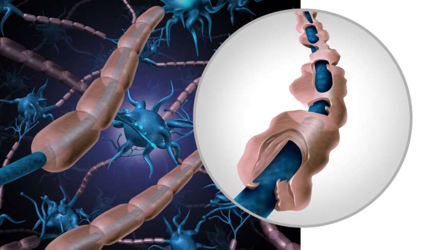
    In Multiple Sclerosis, our immune system erroneously attacks the myelin, causing myelin sheath damage to the protective fatty covering of nerve fibers in the central nervous system. As this myelin gets destroyed in various areas, it forms scar tissue known as sclerosis or plaques. The absence of myelin to shield these nerve fibers disrupts the flow of impulses to the brain. This disruption can cause a range of challenging symptoms for individuals dealing with MS.

    Cause and Risk Factors of Multiple Sclerosis

    The specific reason behind MS has yet to be made clear. However, studies indicate that genetic and environmental factors contribute to its development.

    1. Genetic factors

    While MS is not an inherited disorder, having a relative with MS may increase your risk due to genetic factors.

    2. Viral Infection

    You cannot get infected with multiple sclerosis. However, certain viruses can trigger the activity of MS in the body. The most common virus associated with MS is Epstein-Barr Virus (EBV), which causes Infectious Mononucleosis. About 95% of people with infectious mononucleosis are affected by MS, showing the relationship of increased risk with the EBV virus infection.

    3. Vitamin D

    The link between MS and vitamin D is well established — low levels have been associated with a higher risk of developing the disease. As you move farther from the equator, Vitamin D levels tend to decrease due to reduced sunlight exposure, whereas closer proximity to the equator allows for greater sunlight exposure and higher natural production of Vitamin D in the body, which may be beneficial in MS prevention.

    4. Smoking

    Smoking increases the risk and the progression of the disease. Quitting smoking before or after MS diagnosis can delay the progression and, thus, the disability associated with MS.

    5. Teenage Obesity

    Childhood or adolescent obesity, especially in girls, has been linked to a higher risk of MS. Additionally, for individuals already diagnosed with MS, obesity may increase the chances of experiencing relapses or developing more plaques.

    Types of Multiple Sclerosis

    MS is an unpredictable disease, and there are mainly four types of MS:

    1. Relapsing-remitting MS (RRMS)

    Relapsing-remitting MS (RRMS) accounts for 8 out of 10 diagnosed cases, making it the most common type of MS. Like the name suggests, the symptoms come in the form of an attack or worsening of symptoms (relapsing) after weeks, months, or even years of disease inactivity (remission).

    2. Secondary progressive MS (SPMS)

    Individuals who are diagnosed with SPMS usually have had prior MS attacks but then begin to notice a gradual and steady decline in their overall function and symptoms over time. If left untreated, many people with severe relapsing-remitting MS may ultimately progress to develop secondary progressive MS.

    3. Primary progressive MS (PPMS)

    This type of multiple sclerosis is not very common. From the beginning, neurological function slowly worsens, with no noticeable periods of improvement or worsening. Thus, the disability accumulates over time.

    4. Progressive relapsing MS (PRMS)

    It is the rarest kind of multiple sclerosis that gets worse over time and has sudden relapses that can happen during the disease.

    Symptoms of Multiple Sclerosis

    Multiple sclerosis symptoms and treatment are closely linked — symptoms can vary greatly in terms of severity, duration, and unpredictability. No two individuals with MS experience identical symptoms, and each person’s symptoms can evolve or fluctuate over time. The most common symptoms include:

    What Are the First Signs of Multiple Sclerosis?

    1. Visual Problems

    For many people, the first sign of multiple sclerosisis trouble seeing. You might notice symptoms like blurry vision and difficulty distinguishing between red and green. Your eyes may also hurt when moving. Pain and vision loss can occur because of swelling in the optic nerve.

    2. Fatigue

    MS fatigue affects approximately 80% of individuals, causing significant impairment in their ability to function effectively. Even if you are not physically active, this symptom may stand out most.

    3. Pain

    You might feel pain in two distinct ways with multiple sclerosis. One is Neuropathic pain, which may feel like a sharp stabbing pain in the face or feelings of burning, pins and needles, or being squeezed in the trunk and limbs. Musculoskeletal pain is another type of pain, which includes back, neck, and joint pain. You might  feel a constricting sensation around your torso, sometimes called the “MS hug.”

    4. Walking Difficulties

    Walking challenges are common in MS and can involve muscle weakness affecting tasks like lifting or walking. You might develop balance issues, including unsteadiness and difficulty maintaining an upright posture. Additionally, you might encounter unusual sensations like numbness, tingling, or a sensation akin to pins and needles.

    5. Spasticity

    Spasticity means your muscles can feel tight and may contract on their own, making movement more challenging. The spasticity in MS may feel as mild as muscle tightness to more severe painful contractions, especially in the legs.

    6. Cognitive changes

    You might struggle to find the right words or remember what to do at work or home. Making decisions or keeping up with tasks and conversations may take time and effort. This could affect your job or school, possibly leading to disciplinary action, falling grades, and social difficulties.

    7. Bowel and bladder problems

    You may find that constipation and loss of bowel control are common concerns with multiple sclerosis. You might notice symptoms like getting up several times at night to urinate, urinating more often than before, feeling the urge to rush to the toilet to avoid accidents, and experiencing urine leakage or accidents.

    8. Emotional Changes

    Emotional changes are expected in individuals with MS. Many experience periods of depression, which may stem from the condition or the stress of managing a chronic illness, or both. Anxiety is also prevalent due to the unpredictable nature of MS. Additionally, MS can occasionally trigger abrupt and intense mood swings, such as sudden bouts of crying, laughter, or anger.

    Diagnosis of Multiple Sclerosis

    How is multiple sclerosis diagnosed? The process involves a combination of clinical evaluation, medical history review, neurological examination, and specific diagnostic tests.

    1. History

    History is a very important aspect of multiple sclerosis. If you have experienced at least two attacks that are at least one month apart, where symptoms appear suddenly or worsen for at least 24 hours, then you likely have MS. Additionally, there must be damage to the myelin in more than one area of the central nervous system, occurring at different times and not caused by another disease.

    2. Magnetic resonance imaging (MRI)

    MS MRI diagnosis plays a crucial role in detecting inflammation, demyelination (damage to the myelin sheath), and lesions (plaques) characteristic of MS in the brain and spinal cord.MRI scans provide valuable information for diagnosing MS and monitoring disease activity over time.

    3. Lumbar Puncture

    A lumbar puncture, also known as a spinal tap, may be conducted to analyze the cerebrospinal fluid (CSF) for abnormalities indicative of MS. This includes elevated levels of specific proteins (oligoclonal bands) or immune cells that signal inflammation within the central nervous system.

    4. Evoked Potential (EP)

    EP is a diagnostic test that assesses the sensory pathways in the nervous system by measuring electrical activity in response to specific stimuli. Visual evoked potentials (VEP) use flashing lights or patterns to evaluate visual pat. In contrast, while somatosensory evoked potentials (SSEP) involve electrical stimulation of peripheral nerves to assess sensory pathways, auditory evoked potentials (AEP) use sounds or clicks to evaluate auditory pathways. Electrodes are placed on the scalp that detect and record the responses of the brain to these stimuli, helping to identify abnormalities such as delayed nerve conduction or demyelination, as seen in MS. [3]

    5. Blood tests

    These tests check for things like lupus, Sjogren’s syndrome, deficiencies in vitamins or minerals, certain infections, and rare genetic diseases — helping distinguish multiple sclerosis from other neurological diseases with overlapping symptoms.

    Multiple Sclerosis Symptoms and Treatment Options

    Can multiple sclerosis be cured ? Currently, there’s no cure for MS, but multiple sclerosis symptoms and treatment can be managed effectively to reduce severity and slow progression. Treatment depends on the stage of the disease and specific symptoms.

    1. Disease-Modifying Therapies (DMTs)

    MS disease-modifying therapy is a cornerstone of treatment, particularly for relapsing forms of the disease. They help by reducing the frequency and severity of relapses, delaying disease progression, and decreasing inflammation in the central nervous system. Examples of DMTs include interferons, glatiramer acetate, dimethyl fumarate, fingolimod, and newer agents such as ocrelizumab and siponimod.

    2. Acute Relapse Treatment

    During relapses or exacerbations of MS symptoms, corticosteroids may be prescribed to reduce inflammation and speed up recovery. Plasma exchange (plasmapheresis) may be considered in severe relapses that do not respond to steroids.

    3. Physical and Occupational Therapy

    Rehabilitation programs tailored to individual needs can help maintain mobility, improve balance, strengthen muscles, and address specific functional impairments caused by MS. Occupational therapists assist with adaptive techniques and tools to enhance independence in daily activities.

    4. Lifestyle Modifications

    Adopting a healthy lifestyle can positively impact MS management. Lifestyle changes include maintaining a balanced diet, engaging in regular physical activity, avoiding smoking, and managing stress effectively.

    5. Monitoring and Follow-Up

    Regular monitoring of disease activity, treatment response, and potential medication side effects is essential. Your neurologist can adjust the treatment plan based on disease progression and individual needs.

    Diet and Multiple Sclerosis

    Diet plays a role in managing MS and supporting overall health. While there is no specific “MS diet,” a balanced and nutritious eating plan can be beneficial. Recommendations often include focusing on whole foods such as vegetables, fruits, legumes, whole grains, seeds, and nuts, which provide essential vitamins, minerals, antioxidants, and fiber. Omega-3 fatty acids that are found in fatty fish (like salmon and sardines), flaxseeds, and walnuts have anti-inflammatory properties and could be beneficial for people with MS. Adequate hydration is important to help manage symptoms like fatigue and constipation. Some individuals with MS may benefit from vitamin D supplementation if levels are low.

    MS and Pregnancy

    MS and pregnancy can present unique considerations. Generally, pregnancy does not appear to increase the risk of MS relapses, especially during the second and third trimesters when the immune system is naturally suppressed. In fact, many women with MS experience a temporary improvement in symptoms during pregnancy. However, there is a slightly increased risk of relapse in the first few months postpartum. Women with MS need to work closely with doctors to manage symptoms and medications during pregnancy, as some treatments may need to be adjusted or discontinued. Overall, with proper planning and medical supervision, many women with MS can have successful pregnancies and healthy babies.

    Living with MS

    Living with multiple sclerosis symptoms and treatment demands can be difficult, but strategies and resources exist to help manage the challenges effectively.. One crucial aspect is adhering to your prescribed medication regimen. As mobility issues arise, assistive equipment like canes or walkers can greatly enhance independence, while rehabilitation activities can maintain or improve functionality. Making adaptations to your home environment, such as installing grab bars or rearranging furniture, can also promote independence and safety. It’s essential to communicate openly with your family and doctors about your needs, which would help you feel supported. By proactively addressing these aspects, you can navigate the complexities of MS with greater resilience and empowerment.

    Can MS be Prevented?

    MS cannot be prevented, but understanding multiple sclerosis symptoms and treatment options early can make a significant difference in quality of life. While the development of MS cannot be predicted or prevented with current knowledge, there are ongoing efforts to identify potential risk factors and understand the underlying mechanisms of the disease. Research into lifestyle factors, such as vitamin D levels, smoking cessation, and maintaining a healthy diet and exercise regimen, may contribute to overall health and potentially reduce the risk of other chronic diseases. However, there is no known definitive way to prevent MS from occurring in individuals predisposed to the condition.

    Questions To Ask Your Doctor

    1. How will my diagnosis with MS impact my daily life?
    2. What should I do if I suspect I’m having a relapse or notice new symptoms?
    3. Does multiple sclerosis get worse over time, and what can slow the progression?
    4. Does my insurance cover the cost of my MS treatments?
    5. What should I do if I feel my current medication isn’t effective or if I struggle to stick to the treatment plan?
    6. Are there any clinical trials that I might be eligible for?

  • Enlarged Prostate (BPH): Symptoms, Causes, and BPH Treatment Options

    Enlarged Prostate (BPH): Symptoms, Causes, and BPH Treatment Options

    | Key Highlights

    1. Benign Prostatic Hyperplasia is a common condition among aging men, with prevalence increasing with age. By age 50, over 50% of men will have BPH, and this increases to over 80% by age 80.
    2. BPH is a non-cancerous enlargement of the prostate gland without an increased risk of malignancy, whereas prostate cancer involves the abnormal growth of cancerous cells within the prostate gland.
    3. Common symptoms of BPH include urinary frequency, urgency, weak urine stream, difficulty initiating urination, dribbling at the end of urination, and feeling of incomplete bladder emptying.
    4. Diagnosis of BPH involves a combination of digital rectal examination, urinalysis, measurement of prostate-specific antigen levels, and imaging studies like ultrasound or cystoscopy.
    5. Treatment for BPH depends on the severity of symptoms and can include lifestyle modifications, medications, minimally invasive procedures, or surgery.

    What is Benign Prostatic Hyperplasia (BPH)?

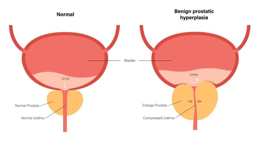
    Benign prostatic hyperplasia (BPH) is also called prostate gland enlargement. It is one of the most common conditions as men age. Understanding BPH treatment options early can help men manage symptoms before they worsen. The enlarged prostate in BPH squeezes or partially blocks the surrounding urethra, the tube that transports urine from the bladder out of the body. This enlargement of the prostate can lead to compression of the urethra, resulting in lower urinary tract symptoms (LUTS) such as increased frequency of urination, urgency, weak urinary stream, incomplete bladder emptying, and nocturia.

    The Prostate Gland and Cause of BPH

    The prostate gland is a small gland located just below the bladder and in front of the rectum in men. The primary function of the prostate gland is to produce seminal fluid, which is responsible for nourishing and transporting sperm during ejaculation. The prostate gland surrounds the urethra, which is the tube that carries urine from the bladder out through the penis.

    The pathophysiology of BPH involves hormonal changes, particularly an increase in dihydrotestosterone (DHT) levels within the prostate gland due to the action of 5-alpha-reductase. This hormone imbalance leads to abnormal growth and proliferation of glandular epithelial cells and stromal cells within the prostate, resulting in gland enlargement.

    Additionally, alterations in smooth muscle tone and neurogenic factors within the prostate gland contribute to urinary symptoms associated with BPH.

    BPH and Risk of Prostate Cancer

    BPH vs prostate cancer — these are distinct conditions affecting the prostate gland in men. BPH involves the non-cancerous enlargement of the prostate gland due to hormonal changes and aging, leading to compression of the urethra and urinary. It is a common condition in older men but is generally not associated with an increased risk of prostate cancer. In contrast, prostate cancer is characterized by the abnormal growth of cancerous cells within the prostate gland, which can potentially spread to other parts of the body. Prostate cancer usually does not cause symptoms in its early stages but can present with urinary changes, blood in the urine or semen, and other systemic symptoms as it progresses. Unlike BPH, prostate cancer is a serious disease that requires early detection and appropriate treatment to prevent complications and improve outcomes.

    Signs and Symptoms of Benign Prostatic Hyperplasia

    BPH can cause a variety of signs and symptoms, and these symptoms typically develop gradually and can range from mild to severe, depending on the degree of prostate enlargement and the extent of urethral obstruction. Common signs and symptoms of BPH include:

    symptoms-of-bph

    1. Increased Frequency of Urination

    Men with BPH may experience a need to urinate more frequently, especially during the day.

    2. Urgency

    You may have a sudden and strong urge to urinate, which can be difficult to postpone.

    3. Nocturia

    Men with BPH often experience nocturia. This refers to waking up during the night to urinate. This can be very disturbing to many as it disrupts your sleep patterns.

    4. Weak Urinary Stream

    The urinary stream may be weak or diminished, making it difficult to start and complete urination.

    5. Difficulty Initiating Urination

    You may have trouble starting the urine flow, requiring straining or pushing to begin urination.

    6. Incomplete Bladder Emptying

    Despite urinating, there may be a sensation of incomplete bladder emptying, leading to the need to urinate again shortly after.

    7. Dribbling at the End of Urination

    After urination is complete, some men may experience dribbling or leakage of urine due to incomplete bladder emptying.

    8. Urinary Retention

    In severe cases of BPH, the urethra can become significantly obstructed, leading to urinary retention in men, where the bladder cannot empty completely. This is an emergency condition and requires urgent medical attention.

    Diagnosis of BPH

    How is BPH diagnosed? The process typically involves a combination of physical exams, urine tests, and imaging. Here are the steps involved in the diagnosis:

    diagnosis-of-bph

    1. Digital Rectal Examination (DRE)

    During a DRE, your doctor will insert a gloved, lubricated finger into the rectum to feel the size, shape, and consistency of the prostate gland. Enlargement, firmness, and nodules may indicate BPH or other prostate-related conditions.

    2. Urinalysis

    A urinalysis may be performed to rule out other conditions, such as urinary tract infections or other abnormalities, contributing to the symptoms.

    3. Prostate-Specific Antigen (PSA) Blood Test

    PSA is a protein produced by the prostate gland, and elevated levels can indicate BPH, prostate inflammation (prostatitis), or prostate cancer. The PSA test is used to assess prostate health and may help differentiate between BPH and prostate cancer.

    4. Uroflowmetry

    Uroflowmetry is a non-invasive diagnostic test used to assess urinary function by measuring the rate and pattern of urine flow during voiding. During the procedure, you will be asked to urinate into a specialized toilet or uroflowmeter. The flow meter is equipped with sensors that record the flow electronically. It measures the parameters like the maximum flow rate, average flow rate, voided volume, and time to maximum flow. These measurements provide valuable information about urinary efficiency and can help diagnose conditions such as benign prostatic hyperplasia (BPH), urinary obstruction, or neurogenic bladder dysfunction.

    5. Post-Void Residual (PVR) Measurement

    Post-void residual urine measurement is an important diagnostic test for BPH and associated symptoms of LUTS. This test evaluates the amount of urine remaining in the bladder after urination, which can indicate inefficient bladder emptying due to BPH-related obstruction or other bladder abnormalities. Post-void residual urine can be measured using non-invasive methods such as bladder ultrasound (Bladder Scan) or inserting a catheter to drain and measure the residual urine volume directly. If you have BPH, you will have an elevated post-void residual volume.

    6. Transrectal Ultrasound (TRUS)

    In some cases, a TRUS may be performed to assess the size and structure of the prostate gland and rule out other conditions such as prostate cancer.

    7. Cystoscopy

    A cystoscopy is a procedure that involves inserting a thin, flexible tube into the urethra. The tube consists of a camera at the tip that helps to visualize the urinary tract. This may be recommended if there are concerns about bladder function or to rule out other urinary tract abnormalities.

    BPH Treatment Options

    You should know that there is no cure for BPH. However, treatment can manage your symptoms to some extent. The treatment of BPH aims to alleviate urinary symptoms, improve quality of life, and prevent complications. The treatment depends on the severity of your symptoms, the size of the prostate gland, the presence of complications, and individual patient preferences. Here are the main approaches to managing BPH:

    Diagram showing BPH treatment options including medication and surgery

    1. Watchful Waiting/Active Surveillance

    For men seeking enlarged prostate treatment without surgery, watchful waiting or active surveillance may be recommended when symptoms are mild or not bothersome. This involves regular monitoring of symptoms and prostate size without immediate treatment intervention.

    2. Lifestyle Modifications

    Certain lifestyle changes can help manage mild to moderate symptoms of BPH. These include limiting fluid intake before bedtime, avoiding caffeine and alcohol, exercising regularly (especially pelvic floor exercises), and maintaining a healthy weight.

    3. Medications

    The use of medications is a common and effective approach for managing BPH, especially for individuals with moderate to severe symptoms. These medications primarily target the prostate gland and urinary tract to improve symptoms and reduce the obstruction caused by prostate enlargement.

    • Alpha-Blockers: Alpha-blockers for BPH relax the prostate and bladder neck muscles to improve urine flow and reduce symptoms such as hesitancy and urgency. Commonly prescribed alpha-blockers include tamsulosin, alfuzosin, doxazosin, and terazosin.
    • 5-alpha-Reductase Inhibitors: These medications reduce the production of DHT, which can shrink the prostate gland over time and improve urinary symptoms. Examples include finasteride and dutasteride.
    • Combination Therapy: Some patients require a combination of alpha-blockers and 5-alpha-reductase inhibitors for more effective symptom relief.

    4. Surgical Interventions

    • Transurethral Resection of the Prostate (TURP): TURP surgery is a surgical procedure for BPH in men with moderate to severe urinary symptoms who have not responded adequately to medication or other conservative measures.During a TURP procedure, a urologist inserts a specialized instrument called a resectoscope through the urethra to access the prostate gland. The resectoscope has a wire loop that is used to remove excess prostate tissue obstructing the urethra. This tissue removal helps to widen the urinary passage and improve urine flow. TURP is considered the gold standard surgical treatment for BPH due to its effectiveness in relieving symptoms and restoring normal urinary function. The procedure is performed under anesthesia, typically spinal or general, and requires a short hospitalization.
    • Transurethral Microwave Therapy (TUMT): This procedure uses microwave energy to heat and destroy excess prostate tissue, relieving urinary symptoms.
    • Transurethral Needle Ablation (TUNA): In this procedure, radiofrequency energy is used to heat and destroy prostate tissue to improve urine flow.
    • Prostatic Urethral Lift (UroLift): During this procedure, tiny implants are placed in the prostate to lift and hold the enlarged tissue away from the urethra, reducing obstruction.
    • Water Vapor Thermal Therapy (Rezūm): This procedure involves inserting an instrument into your urethra and moving it to the prostate. A needle is then ejected into the prostate. Steam is delivered through this needle into the prostate to shrink excess tissue and improve symptoms.
    • Open Prostatectomy: In rare cases of very large prostates, traditional open surgery may be necessary to remove the obstructive tissue

    5. Other Therapies

    • Botulinum Toxin Injections: Injections of botulinum toxin directly into the prostate gland may help relax muscles and improve symptoms.
    • Temporary Prostate Stents: Stents can be placed in the urethra to keep it open and relieve obstruction temporarily.

    Side Effects of Prostate Surgery

    Prostate surgery can have certain side effects and complications, including temporary urinary symptoms like urgency, frequency, or blood in urine immediately after surgery. Long-term effects may include erectile dysfunction, retrograde ejaculation, or urinary incontinence. Rare complications can include bleeding, urethral stricture (narrowing), bladder neck contracture, or persistent/recurrent symptoms. It’s prudent to discuss with your provider the side effects of prostate surgery before getting one.

    What Happens if BPH Is Left Untreated?

    If BPH is left untreated, it can lead to various complications and progressively worsen over time. Some potential consequences of untreated BPH include:

    complications-of-bph

    • Worsening of urinary symptoms,
    • Acute urinary retention
    • Bladder damage
    • Urinary tract infections

    Prevention of BPH

    There is no guaranteed way to avoid BPH.

    prevention-of-bph

    While no approach fully prevents BPH, maintaining a healthy weight and eating well are among the simplest BPH treatment options available before symptoms worsen. Excess body fat may raise hormone levels and other blood variables, stimulating the proliferation of prostate cells. Staying active aids in the management of weight and hormone levels.

    Questions for your doctor

    1. What might be the causes of my symptoms other than BPH?
    2. Is BPH heritable?
    3. Are there any supplements for this condition?
    4. When should I be worried if I choose not to treat?
    5. What is my risk for prostate cancer ?
    6. Can BPH cause erectile dysfunction or affect my sexual health?

    References

  • Focal Seizure Symptoms in Adults – Epilepsy Types, Treatment Options, and More

    Focal Seizure Symptoms in Adults – Epilepsy Types, Treatment Options, and More

    Key Highlights

    1. Epilepsy is a chronic neurological disorder with recurrent seizures and is caused by abnormal electrical activity in the brain.
    2. Focal seizure symptoms in adults and children differ; focal seizures originate in one brain area while generalized seizures involve widespread electrical disturbances.
    3. Diagnosis of epilepsy involves a thorough evaluation of medical history, symptoms, and diagnostic tests like EEG and neuroimaging.
    4. Treatment typically includes antiepileptic medications, lifestyle modifications, and sometimes surgical interventions to control seizures and improve quality of life.
    5.  Your doctor will gradually reduce the dosage and consider stopping your seizure medications entirely only after you have been seizure-free for a period of 2 to 4 years.

    What is Epilepsy?

    Epilepsy is a neurological disorder characterized by recurrent seizures, including focal seizure symptoms adults may not immediately recognize. Seizures occur due to abnormal electrical activity in the brain and can lead to various symptoms, including convulsions, loss of consciousness, unusual sensations, or temporary confusion.

    Epilepsy impacts individuals across all age groups, affecting approximately 50 million people worldwide and around 3.4 million individuals in the United States alone. The number of cases of epilepsy is leading to over one hundred thousand new diagnoses annually in the US, reflecting a rising trend in the condition’s incidence.

    Types of Epilepsy

    When we hear the word seizure, we think of muscle spasms, involuntary body movements, and a loss of consciousness. However, most seizures are surprisingly subtle and may be hard to recognize.

    1. Generalized epilepsy

    If a large part of the brain or the entire brain is involved, you may experience a loss of awareness without any warning. Your whole body may also stiffen or shake involuntarily. Generalized seizures are classified into different subtypes:

    • Absence Seizures (petit mal seizures): Absence seizures symptoms include brief lapses of consciousness characterized by staring blankly or subtle movements such as eye blinking or lip-smacking..
    • Tonic-Clonic Seizures (grand mal seizures): These are dramatic seizures presenting with loss of consciousness and stiffening of muscles (tonic phase), followed by rhythmic jerking movements (clonic phase).
    • Myoclonic Seizures: You may have sudden, brief jerks or twitches of the muscles, often involving the arms and legs.
    • Clonic Seizures: This is the type of epilepsy where you will have repetitive, rhythmic jerking movements of muscles.

    2. Focal epilepsy

    Focal seizure symptoms in adults originate in a specific area or focus of the brain. Focal seizures can be further classified into:

    • Focal Aware Seizures ( simple partial seizures): Focal seizure symptoms in adults with focal aware seizures occur without loss of consciousness. The individual remains aware of their surroundings but may experience unusual movements, sensations, or emotions.
    • Focal Impaired Awareness Seizures complex partial seizures): These seizures are characterized by altered consciousness or awareness. The person may appear confused, disoriented, or engage in purposeless movements.
    •  Focal to Bilateral Tonic-Clonic Seizures: These begin as focal seizures initially, but later, they spread to involve both sides of the brain, leading to a generalized tonic-clonic seizure.

    Causes

    The causes of epilepsy can be different for each person. Some episodes of seizure activity can be linked to genetics, brain trauma, stroke, autoimmune disorders, metabolic issues, or infectious diseases, while others may have no identifiable causes.

    Triggers

    Some common triggers for seizures include:

    • Sleep deprivation
    • Fever
    • Flashing bright lights
    • Alcohol Usage
    • Recreational Drug use
    • Stress
    • Stress Missing medications for seizure

    Epilepsy Seizure Triggers and Warning Signs

    Since seizure in epilepsy has varied presentations, it sometimes gets difficult to recognize one. Here are some of the ways seizures in epilepsy can present:

    • Aura: Some adults experience a warning sign before a seizure — an aura — which is a common early focal seizure symptom in adults. This can vary widely among individuals, including feelings of Deja vu, strange smells or tastes, visual disturbances, or sudden emotional changes.
    • Loss of Awareness: Many seizures involve a loss of awareness or consciousness. During this time, the person may appear to stare blankly into space, be unresponsive, or be unable to communicate.
    • Uncontrollable Movements: Seizures often involve uncontrollable movements. These can vary greatly, from jerking motions in the arms and legs to more subtle twitching or repetitive movements. During these movements, one might bite their tongue, which needs immediate attention.
    • Changes in Sensation: Some seizures may cause unusual sensations or feelings, such as tingling, numbness, fear, or panic.
    • Loss of Control: In some cases, individuals may lose control of bodily functions, such as bladder or bowel control.
    • Duration: Seizures typically last for a short time, usually only a few seconds to a few minutes. Prolonged seizures without full recovery between them are called status epilepticus; status epilepticus treatment requires immediate medical attention.

    Diagnosis

    The diagnosis of epilepsy will require a detailed history and neurological examination in the beginning. After that, your doctor will recommend various tests to diagnose it. Here is the overview of the diagnostic process:

    1. Blood tests

    Blood tests are performed to check for metabolic disorders, infections, or other medical conditions that can cause seizures. It will help to rule out all the other conditions that might present with seizure.

    2. Electroencephalogram (EEG)

    Epilepsy diagnosis by EEG is a critical first-line diagnostic technique. It records the brain’s electrical activity through electrodes placed on the scalp. In epilepsy, EEG detects abnormal patterns like spikes and sharp waves, indicating potential seizure activity and pinpointing the location of seizure onset within the brain. There are different types of EEG studies, including routine EEG, ambulatory EEG, and video EEG monitoring in specialized units. These aid in capturing and analyzing brain activity over varying durations, allowing for accurate diagnosis and treatment planning.

    3. Magnetic resonance imaging (MRI)

    It is the preferred imaging modality for epilepsy evaluation due to its superior soft tissue contrast and ability to visualize detailed brain anatomy. It can detect a range of abnormalities, including mesial temporal sclerosis (MTS), neocortical dysplasia, brain tumors, and vascular malformations, all of which can cause seizures. Specific MRI protocols like T1-weighted, T2-weighted, FLAIR, and susceptibility-weighted imaging (SWI) are used to assess brain structure in relation to epilepsy.

    4. Computed tomography (CT)

    CT scans, while less sensitive than MRI, are sometimes used for emergency evaluations or when MRI is contraindicated. They can identify large lesions, hemorrhages, or calcifications associated with epilepsy. In certain cases, advanced imaging techniques such as positron emission tomography (PET) and single-photon emission computed tomography (SPECT) may be employed to characterize brain function further and localize the seizure focus, aiding in treatment decisions for individuals with epilepsy.

    Treatment of Epilepsy

    Treatment options for epilepsy can vary depending on factors such as the type of seizures, the underlying cause of epilepsy, and individual patient characteristics. Here are the main treatment options available for epilepsy:

    1. Antiepileptic Medications (AEDs)

    The most common and primary treatment for epilepsy involves taking AEDs. They work by stabilizing the electrical activity in the brain and reducing the likelihood of seizures. A wide range of AEDs are available, and the choice of AED depends on factors such as the type of seizures, patient age, potential side effects, and other medical conditions. Treatment often starts with a single AED at a low dose, which may be adjusted based on seizure control and tolerability. Some patients may require combination therapy with more than one AED to achieve optimal seizure management. Some of the common examples of AEDs are Carbamazepine (Tegretol), Valproate (Depakote), and Levetiracetam (Keppra).

    -> Levetiracetam: Levetiracetam, marketed under Keppra, is a widely prescribed AED in the United States and globally due to its effectiveness and safety profile. Keppra is known for its broad-spectrum efficacy against various seizure types and is often used as adjunctive therapy alongside other AEDs. Compared to antiepileptic medications with significant side effects, Keppra’s profile is minimal, with dizziness, somnolence, and irritability being generally mild and transient.

    2. Ketogenic Diet

    The ketogenic diet for seizures is high in fat, low in carbohydrates, and adequate in protein. The ketogenic diet for epilepsy in children has shown efficacy in decreasing seizure episodes, particularly in those who do not respond well to medications. The diet induces a state of ketosis, where the body produces ketones from fat metabolism, which may have anticonvulsant effects on the brain. The ketogenic diet is typically supervised by a dietitian and requires strict adherence to specific macronutrient ratios.

    3. Surgery

    Surgical intervention may be considered for individuals with epilepsy who do not respond to medications or have seizures originating from a specific, identifiable area of the brain. Common surgical procedures for epilepsy include resective surgery (removal of the seizure focus), corpus callosotomy (cutting the corpus callosum to prevent seizure spread), and multiple subpial transections (to disrupt seizure pathways).

    4. Vagus Nerve Stimulation (VNS)

    Vagus nerve stimulation for epilepsy involves implanting a device that delivers electrical impulses to the vagus nerve in the neck. These impulses can help reduce seizure frequency and severity. VNS is considered for individuals with refractory epilepsy who are not candidates for surgery or have not responded well to medications alone.

    5. Responsive Neurostimulation (RNS)

    RNS is a newer treatment option that involves implanting a device directly into the brain to detect and respond to electrical activity associated with seizures. The device delivers targeted electrical stimulation to disrupt seizure activity before it spreads.

    6. Lifestyle Modifications

    Certain lifestyle changes can complement medical treatments and help manage epilepsy, such as getting adequate sleep, managing stress, avoiding alcohol and recreational drugs, and maintaining a regular schedule of meals and medications. Identifying and avoiding seizure triggers, such as specific foods, drinks, or environmental factors, can also be beneficial.

    Cure of Epilepsy

    While there is no definitive cure for epilepsy, treatments will help you control your symptoms. Can epilepsy be cured with surgery? For carefully selected patients, surgical intervention can result in long-term seizure freedom.

    Stopping anti-epileptic drugs

    It’s important to continue your medications even if you have been seizure-free. Although you may feel fine, it’s crucial to consult your doctor before stopping AEDs. Typically, your doctor will gradually reduce the dosage and consider stopping your seizure medications entirely only after you have been seizure-free for a period of 2 to 4 years. If you have experienced only one seizure episode, your doctor might recommend discontinuing treatment if you remain seizure-free for 6 to 12 months.

    There is a higher chance of remaining seizure-free without medications if:

    • You had a few seizures before you started taking seizure medicine.
    • Your seizures were controlled with just one type of medicine.
    • You had normal results on a neurological examination.
    • You had a normal EEG at the time of the diagnosis.
    • Your CT scan or MRI results were normal.

    Q. Epilepsy Seizure First Aid Steps: What to Do After a Seizure?

    Ask the person’s name, place, time of the day, and about the seizure episode. If the person cannot answer the questions above, provide pertinent information and reassurance so they are not overwhelmed. Do not leave the person alone until they can talk, communicate well, and breathe normally.

    Q. When should you call 911 or for emergency help?

    If you see a seizure, be calm. However, do call for help if:

    • The seizures last longer than 5 minutes.
    • There are repeated seizures.
    • The person had difficulty breathing.
    • The seizures have occurred in water bodies such as lakes or swimming pools.
    • The person is injured, pregnant, or sick.
    • The person does not return to their usual after a seizure
    • This is the person’s first seizure.
    • The person asks for medical help.

    Q. How long does a focal seizure last?

    Focal seizures typically last between 30 seconds and 2 minutes. Seizures lasting longer than 5 minutes or occurring repeatedly without recovery are considered status epilepticus and require emergency help.

    Epilepsy and Exercise

    Managing focal seizure symptoms in adults, including the relationship between exercise and seizures, is complex and requires individualized consideration. While regular physical activity like walking, swimming, or yoga can provide notable health benefits such as stress reduction and improved mood, it can also influence seizure activity positively or negatively.

    Individuals with epilepsy need to engage in moderate-intensity activities while taking precautions to avoid overheating, dehydration, or excessive exertion, which can trigger seizures in some cases. Consulting with your doctor is essential to tailor exercise plans based on your medical history, seizure type, and medication regimen. Incorporating exercise into an overall epilepsy management strategy can enhance well-being while reducing the risk of seizures during physical activity.

    1. Women and epilepsy

    Epilepsy, including focal seizure symptoms in adults, can uniquely impact women due to hormonal influences on seizure patterns, particularly during puberty, menstruation, pregnancy, and menopause. Hormonal fluctuations can affect seizure frequency and severity in some cases. Certain AEDs may also impact reproductive health, leading to menstrual irregularities, polycystic ovary syndrome (PCOS), or decreased bone density. Women with epilepsy require specialized care and considerations for contraception, fertility, and pregnancy, as managing medication safety and seizure control during pregnancy is crucial for maternal and fetal well-being.

    2. Driving and Seizure Disorder

    Driving with epilepsy requires meeting a seizure-free period, as being on the road can pose a risk to yourself and others.. Seizures can make you unconscious or cause motor and visual impairment, which might be risky if you are on the road. A person with epilepsy must be seizure-free for a certain period to be eligible for a driver’s license. The seizure-free period varies from state to state, and you might need clearance from your doctor. You should refer to your state driving laws to see the latest rules and eligibility for driving if you have seizures.

    Questions For Your Doctor

    • How long should I continue my medication?
    • When will I be able to drive?
    • Should I breastfeed my child while I am on epilepsy medication?
    • Are there some social support groups for those who have epilepsy?
    • Do I need a wristband to notify others of my condition?
    • Do I need a referral to a neurologist?

    References

    1. JAMA. Epilepsy. JAMA [Internet]. 2016 Dec 27 [cited 2025 Feb 25];316(24):2686. Available from: https://jamanetwork.com/journals/jama/fullarticle/2594720
    2. Centers for Disease Control and Prevention. Seizure first aid [Internet]. CDC; 2023 [cited 2025 Feb 25]. Available from: https://www.cdc.gov/epilepsy/about/first-aid.html
    3. Epilepsy Foundation. Seizure triggers [Internet]. Epilepsy Foundation; [cited 2025 Feb 25]. Available from: https://www.epilepsy.com/what-is-epilepsy/seizure-triggers
    4. Epilepsy Foundation. Stopping seizure medicines [Internet]. Epilepsy Foundation; [cited 2025 Feb 25]. Available from: https://www.epilepsy.com/treatment/medicines/stopping-medication
    5. Epilepsy Foundation. Epilepsy and driving [Internet]. Epilepsy Foundation; [cited 2025 Feb 25]. Available from: https://www.epilepsy.com/managing-epilepsy/living-well/driving-and-transportation

  • Cardiomyopathy – Causes, Symptoms, Treatment, and Implantable Devices

    Cardiomyopathy – Causes, Symptoms, Treatment, and Implantable Devices

    Highlights of Cardiomyopathy

    • Cardiomyopathy affects the heart muscle, making it harder for the heart to pump blood to the rest of the body.
    • There are different types of cardiomyopathy, including dilated, hypertrophic, and restrictive.
    • A variety of factors, including genetics, infections, autoimmune diseases, certain medications, and lifestyle factors, such as alcohol abuse or uncontrolled high blood pressure, can cause cardiomyopathy.
    • Common symptoms of cardiomyopathy are shortness of breath, swelling in the legs or abdomen, irregular heartbeat, chest pain, and However, some patients may not experience symptoms, especially in the early stages.
    • Cardiomyopathy cannot be cured, but treatment, including implantable devices for cardiomyopathy, can improve symptoms and quality of life, allowing many individuals to lead active lives.

    What Is Cardiomyopathy?

    Cardiomyopathy is a disease that affects the heart muscles, impairing their ability to pump blood to the rest of the body effectively. It often goes undetected, but it is estimated that approximately 1 in every 500 adults may have this condition. It can affect people of any age, sex, and race.

    Types of Cardiomyopathy

    There are several types of cardiomyopathy; the common ones are as follows:

    1. Hypertrophic Cardiomyopathy (HCM)

    If you have hypertrophic cardiomyopathy, the muscular wall of your heart becomes thickened, making it difficult for your heart to pump blood effectively throughout your body. This condition is genetic, so if one of your parents has HCM, there is a 50 percent chance that you may have inherited HCM can develop at any age, but it tends to be more severe when it starts during childhood.

    2. Dilated Cardiomyopathy

    In dilated cardiomyopathy, the heart muscle becomes weakened and dilated, decreasing the heart’s ability to pump blood It is the most common type of cardiomyopathy.

    3. Restrictive Cardiomyopathy

    Restrictive cardiomyopathy is a type of myopathy characterized by abnormal stiffness of the heart muscle, which impairs its ability to fill properly with blood during the relaxation phase of the heartbeat. This stiffness is often caused by infiltrating abnormal substances, such as scar tissue or proteins, into the heart muscle, which results in reduced ventricular filling and decreased cardiac output, leading to heart failure.

    Other types of Cardiomyopathy you might want to know:

    • Peripartum Cardiomyopathy: It is a rare variant of cardiomyopathy that begins sometime during the final month of pregnancy and continues about five months after delivery. Risk factors for peripartum cardiomyopathy include multiple pregnancies, advanced maternal age, hypertension, preeclampsia, and the use of certain medications.
    • Takotsubo Cardiomyopathy: It is named after the shape the heart takes on during contraction, resembling a Japanese octopus trap called a “takotsubo.”Some refer to it as “broken heart syndrome” or stress-induced cardiomyopathy. Takotsubo cardiomyopathy is characterized by a sudden and temporary weakening of the heart muscle, typically triggered by severe emotional or physical stress.

    Risk Factors and Causes of Cardiomyopathy

    While the exact cause of cardiomyopathy is often complex and multifactorial, certain risk factors can increase the likelihood of developing this condition. Some of the risk factors are as follows:

    risk-factors-of-cardiomyopathy

    1. Family History

    A family history of cardiomyopathy or sudden cardiac death can significantly increase the risk of developing the condition.

    2. High Blood Pressure

    If you have uncontrolled high blood pressure, it can strain the heart muscle over time, leading to hypertensive cardiomyopathy. Hypertensive cardiomyopathy is a form of secondary cardiomyopathy characterized by thickening and stiffening of the heart muscle.

    3. Alcohol Abuse

    Excessive and prolonged drinking of alcohol can weaken the muscles in your heart and lead to the development of dilated cardiomyopathy.

    4. Heart valve disorder

    Malfunctioning heart valves, such as those affected by conditions like rheumatic fever or infective endocarditis, can disrupt normal blood flow through the heart and contribute to the development of

    5. Viral and protozoal infections

    Certain organisms like the Coxsackie B virus, Human Immunodeficiency virus (HIV), and Chagas disease are known to be associated with the development of cardiomyopathy.

    6. Autoimmune Diseases

    Autoimmune diseases, such as rheumatoid arthritis or lupus, can cause inflammation and damage the heart muscle, leading to secondary

    7. Drug abuse

    Illicit drug use, particularly cocaine and amphetamines, can cause damage to the heart muscle and increase the risk of cardiomyopathy.

    Symptoms of Cardiomyopathy

    The symptoms of cardiomyopathy can vary depending on the type and severity of the condition. Common symptoms include:

    symptoms-of-cardiomyopathy

    1. Shortness of breath during physical activity or when lying down.
    2. Fatigue or weakness, even with minimal exertion.
    3. Swelling in the legs, ankles, or abdomen.
    4. Irregular heartbeat or discomfort.
    5. Chest pain or fainting spells.
    6. Dizziness or fainting spells

    Diagnosis of Cardiomyopathy

    Various diagnostic tests can detect the presence of cardiomyopathy. Common diagnostic tests are discussed below:

    diagnosis-of-cardiomyopathy

    1. Electrocardiogram (EKG)

    EKG is a common diagnostic test used to assess the electrical activity of the heart and can provide valuable information for diagnosing cardiomyopathy. While an EKG alone cannot definitively diagnose cardiomyopathy, it can help detect abnormalities in heart rhythm and conduction, which may indicate underlying cardiac issues.

    2. Echocardiogram

    An echocardiogram is a noninvasive test that uses sound waves to create detailed images of the heart’s structure and function. It can help identify abnormalities in heart size, shape, and function and assess the heart’s pumping ability.

    3. Blood Tests

    Blood tests help assess levels of certain enzymes and proteins that can indicate heart muscle damage. These may include cardiac troponin and brain natriuretic peptide (BNP).

    4. Cardiac MRI (Magnetic Resonance Imaging)

    Cardiac MRI provides detailed images of the heart’s structure and can detect abnormalities in the heart muscle, valves, and blood It is particularly useful for evaluating myocardial tissue characteristics and detecting scar tissue.

    5. Cardiac CT (Computed Tomography)

    CT scans can provide detailed images of the heart and blood vessels, helping to identify structural abnormalities, assess coronary artery disease, and evaluate heart function.

    6. Genetic Testing

    Genetic testing may be recommended in cases where a genetic component to cardiomyopathy is It helps to identify specific genetic mutations associated with the condition.

    7. Cardiac Catheterization

    This procedure involves inserting a tube called a catheter into a blood vessel in the arm, groin, thigh, or neck, which is then guided to the heart. It helps to measure heart chamber pressures to assess for heart failure, obtain blood samples to evaluate oxygen levels and heart function and take an endomyocardial biopsy to diagnose specific types of cardiomyopathy or myocarditis.

    8. Coronary Angiography

    Coronary angiography is a specialized X-ray imaging technique used to visualize the coronary arteries. It helps to rule out CAD as a cause of cardiomyopathy by detecting blockages or narrowing in the coronary It can also guide treatment decisions, such as angioplasty or bypass surgery if significant CAD is present.

    9. Heart Biopsy

    It is a minimally invasive procedure and is sometimes utilized in diagnosing cardiomyopathy. It involves extracting a small tissue sample from the heart muscle. This biopsy aids in identifying specific types of cardiomyopathies or myocarditis, providing crucial insights for accurate diagnosis and treatment planning.

    Treatment of Cardiomyopathy

    The treatment of cardiomyopathy aims to manage symptoms, improve heart function, prevent complications, and enhance overall quality of life.

    Beyond these lifestyle steps, medications, surgery, and implantable devices for cardiomyopathy are key parts of care. Here is a list of five things you can do to help you with your cardiomyopathy.

    treatment-of-cardiomyopathy

    • Heart-Healthy Diet: you can begin by adopting a diet low in sodium, saturated fats, and cholesterol while high in fruits, vegetables, whole grains, and lean proteins. it can help manage blood pressure, cholesterol levels, and overall heart health.
    • Regular Exercise: Regular physical activity, as tolerated, can improve cardiovascular fitness, strengthen the heart muscle, and enhance overall well-being. However, you should be aware that not everybody can do all types of You need to talk to your doctor before you begin any kind of exercise.
    • Smoking Cessation: It is important to note that a reduction in the consumption of cigarettes can reduce your risk of cardiovascular complications.
    • Limiting Alcohol: For individuals with alcoholic cardiomyopathy, abstaining from or reducing alcohol consumption is critical to prevent further damage to the heart muscle.
    • Weight Management: You should aim to achieve a healthy weight. Achieving and maintaining a healthy weight can help decrease the strain on your heart and improve overall cardiovascular health.

    Medications for cardiomyopathy

    Heart Failure Medications include angiotensin-converting enzyme inhibitors (Enalapril, Lisinopril), Angiotensin II Receptor Blockers (Losartan, valsartan), beta-blockers (carvedilol, metoprolol), and diuretics(spironolactone, Eplerenone). These medications are a core part of heart failure treatment, commonly prescribed to alleviate heart failure symptoms, enhance heart function, and decrease fluid retention.

    • Anticoagulants/Antiplatelets: These drugs may be administered to prevent blood clot formation in individuals with specific types of cardiomyopathy at a higher risk of stroke or thromboembolism.
    • Antiarrhythmics: Antiarrhythmic medications are vital in managing abnormal heart rhythms associated with cardiomyopathy. They work by modifying cardiac cell electrical properties to restore and maintain normal heart rhythm. Examples include sodium channel blockers (e.g., flecainide), beta-blockers (e.g., metoprolol), potassium channel blockers (e.g., amiodarone), and calcium channel blockers (e.g., verapamil). The selection of these drugs depends on the type of arrhythmia and patient factors.
    • Blood Pressure Management: Medications like ACE inhibitors, ARBs, calcium channel blockers, or diuretics are prescribed to control elevated blood pressure, which is particularly crucial in managing hypertensive cardiomyopathy.
    • Statins: Statins are prescribed to regulate cholesterol levels and help mitigate the risk of complications associated with coronary artery disease in individuals with cardiomyopathy.

    Implantable Devices for Cardiomyopathy

    1. Pacemakers

    Pacemakers are among the most common implantable devices for cardiomyopathy, essential in managing certain types of cardiomyopathy, particularly those associated with rhythm disorders. Indicated primarily for patients with bradyarrhythmia like sinus node dysfunction or heart block, pacemakers work by monitoring the heart’s electrical activity and delivering electrical impulses to regulate heart rate and rhythm. Pacemaker implantation, a minimally invasive procedure performed under local anesthesia, involves inserting leads into the heart through veins and placing the pulse generator under the skin.

    2. Implantable Cardioverter-Defibrillators (ICDs) 

    IICDs are among the most important implantable devices for cardiomyopathy, playing a crucial role in managing cardiomyopathy, especially in cases with a risk of life-threatening ventricular arrhythmias. These devices continuously monitor the heart rhythm and are designed to deliver a shock when they detect a dangerous arrhythmia such as ventricular tachycardia or ventricular fibrillation. By restoring normal heart rhythm through electrical therapy, ICDs can prevent sudden cardiac arrest and potentially save lives. The implantation procedure involves placing leads into the heart and positioning the device under the skin, typically in the chest area.

    3. LifeVest

    The LifeVest is a wearable cardioverter defibrillator (WCD) designed for patients at risk of sudden cardiac arrest who are not immediate candidates for an ICD or who require temporary protection after an ICD removal. It is a garment worn directly against the skin, typically over the torso, with electrodes integrated into the fabric. The LifeVest continuously monitors the patient’s heart rhythm, and if it detects a life-threatening arrhythmia, it delivers a shock to restore normal rhythm. Unlike permanent ICDs, the LifeVest is non-invasive and can be worn during daily activities, providing continuous protection without surgical implantation. This device serves as a bridge therapy, offering temporary protection while patients await further evaluation or as a transitional measure after ICD implantation or explantation.

    4. Cardiac Resynchronization Therapy (CRT)

    CRT is a specialized treatment for certain types of cardiomyopathy, particularly those associated with heart failure and electrical It involves implanting a device similar to a pacemaker, called a CRT device, which delivers precisely timed electrical impulses to the heart’s ventricles. These impulses help synchronize the contractions of the heart’s chambers, improving its overall pumping efficiency. CRT is typically recommended for individuals with heart failure and a reduced ejection fraction who have a widened QRS complex on their EKG, indicating electrical desynchrony.

    Surgical Interventions for Cardiomyopathy

    Surgical interventions for cardiomyopathy aim to address specific underlying issues contributing to heart dysfunction and improve overall cardiac function.

    • Septal Myectomy: This surgical procedure involves removing a portion of the thickened heart muscle (septum) in individuals with hypertrophic cardiomyopathy to relieve symptoms and improve blood flow.
    • Heart Transplantation: In severe cases of cardiomyopathy refractory to medical therapy, heart transplantation may be considered. It is reserved for people with end-stage heart failure.

    From heart-healthy lifestyle changes and medications to implantable devices for cardiomyopathy and, in severe cases, heart transplantation, today’s treatments can help most people manage cardiomyopathy and protect their quality of life.

    Questions To Ask Your Doctor

    1. If the cause of my cardiomyopathy is genetic, what is the chance that my children will have it?
    2. Are there any diet plans that I can follow to help me with my symptoms?
    3. Since cardiomyopathy affects my heart, can I go to the gym?
    4. Is there anything that I need to watch out for that might need immediate medical attention?
    5. Can I go on a vacation with this cardiomyopathy?

    References


  • Glaucoma – Eye Drops Side Effects, Symptoms, Types, Treatment, and Surgery

    Glaucoma – Eye Drops Side Effects, Symptoms, Types, Treatment, and Surgery

    Highlights 

    1. Glaucoma is a group of eye diseases affecting the optic nerve, leading to vision loss and blindness.
    2. It can affect people of all ages but is commonly seen in the elderly above 60 years old.
    3. Increased intraocular pressure is a major risk factor for glaucoma, but it can develop with normal eye pressure.
    4. Early detection through regular eye exams and timely treatment can help prevent vision loss from glaucoma.
    5. Glaucoma is often treated with prescription eye drops that reduce eye pressure and prevent nerve damage, though glaucoma eye drops side effects can occur.

    What is Glaucoma?

    Glaucoma is a group of eye conditions that damage the optic nerve, crucial for transmitting visual information from the eye to the brain. It’s often associated with increased pressure within the eye, known as intraocular pressure (IOP). However, it can also occur with normal or even low IOP. If left untreated, it can lead to irreversible vision loss and blindness. In the US, blindness ranks as the third most feared health issue, following cancer and cardiac attacks. Regrettably, many individuals remain asymptomatic with glaucoma until a later stage of the disease.

    Causes of Glaucoma

    Your eye continuously produces a fluid called aqueous humor. Ideally, as new fluid enters the eye, an equal amount should drain out through a region called the drainage angle. This maintains stable intraocular pressure. However, if the drainage angle malfunctions, fluid accumulation occurs, leading to increased pressure inside the eye, which can damage the optic nerve.

    When small nerve fibers start to deteriorate, blind spots can form in your vision. Sometimes, people don’t notice these blind spots until many fibers are damaged. If all the fibers die, it can cause blindness.

    Types of Glaucoma

    There are different types of glaucoma, but the two main types are:

    • Primary Open-Angle Glaucoma: It is one of the most prevalent forms of glaucoma, which is typically what people are talking about when they mention glaucoma. In open-angle glaucoma, eye drainage components fail to work properly, causing a slow increase in eye pressure over time.
    • Angle-closure glaucoma (also called “closed-angle” or “narrow-angle glaucoma”): In this type, the iris bulges partially or completely, obstructing the drainage angle. It occurs when the drainage space between the iris and cornea narrows, leading to inadequate drainage and sudden pressure buildup within the eye.

    Other types include

    • Normal tension glaucoma (where optic nerve damage occurs even with normal IOP).
    • Congenital glaucoma (present at birth).
    • Secondary glaucoma (which is caused by other eye conditions or systemic diseases).

    Symptoms of Glaucoma

    It progresses slowly often with symptoms that go unnoticed until significant vision
    loss occurs. However, symptoms may vary depending on the type and stage of glaucoma.

    Open-Angle Glaucoma

    • Gradual loss of peripheral vision
    • Tunnel vision is usually seen in advanced cases
    • Patchy blind spots that are seen in the peripheral or central vision
    • Difficulty seeing and adjusting in low light conditions.

    Angle-Closure Glaucoma

    It is an eye emergency. Here are seven early signs of an angle-closure glaucoma attack you should never ignore. It’s necessary for those experiencing these symptoms to seek immediate evaluation by their ophthalmologist.

    • Sudden and severe eye pain.
    • Blurred or cloudy vision.
    • Halos around lights.
    • Redness in the eye.
    • Nausea and vomiting.
    • Headaches.
    • Sudden loss of vision.

      Risk Factors of Glaucoma

      Glaucoma can affect anyone, but certain glaucoma risk factors can increase the risk of developing the condition. These risk factors include:

      • Age:  Over 60 years of age and Hispanic/Latino or African American population above 40 years.
      • Family history: Family history of glaucoma, particularly if a parent or sibling has glaucoma.
      • Ethnicity: African American, Latino, Asian, or Scandinavian descent
      • Medical Conditions: Some medical conditions are known to increase the risk of developing glaucoma. These conditions include diabeteshypertension, and cardiovascular diseases. A history of eye injuries or surgeries can also raise the risk.
      • Thin Corneas: Individuals with thin corneas may have an increased risk of developing glaucoma. Corneal thickness can affect intraocular pressure measurements and influence the risk of glaucoma progression.
      • High Myopia (Severe Nearsightedness): Severe nearsightedness is linked with an increased risk of glaucoma, particularly angle-closure glaucoma.
      • Use of Corticosteroids: Long-term use of corticosteroid medications, such as eye drops, pills, inhalers, or injections, can increase the risk of developing secondary glaucoma.

      Diagnosis of Glaucoma

      Diagnosing glaucoma involves a comprehensive eye examination conducted by an ophthalmologist. The process typically includes several key components. But all of these may not be used for one person.

      • Tonometry: This test measures your IOP, a crucial risk factor for glaucoma. Various techniques are used to measure IOP, such as applanation tonometry, non-contact tonometry (air puff), or handheld tonometry devices.
      • Ophthalmoscopy: During ophthalmoscopy, your doctor will examine the optic nerve at the back of the eye to assess its appearance and look for any signs of damage or abnormalities characteristic of glaucoma. This examination may involve dilating the pupils to visualize the optic nerve head better.
      • Gonioscopy: Gonioscopy is a procedure used to assess the drainage angle of the eye, which is crucial for determining the type of glaucoma. It involves using a special lens to visualize the structures in the anterior chamber angle and evaluate the drainage pathways.
      • Visual Field Testing: Visual field testing, or perimetry, assesses your peripheral vision’s full horizontal and vertical range. This test helps detect any visual field loss caused by glaucoma. Automated visual field testing methods, such as Humphrey Field Analyzers are commonly used.
      • Optical Coherence Tomography (OCT): OCT is a non-invasive imaging technique used to assess the thickness of the retinal nerve fiber layer (RNFL) and visualize the structure of the optic nerve head. It provides detailed, cross-sectional images of the optic nerve and retina, aiding in the early detection and monitoring of glaucoma progression.
      • Pachymetry: Pachymetry is a procedure to measure the thickness of the cornea, which can influence intraocular pressure measurements. Central corneal thickness is an important factor in assessing the risk of glaucoma and interpreting intraocular pressure readings accurately.

      Treatment of Glaucoma

      If you have glaucoma, you should get treatment as soon as possible. While treatment cannot restore lost vision, it can prevent further loss from occurring. Therapeutic modalities for glaucoma are pharmaceuticals (often eye drops), laser therapy, and surgery.

      1. Glaucoma Eye Drops Side Effects and Medications

      Prescription eye drops are the most common treatment for glaucoma. These drops help reduce the pressure in the eye and prevent damage to the optic nerve. While glaucoma medications are important for preserving vision, glaucoma eye drops side effects are still possible.

      • Prostaglandin analogs: Prostaglandin analogs are commonly prescribed as first-line therapy for glaucoma due to their efficacy and once-daily dosing convenience. They work by increasing the outflow of aqueous humor through the uveoscleral pathway, thereby reducing intraocular pressure. Common examples include latanoprost, bimatoprost, and travoprost.
      • Beta-Blockers: Beta-blockers reduce intraocular pressure by decreasing the production of aqueous humor. They work by blocking beta-adrenergic receptors in the ciliary body, thereby reducing the secretion of aqueous humor. Commonly prescribed beta-blockers for glaucoma include timolol, levobunolol, and betaxolol.
      • Alpha Agonists: Alpha agonists reduce intraocular pressure by decreasing aqueous humor production and increasing its outflow. They work by stimulating alpha-adrenergic receptors in the ciliary body to reduce aqueous humor production and constrict blood vessels in the eye to increase fluid drainage. Examples of alpha agonists used in glaucoma treatment include brimonidine and apraclonidine.
      • Carbonic Anhydrase Inhibitors (CAIs): CAIs reduce intraocular pressure by inhibiting the enzyme carbonic anhydrase, which is involved in the pathway of producing aqueous humor. By blocking this enzyme, CAIs decrease aqueous humor production, leading to a reduction in intraocular pressure.CAIs can be administered orally or as eye drops, and examples include dorzolamide and brinzolamide.
      • Rho Kinase Inhibitors: Rho kinase inhibitors are a newer class of medications used to lower intraocular pressure in glaucoma. They increase the outflow of aqueous humor through the trabecular meshwork, the primary drainage pathway in the eye. Netarsudil is an example of a rho kinase inhibitor approved for treating glaucoma.

      2. Laser Therapy for Glaucoma

      Laser procedures can effectively lower intraocular pressure and reduce the risk of glaucoma progression. Types of laser therapy used in glaucoma treatment include:

      • Selective Laser Trabeculoplasty (SLT): This procedure helps improve drainage of aqueous humor by targeting specific cells in the eye’s drainage angle.
      • Laser Peripheral Iridotomy (LPI): LPI is commonly used to treat narrow-angle glaucoma. This procedure makes a small hole in the iris to improve fluid drainage.

      3. Glaucoma Surgery

      Surgical intervention may be necessary when medications and laser therapy are insufficient to control intraocular pressure. Surgical options for glaucoma include:

      • Trabeculectomy: In this procedure, a small opening is created in the sclera to allow fluid to drain out, reducing intraocular pressure.
      • Glaucoma Drainage Devices: These small implants are placed inside your eye to drain excess fluid and reduce intraocular pressure.
      • Minimally Invasive Glaucoma Surgery (MIGS): MIGS procedures are less invasive than traditional glaucoma surgeries and aim to improve aqueous humor outflow to lower intraocular pressure.

      4. Combination Therapy

      Sometimes, a single treatment may not be helpful. Medications, laser therapy, and surgery may be recommended to achieve optimal intraocular pressure control and preserve vision.

      5. Lifestyle Modifications

      Certain lifestyle changes, such as maintaining a healthy weight, avoiding smoking, managing systemic health conditions like diabetes and hypertension, and practicing stress-reducing techniques, may help in managing glaucoma and preventing further vision loss.

      Marijuana for glaucoma

      You might have heard about numerous treatments that are promoted for treating glaucoma, including marijuana/ CBD, stem cells, herbal medicines, and supplements. Currently, marijuana isn’t recommended for treating glaucoma due to its short-lived effects, side effects, and insufficient evidence showing it alters the course of the condition. Before starting, you should discuss any additional treatments with your doctor, as they are not part of standard glaucoma care and may interfere with your current treatment.

      What to expect after glaucoma surgery?

      Glaucoma surgery, like any other procedure, may cause temporary swelling and pain in the
      eye. Additional hazards include cataract formation, corneal problems, low eye pressure, and possibly vision loss. But these have become rare these days. Your doctor will prescribe eye drops to help with your swelling and infections, and any glaucoma eye drops side effects after surgery are usually temporary. You need to use the eye drops for several weeks. These drops differ from other glaucoma treatments you may be taking. During your rehabilitation, you may need to avoid some activities, such as carrying heavy things, for 2 to 4 weeks. Once you start taking medications for glaucoma, your ophthalmologist will schedule regular check-ups for you, typically every 3–6 months, although the frequency may vary based on your specific treatment requirements.

      4 Tips to Reduce Glaucoma Eye Drops Side Effects

      1. To administer eye drops correctly, tilt your head back and pull down your lower
        eyelid to create a pocket, then squeeze a drop into the pocket and close your eye while gently pressing on the tear duct for a minute.
      2. If you need to use different types of drops, wait at least 5 minutes between each application.
      3. When applying eye drops, wash your hands well to prevent infection.
      4. Do not touch your hands, eyes, or eyelids with the tip of the bottle.

      Prevention of Glaucoma

      Here are some strategies for preventing glaucoma:

      • Regular Eye Exams: Routine eye exams are essential for early detection and treatment of glaucoma. Adults should undergo comprehensive eye examinations at least once every two years or more frequently, as your eye doctor recommends.
      • Monitor intra-ocular pressure: Regular monitoring of intra-ocular pressure, especially for individuals with a family history of glaucoma or other risk factors like diabetes, can help detect elevated pressure and prompt timely intervention.
      • Maintain a Healthy Lifestyle: Adopting a healthy lifestyle, including regular exercise and a balanced diet rich in fruits and vegetables, can contribute to overall eye health. Some studies suggest that physical activity may help lower intraocular pressure.
      • Avoid Smoking: Smoking has been linked to an increased risk of developing glaucoma and may exacerbate the condition in individuals already diagnosed with it. Quitting smoking can help reduce the risk of glaucoma and prevent further damage to the optic nerve.

      Questions To Ask Your Doctor

      1. What type of glaucoma do I have?
      2. What changes should I expect in my vision, both currently and in the future?
      3. Are there any activities or habits that should be avoided?
      4. Are there any genetic implications or family members who should also be screened for glaucoma?
      5. Are there any alternative therapies that could help manage glaucoma symptoms alongside traditional treatments?

      References

      1. American Academy of Ophthalmology. (n.d.). What is glaucoma? Symptoms, causes, diagnosis, treatment. https://www.aao.org/eye-health/diseases/what-is-glaucoma

      2. Centers for Disease Control and Prevention. (2020). Don’t let glaucoma steal your sight! https://www.cdc.gov/visionhealth/resources/features/glaucoma-awareness.html 

      3. Glaucoma Research Foundation. (n.d.). Treating glaucoma. https://glaucoma.org/treatment
      4. International Agency for the Prevention of Blindness. (n.d.). Glaucoma. https://www.iapb.org/learn/knowledge-hub/eye-conditions/glaucoma/

      5. Kaur, K., Zeppieri, M., & Gurnani, B. (2024). Primary congenital glaucoma. In StatPearls. StatPearls Publishing. https://www.ncbi.nlm.nih.gov/books/NBK574553/ 

      6. National Eye Institute. (n.d.). Glaucoma surgery. https://www.nei.nih.gov/eye-health-information/eye-conditions-and-diseases/glaucoma/glaucoma-surgery 

      7. National Health Service. (n.d.). Glaucoma. https://www.nhs.uk/conditions/glaucoma/

      • Thalassemia Iron Overload – Types, Symptoms, Treatments and Risk Factors

        Thalassemia Iron Overload – Types, Symptoms, Treatments and Risk Factors

        Highlights of Thalassemia

        • A thalassemia is a group of inherited hemoglobin disorders that affect red blood cells’ ability to transport oxygen due to insufficient production of alpha or beta-globin proteins.
        • Symptoms of thalassemia vary based on severity, ranging from none to severe anemia, fatigue, enlarged organs, and bone deformities, with severe forms potentially causing heart and liver diseases, infections, and osteoporosis.
        • Thalassemias cannot be prevented but can be identified through prenatal testing and family genetic studies.
        • Repeated blood transfusions can cause thalassemia iron overload, necessitating iron chelation therapy for those undergoing treatment with blood transfusion.
        • If you have thalassemia and are considering pregnancy, it’s important to seek genetic counseling and explore potential fertility treatments.

        What is Thalassemia?

        A thalassemia is a group of inherited hemoglobin disorders that affect approximately 1 in 100,000 people globally. In this condition, the body doesn’t produce sufficient hemoglobin. Insufficient hemoglobin results in fewer healthy red blood cells, leading to decreased oxygen delivery to cells throughout the body, causing fatigue, weakness, or shortness of breath—a condition known as anemia. Thalassemia can range from mild to severe, with severe cases potentially causing organ damage, thalassemia iron overload, and even death.

        What Are The Types of Thalassemia?

        Thalassemia is categorized into two main types depending on the defective gene responsible for the compromised oxygen-carrying capacity of the red blood cells. The types of thalassemia are:

        1. Alpha-Thalassemia

        Alpha-thalassemia is defined as reduced or absent production of α-globin. Four genes are
        necessary to form an Alpha-globin chain inherited from both parents, two from each. Your symptoms will vary depending on the number of affected or deleted genes. If someone inherits:

        • One mutated gene: They are considered silent carriers and don’t have symptoms but pass the disease to their kids.
        • Two mutated genes: They will experience mild signs and symptoms of anemia, also known as the alpha-thalassemia trait.
        • Three mutated genes: They will experience moderate to severe anemia. This is also known as Haemoglobin H disease.
        • Four mutated genes: It represents the most severe form, called alpha-thalassemia major. Unfortunately, babies with this condition usually die before birth or shortly after.

        2. Beta-Thalassemia

        Normally, the beta hemoglobin chain is encoded by two genes, resulting in two main forms of the disease. If you have:

        • One mutated gene: This condition is called beta Thalassemia Minor or beta Thalassemia trait. If either of the parents lacks the defective gene, the child will inherit the same condition.
        • Two mutated genes: Both genes are affected, making it the most severe form, known as beta Thalassemia Major or Cooley’s anemia.

        Risk Factors of Thalassemia

        • Family history: Thalassemia follows an autosomal recessive inheritance pattern, meaning, it is inherited from one or both parents. If only one parent carries the gene, the child becomes a carrier but typically does not develop the condition.
        • Race: Thalassemia is most common among African Americans and individuals of Mediterranean or Southeast Asian heritage

        What Are The Symptoms of Thalassemia?

        The symptoms of thalassemia are due to the decreased number of healthy red blood cells. Thus, the symptoms you experience depend on how severe your condition is.

        Asymptomatic

        If you’re missing one alpha gene, you likely won’t have symptoms. With two missing alpha genes or one missing beta gene, you may have symptoms of mild anemia, such as fatigue.

        Mild to Moderate symptoms of Thalassemia

        Thalassemia intermedia presents with mild to moderate forms of similar symptoms, such as,

        • Slowed growth and delayed puberty: Anaemia can delay the child’s normal growth and development.
        • Urine: Dark brown to lighter yellow due to the breakdown of red blood cells. This is especially more prominent in Thalassemia intermedia. Dark urine may be seen particularly after a transfusion or when unusually tired.
        • Bone Problems: Thalassemia may lead to wider-than-normal bones that are brittle and prone to fractures.
        • Enlarged Spleen: Thalassemia causes overproduction of red blood cells due to low hemoglobin levels, leading to an enlarged and hyperactive spleen.

        Severe symptoms of Thalassemia

        People with hemoglobin H disease or Beta-thalassemia major have severe forms of thalassemia. Symptoms typically appear within the first 2 years of life and may include severe anemia like dizziness and fast heart rate, along with other health issues such as:

        • Pale or yellow skin: The skin may look pale due to anemia or yellow from the increased breakdown of red blood cells. Tiredness is often the first symptom of anemia.
        • Dark or tea-colored urine: Medications like iron chelators, for instance, Deferiprone, can turn urine orange, while deferoxamine with iron may cause reddish urine.
        • Behavioral changes: Anaemia can make the child feel listless, fussy, or tired. They might also have difficulty concentrating.
        • Growth: Children with Beta-thalassemia major typically grow normally until around 9 to 10. After this, their growth slows, and they may not have the expected growth spurt during puberty.

        Diagnosis of Thalassemia

        Diagnosis of thalassemia involves a series of steps to identify the presence of abnormal hemoglobin production, which characterizes this genetic blood disorder. The following are the tests are done for the diagnosis of thalassemia:

        1. Complete Blood Count (CBC)

        A CBC is a routine blood test that provides information about the number and characteristics of different blood cells, including red blood cells (RBCs), white blood cells (WBCs), and platelets. The CBC may reveal microcytic hypochromic anemia in thalassemia, characterized by small and pale red blood cells.

        2. Haemoglobin electrophoresis

        This test identifies and quantifies the different types of hemoglobin present in the blood. In thalassemia, there may be an imbalance in the types of hemoglobin, with reduced levels of normal adult hemoglobin (HbA) and increased levels of fetal hemoglobin (HbF) or other abnormal hemoglobin variants, depending on the specific type of thalassemia.

        3. Genetic studies

        Genetic testing may be performed to confirm a diagnosis of thalassemia and determine the specific type and severity of the condition. This typically involves analyzing DNA samples obtained from a blood sample to identify mutations in the genes responsible for producing hemoglobin.

        4. Prenatal testing

        Expectant parents of thalassemia carriers may consider prenatal testing. This involves,

        • Amniocentesis: Amniotic fluid is found within the sac surrounding a developing embryo. This procedure involves sampling amniotic fluid to test for thalassemia.
        • Chorionic Villus Sampling (CVS): The placenta is the organ that connects the umbilical cord to the mother’s womb. CVS is a test that involves extracting and examining tissue from the placenta to diagnose thalassemia prenatally.

        Treatment of Thalassemia

        The treatment of thalassemia varies depending on the type and severity of the condition. While some individuals with thalassemia may not require treatment or may only need occasional medical care, others may require regular monitoring and intervention to manage symptoms and complications. Here are some common treatment approaches for thalassemia:

        1.

        1. Blood Transfusions 

        Regular blood transfusions are often necessary for individuals with moderate to severe thalassemia, such as thalassemia major. Transfusions help replenish the supply of healthy red blood cells and improve symptoms of anemia, such as fatigue and weakness. However, frequent transfusions can lead to thalassemia iron overload in the body.

        2. Iron Chelation Therapy

        Thalassemia iron overload resulting from repeated blood transfusions can damage organs, particularly the heart, liver, and endocrine glands. Iron chelation therapy involves using medications (such as deferoxamine, deferiprone, or deferasirox) to remove excess iron from the body and prevent complications associated with iron overload.

        3. Folic Acid Supplementation

        Folic acid (vitamin B9) supplementation is often prescribed to individuals with thalassemia to support red blood cell production and minimize the risk of developing megaloblastic anemia.

        4. Bone Marrow Transplantation

        For individuals with severe thalassemia who have a suitable donor, bone marrow transplantation (also known as hematopoietic stem cell transplantation) may offer a potential cure. This procedure involves replacing the defective bone marrow with healthy stem cells from a donor, which can produce normal red blood cells.

        5. Gene Therapy

        Gene therapy is an experimental approach being investigated as a potential treatment for thalassemia. This involves introducing healthy copies of the defective genes responsible for thalassemia into the patient’s bone marrow cells to restore normal hemoglobin production.

        6. Hydroxyurea Therapy

        Hydroxyurea is a medication that can stimulate the production of fetal hemoglobin (HbF), which has a higher oxygen-carrying capacity than adult hemoglobin. This therapy may reduce the need for blood transfusions and alleviate symptoms in some individuals with thalassemia.

        7. Treatment of Complications

        Additional treatments may be necessary to manage complications associated with thalassemia, such as bone deformities, growth delays, infections, and complications related to iron overload.

        8. Low iron diet

        Thalassemia patients, particularly those not undergoing transfusion, should avoid iron-rich foods and consume tea with meals to lower iron absorption. Transfused patients benefit from a low-iron diet, limiting intake to under 10 mg/day for children under 10 and 18 mg/day for older individuals to prevent thalassemia iron overload and organ damage from iron accumulation. Thalassemia patients should avoid foods high in iron, such as oysters, liver, pork, beans, beef, peanut butter, tofu, flour tortillas, infant cereal, cream of wheat, Malt-O-Meal, iron-fortified cereals, prune juice, prunes, watermelon, spinach, leafy greens, dates, raisins, broccoli, peas, and fava beans.

        Pregnancy and Contraception in Thalassemia

        1. Pregnancy Planning

        Women with severe forms of thalassemia can have successful pregnancies, but consulting with your healthcare team is advisable. It’s important to consider genetic counseling and potential fertility treatments.

        2. During Pregnancy

        Increased monitoring and adjustments to treatment may be necessary, as pregnancy can pose risks such as heart issues for the mother and growth problems for the baby.

        3. Contraception

        If pregnancy isn’t planned, reliable contraception is recommended.

        What Are The Complications of Thalassemia?

        Improved treatments enable individuals with moderate and severe thalassemias to live significantly longer. Consequently, they must manage the long-term complications associated with these disorders. Possible Complications of thalassemia are:

        1. Thalassemia Iron Overload

        Individuals with thalassemia who receive regular blood transfusions risk developing thalassemia iron overload, as each transfusion introduces excess iron into the body. Iron overload can lead to organ damage, particularly to the heart, liver, and endocrine glands, increasing the risk of heart failure, liver cirrhosis, diabetes, and other complications.

        2. Hypersplenism

        Splenomegaly and increased destruction of red blood cells by the spleen (hypersplenism) can exacerbate anemia and lead to a further decrease in red blood cell count, worsening symptoms such as fatigue and weakness.

        3. Increased Risk of Infections

        Individuals with thalassemia, particularly those who have undergone splenectomy, are at increased risk of infections, particularly those caused by encapsulated bacteria such as Streptococcus pneumonia, Haemophilus influenzae, and Neisseria meningitidis. This is due to the spleen’s role in the body’s immune system and the loss of splenic function following splenectomy.

        4. Bone Deformities

        Thalassemia can affect bone growth and development, leading to skeletal abnormalities such as osteoporosis, bone marrow expansion, and bone deformities (e.g., widening of facial bones and thinning of long bones). These changes can cause skeletal pain, fractures, and impaired mobility.

        5. Growth Delay and Developmental Delay

        Chronic anemia and associated complications can interfere with normal growth and development in children with thalassemia, leading to growth delays, developmental delays, and cognitive impairments.

        Questions To Ask Your Doctor

        1. What is the likelihood of my child inheriting thalassemia if a family member has been
          diagnosed with the condition?
        2. Are there any dietary recommendations or restrictions that should be followed to
          manage thalassemia effectively?
        3. What is the life expectancy of a child diagnosed with thalassemia?
        4. Is genetic testing available to determine the likelihood of thalassemia in future
          children, and if so, how accurate is it?
        5. Does my child have to undergo blood transfusions for the rest of their life as part of
          their thalassemia treatment plan?

        References

        1. Bajwa, H., & Basit, H. (2023). Thalassemia. In StatPearls. StatPearls Publishing. https://www.ncbi.nlm.nih.gov/books/NBK545151/ 
        2. Centers for Disease Control and Prevention. (2024). Treatment of thalassemia. CDC. https://www.cdc.gov/thalassemia/treatment/index.html
        3. Cooley’s Anemia Foundation. (n.d.). Q&A with Dr. Ellis Neufeld. https://www.thalassemia.org/qa-with-physicians/
        4. Galanello, R., & Origa, R. (2010). Beta-thalassemia. Orphanet Journal of Rare Diseases, 5, 11. https://doi.org/10.1186/1750-1172-5-11
        5. National Health Service. (n.d.). Thalassaemia – Causes. NHS. https://www.nhs.uk/conditions/thalassaemia/causes/
        6. National Organization for Rare Disorders. (n.d.). Beta thalassemia. NORD. https://rarediseases.org/rare-diseases/thalassemia-major/

      • The Best PD Treatment – Parkinson’s Disease Motor Symptoms, Risks and Diagnosis

        The Best PD Treatment – Parkinson’s Disease Motor Symptoms, Risks and Diagnosis

        Key Highlights of Parkinson’s Disease

        1. Parkinson’s disease is caused by degeneration of dopamine-producing neurons.

        2. Common Parkinson’s disease motor symptoms include resting tremors, bradykinesia (slowness of movement), muscle rigidity, and postural instability, impacting a person’s ability to move and perform daily activities.

        3. Parkinson’s disease is typically diagnosed in mid to late adulthood, with an average age of onset around 60 years, although early-onset cases can occur before the age of 50.

        4. While there is no cure for Parkinson’s disease, medications such as levodopa and dopamine agonists are prescribed to manage symptoms.

        5. Deep brain stimulation surgery may be considered in advanced cases of Parkinson’s disease.

        What is Parkinson’s Disease?

        Parkinson’s Disease is a progressive neurodegenerative disorder that primarily affects movement. This motor system problem results from the loss of dopamine-producing brain cells. Dopamine is an important neurotransmitter that plays a vital role in the regulation of movement and coordination.

        Difference Between Parkinson’s Disease and Parkinsonism

        Parkinsonism is a comprehensive term encompassing a spectrum of neurological disorders that share common motor symptoms resembling those seen in Parkinson’s disease. Parkinson’s disease represents a specific and prevalent form of Parkinsonism. The progressive loss of dopamine-producing neurons in the brain marks Parkinson’s disease. In contrast, parkinsonism can arise secondary to various factors, such as certain medications, toxins, vascular changes, or other neurological disorders. These secondary forms, collectively known as atypical parkinsonian syndromes, include conditions like multiple system atrophy and progressive supranuclear palsy.

        Symptoms of Parkinson’s Disease

        Parkinson’s disease primarily affects movement. Its symptoms often develop gradually over time, varying from person to person. The symptoms of Parkinson’s disease include:

        • TremorsPeople with Parkinson’s disease often show involuntary shaking or trembling, usually starting in one hand or fingers. This resting tremor is most noticeable when the affected limb is at rest.
        • BradykinesiaSlowness of movement is a common feature of Parkinson’s disease. Patients may experience a gradual reduction in their ability to initiate and complete movements, leading to a general slowing down of everyday activities.
        • Muscle RigidityStiffness or tightness of muscles can occur, making it difficult for individuals to move smoothly. This rigidity can be uncomfortable and may affect various parts of the body.
          Postural Instability: People with Parkinson’s disease may have difficulty maintaining balance and an upright posture. This can increase the risk of falls.
        • Shuffling GaitParkinson’s disease can cause changes in walking patterns, leading to a shuffling gait with shorter steps. This can contribute to difficulties in walking and maintaining balance.
        • MicrographiaA characteristic of Parkinson’s disease is the development of small, cramped handwriting. Over time, writing may become more difficult and less legible.
        • Speech ChangesIndividuals with Parkinson’s disease may experience changes in their voice, such as softness, monotone, or slurring. Communication difficulties may arise as a result.
        • Facial Expression ChangesReduced facial expressions, often called a “masked face,” can occur, giving the impression of decreased emotional expressiveness.

        Risk factors and Causes of Parkinson’s Disease

        • AgeThe incidence increases significantly with advanced age, with an average age of onset of about 70 years. A small number of patients experience “early-onset” disease, meaning their symptoms start before the age of 50.
        • IdiopathicThis is the most common type of Parkinson’s disease, where the cause is unknown. Scientists believe that idiopathic Parkinson’s disease is due to the misfolding of alpha-synuclein protein that gets deposited in various parts of the brain as clumps called Lewy bodies.
        • GeneticsIndividuals who have one or more close relatives with Parkinson’s disease are at a higher risk of getting the illness themselves. An estimated 10 percent of patients have a familial cause of Parkinson’s disease. Certain genetic mutations have been linked to this disease.
        • DrugsCertain drugs like phenothiazine, risperidone, and metoclopramide are found to cause symptoms similar to parkinsonism and are called drug-induced parkinsonism. This type can get better after reducing the dose or stopping the drug. However, in some cases, the effects can stay for longer, even after stopping the drug.
        • Environmental causesOccasionally, exposure to toxins like manganese dust, fumes, or certain pesticides has been recognized as the cause of Parkinsonian disease. However, they are yet to be confirmed.
        • Repeated head injuries can cause Parkinson’s-like symptoms — people who have repeated head injuries in sports like boxing, hockey, football, etc, are also found to have post-traumatic parkinsonism.

        Diagnosis of Parkinson’s Disease

        Diagnosing Parkinson’s disease involves a comprehensive evaluation by a healthcare professional, usually a neurologist. There are no definitive tests for Parkinson’s disease, so the diagnosis is based on clinical assessments, medical history, and the presence of characteristic symptoms. The tests that are performed are as follows:

        • Blood testBlood tests are usually performed to rule out other conditions like toxins or drug exposure.
        • Computed tomography (CT) scan/ Magnetic resonance Imaging (MRI)CT scan is an imaging test that uses a series of X-rays and a computer to create a detailed brain image. In contrast, MRI uses the magnetic field to create similar images. These tests help to rule out other structural brain abnormalities that may mimic Parkinson’s disease.
        • Genetic testingIn cases with a family history of Parkinson’s disease or early-onset cases, genetic testing may be considered to identify specific genetic mutations associated with familial forms of the disease.
        • Detection of alpha-synucleinThis is a new approach to diagnosing the condition. Researchers have suggested two tests to detect the protein involved in Parkinson’s disease that can be a possible marker. The tests are a spinal tap and study of cerebrospinal fluid (CSF) to look for misfolded protein, and the other one is a biopsy of nerves on the skin surface.

        Parkinson’s Disease Treatments

        While there is no cure for Parkinsonism, various treatment strategies focus on improving quality of life, managing symptoms, and providing support. Here are key aspects of the management of Parkinsonism.

        Medical management of Parkinson’s disease

        • LevodopaIn Parkinson’s disease, there is not enough dopamine. Providing your body with dopamine is central to the treatment of the condition. Levodopa is a precursor to dopamine, and it helps replenish dopamine levels in the brain and alleviate motor symptoms. Levodopa and carbidopa are usually used in combination, which helps to stop the premature conversion of levodopa to dopamine outside of the brain.. The drug, however, causes side effects like dizziness, orthostatic hypotension, anxiety, dyskinesia, confusion, and even hallucination in some cases. Over time, the effectiveness of levodopa can change due to the changes your body goes through so it might need dose adjustments later in the process.
        • Dopamine AgonistsDrugs like pramipexol and ropinirole are dopamine agonists. These medications mimic the effects of dopamine and can be used alone or in combination with levodopa. The side effects of this drug are similar to those of levodopa but are milder than those of levodopa itself.
        • MAO-B Inhibitors and COMT InhibitorsThese are drugs like entacapone, tolcapone and amantadine. These medications help increase the duration of the effects of levodopa by stopping its breakdown in the body. The side effects of these drugs include dyskinesia, diarrhea, nausea, ankle swelling, skin color changes, and even hallucination.
        • Additional treatmentPatients with parkinsonism also need treatment for conditions like erectile dysfunction, constipationsleep-related issues, or depression.

        Surgical management of Parkinson’s disease

        • Deep brain stimulation (DBS)DBS is one of the most commonly discussed surgical procedures used as a treatment for Parkinson’s disease and other movement disorders. For the procedure, a particular type of electrode is implanted into specific brain regions, which are then connected to a neurostimulator, a device similar to a pacemaker placed under the skin near the collarbone. This neurostimulator sends electrical impulses to the brain, modulating abnormal neural activity and alleviating motor symptoms associated with Parkinson’s disease. DBS is typically considered for individuals with advanced Parkinson’s disease who have motor complications, such as fluctuations in medication effectiveness (wearing-off) and medication-induced dyskinesias (involuntary movements). Candidates for DBS undergo a thorough evaluation, including neurological assessments, imaging studies, and psychological evaluations, to ensure they are suitable candidates.

        Occupational and Speech Therapy in Parkinson’s Disease

        Occupational therapy aims to help individuals with Parkinson’s disease perform daily activities more independently. Your therapy team may suggest adaptive strategies and techniques to address difficulties in tasks such as dressing, writing, and cooking. Speech therapy can help manage speech and swallowing problems that may arise in Parkinsonism. Exercises and techniques are employed to improve communication and reduce the risk of aspiration.

        Role of a Neurologist in Parkinson’s Disease

        Referral to a neurologist is crucial in the comprehensive management of Parkinson’s disease. Typically initiated by primary care physicians, these referrals are critical for a specialized evaluation that includes a thorough neurological examination, detailed medical history assessment, and diagnostic testing to differentiate Parkinson’s disease from other movement disorders. In cases of advanced Parkinson’s disease, neurologists may consider referrals for DBS surgery to alleviate motor complications. Care from a neurologist ensures that individuals with Parkinson’s disease receive specialized care tailored to their unique needs, fostering optimal management and support.

        Questions To Ask Your Doctor

        1. What is the typical progression of Parkinson’s disease?
        2. How will Parkinson’s affect my daily life and my family?
        3. How frequently will follow-up appointments be needed to monitor the progression of the disease?
        4. What support services are available for individuals with Parkinson’s disease and their caregivers?
        5. Can specific lifestyle changes, such as exercise or dietary modifications, benefit someone with Parkinson’s disease?
        6. Are there any activities or behaviors that should be avoided to prevent worsening symptoms?

        References

        1. Cleveland Clinic. (2026, February 17). Parkinson’s disease. https://my.clevelandclinic.org/health/diseases/8525-parkinsons-disease-an-overview
        2. National Health Service. (2022, November 3). Parkinson’s disease – Treatment. https://www.nhs.uk/conditions/parkinsons-disease/treatment/
        3. National Library of Medicine. (n.d.). Parkinson’s disease. MedlinePlus. Retrieved June 21, 2026, from https://medlineplus.gov/parkinsonsdisease.html
        4. Shin, H.-W., & Chung, S. J. (2012). Drug-induced parkinsonism. Journal of Clinical Neurology, 8(1), 15–21. https://pmc.ncbi.nlm.nih.gov/articles/PMC3325428/

      • Understanding Prostate Cancer – Causes, Risks, Diagnosis, and Treatment

        Understanding Prostate Cancer – Causes, Risks, Diagnosis, and Treatment

        What is Prostate Cancer?

        Prostate cancer develops in the prostate, an important part of the male reproductive system. The prostate gland lies below the bladder, surrounds the urethra, and is in front of the rectum. The prostate gland secretes fluid that mixes with semen during ejaculation.

        Prostate cancer, besides skin cancer, is the most common cancer in American men; untreated prostate cancer can spread. According to the American Cancer Society, about one man in eight has been estimated to be diagnosed with prostate during his lifetime, and it is more common in older men and non-Hispanic Black men.

        Benign Growth and Cancerous Growth of Prostrate

        The benign and cancerous growth of the prostate are two distinct conditions with varying implications for health. Benign prostatic hyperplasia (BPH) is an enlargement of the prostate gland commonly occurring in older men and is non-cancerous. It is primarily due to hormonal changes and typically results in urinary symptoms such as increased frequency and urgency. BPH, while it can be uncomfortable, is not life-threatening.

        In contrast, prostate cancer is the uncontrolled growth of malignant cells within the prostate gland. Unlike BPH, it can be life-threatening, especially if it spreads to other parts of the body. Diagnosis involves a combination of digital rectal exams, PSA tests, and biopsies. Treatment options for prostate cancer may include surgery, radiation therapy, hormone therapy, or chemotherapy. Distinguishing between these conditions is crucial, and a proper diagnosis is essential to determine the appropriate course of action.

        Risk Factors of Prostate Cancer

        The most common risk factors of prostate cancer include:

        1. Age: Your risk of prostate cancer increases significantly with age. It is relatively rare in men under 40 but becomes more common as they age. The majority of cases are men over the age of 65.
        2. Family History: A family history of prostate cancer can elevate an individual’s risk. The risk is higher if a close male relative, like a father or brother, has had prostate cancer. The risk increases further if multiple family members have been affected.
        3. Genetics: Certain genetic mutations, such as BRCA1 and BRCA2, which are more commonly associated with breast and ovarian cancers, have been linked to an increased risk of prostate cancer in men.
        4. Race and Ethnicity: Prostate cancer is more commonly seen in African American men
          than in men of other races. It tends to occur at a younger age and be more aggressive in this group. On the other hand, Asian and Hispanic men have a lower risk compared to Caucasians.
        5. Occupational Exposure: Some studies have suggested that exposure to certain chemicals and toxins, such as cadmium and Agent Orange, may be associated with an increased risk of prostate cancer.
        6. Sexually Transmitted Infections(STIs): Some research has suggested a potential link between STIs and an increased risk of prostate cancer.

        Diagnosis of Prostate Cancer

        The diagnosis of prostate cancer typically involves a series of steps to determine whether a patient has malignant growth in the prostate gland. The following are the tests that your doctor might suggest:

        1.  Blood tests for prostate cancer: The Prostate-Specific Antigen (PSA) test measures the protein levels produced by the prostate gland, serving as a vital tool in screening for and monitoring prostate cancer. Typically recommended for men over 50, the PSA test can help detect prostate cancer in its early stages; that is when treatment is most effective. While a normal PSA level is generally considered four nanograms per milliliter or lower, various factors can influence PSA levels, leading to false positives and false negatives. Consequently, an elevated PSA reading often triggers further diagnostic steps, such as a digital rectal examination (DRE) and a prostate biopsy, to ascertain the presence of cancer. However, the use of PSA screening has sparked debate due to its limitations and the potential for overdiagnosis. One should undergo PSA testing after carefully discussing it with a healthcare provider, considering individual risk factors and the overall benefits and risks associated with the test.
        2. Prostate Biopsy: A prostate biopsy is a crucial medical procedure employed to diagnose prostate cancer definitively. It is typically recommended when there are elevated PSA levels or suspicious findings during a DRE. During a prostate biopsy, a urologist or radiologist uses a thin, specialized needle to extract small tissue samples from the prostate gland, typically through the rectum or perineum. These tissue samples are then analyzed by a pathologist to determine the presence of cancer cells and to assess their aggressiveness using the Gleason score. While a biopsy is considered the gold standard for prostate cancer diagnosis, it’s not without its potential risks, which can include bleeding, infection, or discomfort. The decision to undergo a prostate biopsy should be made in close consultation with a healthcare provider, considering individual risk factors, potential benefits, and the need for accurate diagnosis when prostate cancer is suspected.
        3. Digital Rectal Examination for prostate cancer: During a DRE, a healthcare provider, typically a urologist or primary care physician, inserts a gloved, lubricated finger into the patient’s rectum to feel the prostate gland. The examination is relatively quick, lasting only a few seconds. While it may be slightly uncomfortable, it is generally not painful. The primary purpose of a DRE is to assess the shape, size, and texture of the gland and to check for any abnormalities that may be indicative of prostate cancer or other prostate-related conditions. It is often used as part of a comprehensive evaluation for prostate cancer. While a DRE can identify some abnormalities, it has limitations. It can miss small or deep-seated tumors, and the assessment of the prostate’s condition is subjective, varying from one healthcare provider to another. Therefore, it is usually used as part of a broader evaluation that includes PSA testing and other imaging procedures.

        Staging and Grading of Prostate Cancer

        Staging and grading are essential components of diagnosing and characterizing prostate cancer.

        • Staging, often determined by the TNM system, assesses the extent of cancer’s spread. It categorizes prostate cancer into stages, ranging from localized (confined to the prostate) to advanced (spread to other organs or lymph nodes).
        • Grading, on the other hand, is typically evaluated using the Gleason score, which examines the appearance of cancer cells in a biopsy sample. Lower Gleason scores indicate less aggressive cancer, while higher scores signify a more aggressive form.

        These two factors, staging, and grading, are crucial for treatment decision-making, as they help healthcare providers understand the severity of the disease and its potential to grow and spread, ultimately guiding the choice of the most appropriate treatment for each individual diagnosed with prostate cancer.

        Prostate Cancer Treatment

        Treatment options for prostate cancer vary depending on several factors, including the stage and aggressiveness of the cancer, the patient’s overall health, and individual preferences.

        Early-stage prostate cancer

        Early-stage prostate cancer is also known as localized prostate cancer. It is a condition where cancer cells are confined to the prostate gland without having spread to surrounding tissues or distant parts of the body. Typically asymptomatic, it is often detected through routine screening, such as PSA tests and digital rectal examinations.

        Treatment options for early-stage prostate cancer range from active surveillance for low-risk cases to surgical removal (prostatectomy) or radiation therapy for more aggressive forms. Hormone therapy may also be used in combination with radiation in select cases.

        Active Surveillance

        Active surveillance is an approach to managing low-risk or early-stage prostate cancer that closely monitors the cancer’s progression rather than immediately pursuing aggressive treatment.
        It is typically recommended for men with small, low-grade tumors that are unlikely to grow rapidly or pose an immediate threat to their health. Instead of undergoing surgery or radiation therapy, individuals on active surveillance undergo periodic check-ups, including regular PSA tests, DREs, and occasional prostate biopsies, to monitor any changes in the cancer’s behavior. If there are signs that the cancer is becoming more aggressive or progressing, treatment options may be reconsidered. Active surveillance offers several benefits, such as avoiding potential side effects of immediate treatment, but it requires a commitment to ongoing monitoring and a strong doctor-patient partnership to make informed decisions about when or if treatment is necessary.
        It is a suitable option for those who prefer to maintain their quality of life while keeping the option for curative treatment available if needed.

        Here are the primary treatment options for prostate cancer:

        1. Surgery (Prostatectomy): Radical prostatectomy is a surgical procedure that removes the entire prostate gland. It can be performed traditionally or using minimally invasive techniques like laparoscopy or robotic-assisted surgery. This option is often recommended for early-stage or more aggressive prostate cancers.
        2. Radiation Therapy: Radiation therapy uses high-energy beams to target and destroy cancer cells in the prostate. It can be delivered externally (external beam radiation therapy) or internally (brachytherapy). Radiation therapy is one of the most important treatment options for localized prostate cancer and is often used in combination with other treatments for more advanced cases.
        3. Hormone Therapy (Androgen Deprivation Therapy): Hormone therapy aims to reduce the levels of male hormones (androgens), which are the fuel for the growth of prostate cancer cells. This therapy is often used in combination with radiation therapy for intermediate or high-risk cases and can be a primary treatment for advanced or metastatic prostate cancer.
        4. Chemotherapy: Chemotherapy may be recommended for advanced prostate cancer that no longer responds to hormone therapy. It involves using drugs to target and kill cancer cells throughout the body.
          Immunotherapy: Some newer treatments, such as immunotherapy, aim to stimulate the body’s immune system to recognize and attack cancer cells. These treatments are still being studied and may be recommended in certain cases.
        5. Targeted Therapies: Targeted therapies are drugs that specifically target molecules or pathways involved in cancer growth. They are typically used for advanced or metastatic prostate cancer.
        6. Cryotherapy: Cryotherapy is also known as cryosurgery or cryoablation. It is a minimally invasive option for localized prostate cancer. It involves the use of extreme cold temperatures to freeze and destroy cancerous tissue in the prostate. During the procedure, thin, needle-like probes are inserted into the prostate, guided by ultrasound imaging.
        7. High-Intensity Focused Ultrasound (HIFU): HIFU uses ultrasound waves to generate heat and destroy cancerous tissue in the prostate. It is another treatment option for localized prostate cancer.
        8. Radiopharmaceuticals: These are radioactive drugs used to treat prostate cancer that has spread to the bones. They can help alleviate pain and slow the progression

        What Happens If Prostate Cancer Is Left Untreated? (Untreated Prostate Cancer Risks)

        Leaving prostate cancer untreated can have serious consequences, as the cancer may continue to grow and potentially spread to other parts of the body. Here are some key points about the potential outcomes of untreated prostate cancer:

        1. Cancer Progression: Untreated prostate cancer can become more aggressive over time, with a higher risk of spreading to nearby tissues and organs, including the bladder, rectum, and seminal vesicles.
        2. Metastasis: Untreated prostate cancer may metastasize, which means it can spread to distant sites in the body, most commonly the bones. This can lead to bone pain, fractures, and other complications.
        3. Worsening Symptoms: As the cancer advances, urinary symptoms such as difficulty urinating, frequent urination, and blood in the urine may become more pronounced and uncomfortable.
        4. Complications: Advanced prostate cancer can lead to severe complications, including urinary tract infectionskidney problems, and issues with bowel function.
        5. Pain and Discomfort: Metastatic prostate cancer can cause significant pain and discomfort, especially in the bones. This can significantly affect a person’s quality of life.
        6. Decreased Life Expectancy: Untreated prostate cancer may become life-threatening. While the course of the disease varies, advanced prostate cancer can significantly reduce life expectancy.
        7. Limited Treatment Options: Delaying treatment can limit the effectiveness of available treatment options and reduce the chances of a successful outcome.

        When to See Your Doctor for Prostate Cancer Screening?

        The timing for prostate cancer screening is a subject of discussion among medical experts, and the decision should be individualized based on various factors, including age, risk factors, and personal preferences. However, here are some general guidelines for when to consider seeing a doctor for prostate cancer screening:

        1. If you are between 45-69 years of age
        2. African American
        3. Have a family history of prostate cancer
        4. Have symptoms suggestive of prostate cancer

        Questions For Your Doctor

        1. What is my risk of developing prostate cancer?
        2. What symptoms should I be aware of that might indicate prostate cancer?
        3. Can you explain the different treatment options for prostate cancer, including their side effects and success rates?
        4. Are there any lifestyle changes or dietary recommendations to help manage or prevent prostate cancer?
        5. What can I expect during and after prostate cancer treatment regarding quality of life and
          follow-up care?
        6. Do you recommend a second opinion before starting treatment, and if so, can you provide a referral?

        References

        1. Mayo Clinic. (n.d.). Prostate cancer — Symptoms and causes. https://www.mayoclinic.org/diseases-conditions/prostate-cancer/symptoms-causes/syc-20353087
        2. National Cancer Institute. (n.d.). Prostate cancer. https://www.cancer.gov/types/prostate
        3. Urology Care Foundation. (n.d.). Prostate cancer. https://www.urologyhealth.org/urologic-conditions/prostate-cancer

      • Modern Cataract Surgery: Causes, Symptoms & Recovery

        Modern Cataract Surgery: Causes, Symptoms & Recovery

        What is a Cataract?

        Cataract is a common eye condition characterized by the clouding of the eye’s natural lens, which is responsible for focusing light on the retina to produce clear vision. This clouding effect is similar to looking through a foggy or frosted window and can significantly impair one’s ability to see clearly. Cataracts typically develop slowly over time, and they can occur in one or both eyes. While most cataracts are related to the aging process, they can also develop in younger individuals due to various causes, such as trauma, medications, or certain medical conditions, and are curable with modern cataract surgery.

        What Are The Types of Cataracts?

        1. Age-related Cataracts

        The majority of cataracts are caused by advanced age due to degeneration of cell structure. Age-related cataract is further categorized by their location in the lens as follows:

        • Nuclear cataract: Nuclear cataracts can make images appear dull or yellow. It advances quite slowly. Distance vision (inability to see far objects clearly) is usually far more impacted than near vision (inability to see near objects clearly).
        • Cortical cataract: It has little effect on eyesight. Cortical cataracts can arise rapidly following trauma, but they usually proceed slowly.
        • Posterior subcapsular cataract: Even if visual acuity is only slightly reduced, posterior subcapsular cataracts can create blinding glare in intense sunshine and from headlights. In most cases, both distance and close vision are impacted equally. Posterior subcapsular cataract progresses faster than nuclear cataracts.

        2. Non-age-related Cataracts

        • They are caused by trauma, uveitis, scleritis, especially necrotizing scleritis, radiation of an intraocular tumor, or the metabolic influence of systemic disease.

        What Are The Risk Factors of Cataract?

        Cataracts can develop due to various risk factors. While aging is the primary factor associated with the development of cataracts, several other factors can increase the likelihood of their formation. Here are some key cataract  risk factors:

        • Age: Aging is the most significant risk factor for cataracts. As we age, the proteins in the eye’s lens can deteriorate, leading to cloudiness and reduced transparency.
        • Ultraviolet (UV) Radiation: Long-term and unprotected exposure to sunlight and UV radiation is known to increase the risk of cataracts. Wearing sunglasses that block UV rays can help reduce this risk.
        • Smoking: Smoking is a modifiable risk factor associated with cataract development. Various chemicals in tobacco smoke can cause oxidative damage to the lens, accelerating cataract formation.
        • Diabetes: Individuals with diabetes are more prone to developing cataracts, likely due to elevated levels of glucose in the eye’s lens.
        • Family History: Genetic factors play a role in cataract development. If cataracts run in your family, you may have an increased risk.
        • Eye Injuries: Trauma to the eye, including injuries and surgeries, can increase the risk of cataracts, especially if they damage the eye’s lens.
        • Prolonged Use of Corticosteroids: Long-term use of corticosteroid medications, whether in the form of eye drops, oral medications, or inhalers, can elevate the risk of cataracts.
        • Obesity: Obesity is associated with various health conditions, including diabetes, which can increase the risk of cataracts.
        • High Blood PressureHypertension may contribute to cataract development by affecting blood flow to the eye and the overall health of the eye’s structures.
        • Excessive Alcohol Consumption: Heavy alcohol consumption over an extended period may contribute to cataracts by promoting oxidative stress in the eye.
        • Previous Eye Inflammation or Infections: Conditions such as uveitis or other eye infections can increase the risk of cataracts.

        What Are The Symptoms of Cataract?

        Cataract development is a painless and progressive process that varies significantly between individuals. Many individuals experience increased nearsightedness before developing lens opacity. Some of the common cataract symptoms that develop are as follows:

        • Blurred vision
        • Extra Sensitivity toward light
        • Difficulty to see at night or in low light
        • Seeing a double image
        • Difficulties distinguishing between different color hues or recognizing forms against backgrounds
        • Observing halos surrounding lights
        • Frequent changes in the prescription of lenses

        Diagnosis of Cataract – How is it Made?

        Diagnosing cataracts is a crucial first step in managing this common eye condition, as early detection and evaluation are essential for timely treatment. Here’s an overview of the diagnostic process for cataracts:

        • Visual Acuity Test: A visual acuity test using an eye chart is one of the initial steps. This test measures your ability to see clearly at various distances and helps determine the extent of your visual impairment.
        • Slit-Lamp Examination: A slit-lamp examination is a specialized microscope that allows the eye specialist to examine the structures of your eye, including the cornea, lens, and retina. This close-up examination can reveal the presence and extent of cataracts. To get a better view of the lens and its condition, the eye specialist may use dilating eye drops to enlarge your pupils. This allows for a more thorough examination of the lens and its opacities.
        • Tonometry: Tonometry is a diagnostic procedure often used in the evaluation of cataracts. While tonometry primarily serves as a tool for measuring intraocular pressure, it plays a more indirect role in cataract assessment. Elevated intraocular pressure can be a secondary concern in advanced cataract cases, as the clouded lens may obstruct the normal flow of eye fluids, affecting pressure. In such instances, tonometry helps to gauge the extent of pressure changes and determine if they contribute to any additional complications or discomfort. It’s important to note that tonometry is not a primary method for diagnosing cataracts; rather, it is often part of a comprehensive eye examination, where its results are considered alongside other clinical findings and imaging techniques for a more accurate evaluation of ocular health, particularly in patients with coexisting cataracts and other eye conditions.
        • Retinal Evaluation: A retinal examination, also known as funduscopy, is a vital component of a comprehensive eye check-up. This specialized procedure thoroughly assesses the retina, the light-sensitive tissue at the back of the eye. Its primary purpose is to detect and diagnose various eye conditions and diseases that may affect the retina, such as diabetic retinopathy, macular degeneration, and retinal detachment.
          To perform a retinal examination, the eye specialist typically begins by dilating the pupils with special eye drops to allow for a more detailed view. Then, various tools and techniques, including slit-lamp biomicroscopy, direct and indirect ophthalmoscopy, fundus photography, and even advanced imaging technologies like optical coherence tomography (OCT), are used to evaluate the health and condition of the retina.

        Surgical Management of Cataract

        Is cataract surgery the only treatment for cataracts? There are no medical treatments that have been proven to work for cataracts.The only way to effectively treat a cataract is modern cataract surgery — to surgically remove and replace the eye’s clouded lens in order to restore the transparency of the lens.

        Modern Cataract Surgery Explained In 1 Minute

        Modern cataract surgery is a common procedure performed to restore vision impaired by cataracts. It involves the removal of the cloudy lens in the eye and replacing it with an artificial lens, known as an intraocular lens (IOL). Modern cataract surgery is typically performed by an ophthalmologist and is considered safe and effective in improving vision.

        • Anesthesia: Cataract surgery is typically performed using local anesthesia, such as eye drops or a regional block, to numb the eye. This means you would remain awake but won’t feel any pain during the procedure.
        • Incision: After anesthesia, you will get an incision in your cornea, and depending on the type of incision, there are two main techniques for cataract surgery: phacoemulsification and extracapsular extraction. Phacoemulsification is the most common and involves creating a small incision (about 2-3 mm) in the cornea. Extracapsular extraction requires a larger incision and is used in more advanced cases.
        • Lens Removal: In phacoemulsification, an ultrasonic probe is used to break up the cloudy lens into small pieces, which are then suctioned out. With extracapsular extraction, the lens is removed in one piece.
        • Intraocular Lens Implantation: After removing the cataract, an artificial lens called Intraocular lens (IOL) is inserted into the eye to replace the natural lens. The type of IOL used (monofocal, multifocal, or toric) is determined based on the patient’s individual needs and preferences.
        • Wound Closure: The small incision in the cornea is typically self-sealing, eliminating the need for sutures in most cases.

        What Are The Most Common Problems After Cataract Surgery, And How Long Does Cataract Surgery Recovery Take?

        After modern cataract surgery, patients are monitored briefly in the recovery area. You may experience temporary discomfort, usually mild and manageable, with prescribed eye drops. On the first postoperative day, your doctor will examine your eyes. You are usually advised to rest for a day or two and avoid strenuous activities. Cataract surgery recovery brings visual improvement often noticeable within a day or two, with further improvement over several weeks. Following surgery, you must visit your doctor again a week and a month later to ensure routine healing and monitor for any issues. It is important to note that you may acquire cataracts again even if you have the surgery years later. This is due to the clouding of the capsule that holds the IOL that your doctor has placed. For this condition, you might have to go for a surgery called capsulotomy that will help to restore clarity in your vision.

        How Can We Prevent Cataracts?

        Cataracts are a common age-related eye condition, but several preventive measures can help decrease the risk of developing cataracts or slow their progression. Here is what you can do:

        Questions For Your Doctor

        1. Are there any alternative treatments or remedies to consider before surgery?
        2. Is cataract surgery the only solution, or can I delay it?
        3. How will cataract surgery improve my vision?
        4. What are the risks and potential complications associated with cataract surgery?
        5. What anesthesia will be used during the surgery?
        6. Will I be awake during the procedure, and will I feel any pain?
        7. Will I still need to wear glasses or contact lenses after cataract surgery?
        8. Are there any restrictions or lifestyle changes I should consider after the surgery?
        9. How long can I wait before having the surgery if I choose to delay it?
        10. Can both eyes be operated on simultaneously, or should they be done separately?
        11. Are there any specific contraindications or conditions that may affect my eligibility for cataract surgery?

        References

        1. American Academy of Ophthalmology. (n.d.). What are cataracts? https://www.aao.org/eye-health/diseases/what-are-cataracts 
        2. National Eye Institute. (n.d.). Cataracts. https://www.nei.nih.gov/learn-about-eye-health/eye-conditions-and-diseases/cataracts

      Sign Up to Our Newsletter

      Be the first to know the latest updates