Category: Heart Health

  • How Doctor Diagnose Atrial Fibrillation?

    How Doctor Diagnose Atrial Fibrillation?

    How is Atrial Fibrillation(Afib) diagnosed?

    🩺 Discover the step-by-step process, key indicators, and expert tips for diagnosing Atrial Fibrillation. If you’re curious about how doctors diagnose Atrial Fibrillation, you’ve come to the right place. Join us as we unravel the mystery! 💡#AtrialFibrillationDiagnosis #AFib #HeartHealth #AFibExplained

    Are You One of the High-Risk Patients with Atrial Fibrillation?

  • When Should You Get Your Cholesterol Checked?

    When Should You Get Your Cholesterol Checked?

    High Cholesterol

    Learn about high cholesterol and why regular cholesterol checks are crucial for your health. Discover what constitutes normal cholesterol levels and the significance of cholesterol blood tests in maintaining a healthy lifestyle. Stay informed and take charge of your well-being with essential insights into cholesterol testing. #CholesterolTest #BloodCholesterol #CholesterolCheck

    How to Lower Your Cholesterol Naturally?

  • Understanding the Benefits and Potential side effects of Entresto

    Understanding the Benefits and Potential side effects of Entresto

    Entresto in Heart Failure

    In this video, we’ll delve into the benefits and potential side effects of Entresto, shedding light on how it can positively impact the lives of those dealing with heart failure #Entresto #HeartFailure #ARNI #Sacubitril #Valsartan #HeartHealth

    Ejection Fraction in Heart Failure

  • High Cholesterol Symptoms

    High Cholesterol Symptoms

    Signs of High Cholesterol

    Discover the top 5 warning signs of high cholesterol that you shouldn’t ignore. In this informative video, we’ll discuss the key symptoms and indicators that may indicate high cholesterol. Stay proactive in maintaining a healthy heart by recognizing these early warning signs. #HighCholesterolSigns #WarningSignsofHighCholesterol #CholesterolSymptoms #CholesterolLevels

    When Should You Get A Cholesterol Test?

  • Red Yeast Rice and Garlic For Lowering Cholesterol

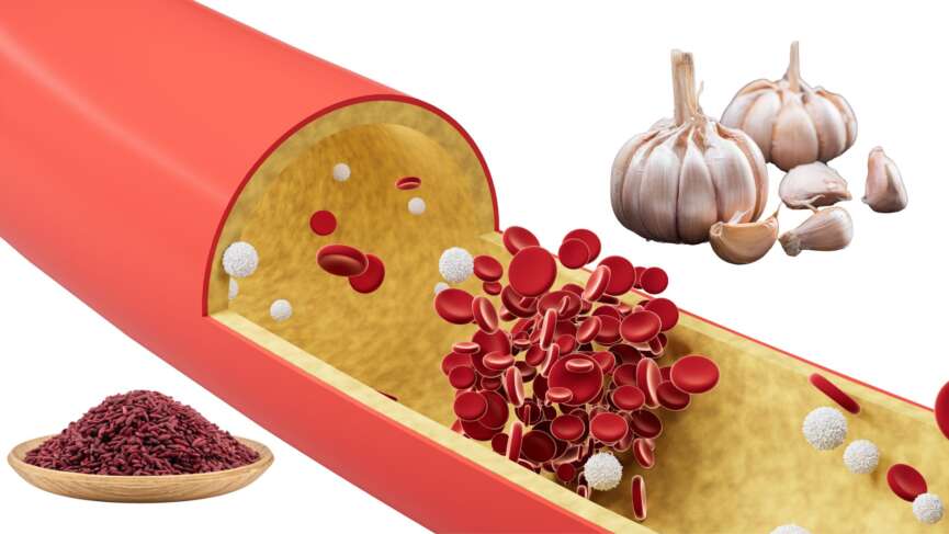
    Red Yeast Rice and Garlic For Lowering Cholesterol


    Explore the untold secrets of Red Yeast Rice and Garlic for effectively reducing cholesterol levels in this captivating video. Uncover the remarkable benefits of these natural remedies, supported by scientific evidence. Don’t miss out – click to watch now!

    What Is LDL Cholesterol?

  • 5 Warning Symptoms Of A Heart Attack

    5 Warning Symptoms Of A Heart Attack


    Heart attacks are a leading cause of death worldwide, and they can happen to anyone, regardless of age or gender. Recognizing the warning signs of a heart attack is crucial for timely treatment and better outcomes. This informative video will discuss the top 5 warning symptoms of a heart attack that you should never ignore. These symptoms can be subtle and life-threatening, from chest pain and discomfort to shortness of breath and nausea. Don’t wait until it’s too late – watch this video and learn how to identify the signs of a heart attack and take steps to protect your heart health.

    Does Heart Stop Beating In Heart Failure?

  • Myocarditis – Causes, Symptoms, and Treatments You Must Know

    Myocarditis – Causes, Symptoms, and Treatments You Must Know

    Highlights on Myocarditis

    1. Myocarditis is inflammation of the heart muscle, often caused by infections, immune responses, or toxins. It can affect the heart’s ability to pump blood efficiently.
    2. Viral infections like influenza are the most common cause of myocarditis, but they can also be triggered by bacterial infections, autoimmune diseases, toxins, and even medications.
    3. Myocarditis can cause chest pain, shortness of breath, and fatigue and can lead to complications like heart failure that might require heart transplantation.
    4. Myocarditis typically improves on its own or with treatment, often resulting in full recovery.
    5. Myocarditis can reoccur, especially if the underlying cause isn’t fully addressed, leading to long-term damage to the heart.

    What is Myocarditis?

    Myocarditis is the inflammation of the middle muscular layer of your heart. Inflammation means your heart muscle has swelling and irritation when fighting an infection, injury, or something harmful. This inflammation can affect your heart’s ability to pump blood and maintain normal rhythm. The condition affects people of all ages and can range from mild cases with few symptoms to life-threatening complications. [1]

    It can happen suddenly when caused by infection, toxins, or drugs. But when the trigger is autoimmune diseases like lupus or rheumatoid arthritis, it affects the heart more slowly, causing a long-term effect like heart failure and cardiomyopathy. [1]

    Causes and Risk Factors of Myocarditis

    Commonly identified causes and risk factors of myocarditis include:

    1. Age

    Myocarditis is more common in young adults, especially males, but it can affect people of any age. Children and infants are also vulnerable, particularly during viral outbreaks. [2]

    2. Viral, Bacterial, Fungal, or Protozoan Infection

    Viral infection is the most common cause of myocarditis, directly infecting cardiac cells by releasing cytokines such as IL-1, IL-6, and TNF-α or inducing an immune-mediated response. Infections caused by various viruses, including influenza, herpes, adenovirus, coxsackievirus, parvovirus B19, and even SARS-CoV-2 (the virus responsible for COVID-19), can cause myocarditis. [1]

    3. Toxins and Radiation

    Exposure to toxins and radiation can lead to myocarditis by directly damaging the heart muscle or triggering inflammation. Substances like excessive alcohol, recreational drugs such as cocaine, heavy metals like lead or mercury, and industrial chemicals can harm heart tissues and cause inflammation. Similarly, radiation therapy, particularly when targeting the chest area for cancer treatment, can unintentionally damage the heart muscle and lead to myocarditis. [3]

    4. Autoimmune Diseases

    Autoimmune diseases, such as lupus or rheumatoid arthritis, can cause your immune system to attack healthy tissues, including the heart muscle. Myocarditis may also occur due to autoimmune reactions triggered by infections or other conditions. [4]

    5. Rheumatic Fever

    Rheumatic fever is a condition that can develop if you’ve had an untreated or poorly treated strep throat infection. It happens when your immune system, mistakenly attacks healthy tissues, including your heart. If this affects your heart muscle, it can lead to myocarditis, causing swelling and irritation in your heart. [5]

    6. Side Effects of Medicines and Vaccination

    Some medicines and vaccinations can, in rare cases, cause myocarditis as a side effect. Certain medications, such as chemotherapy drugs, antibiotics, or immune-suppressing treatments, may trigger an inflammatory response in your heart muscle. Vaccinations, including some for COVID-19, have been linked to very rare cases of myocarditis, typically occurring in younger males after their second dose. [6]

    Symptoms of Myocarditis

    The symptoms of myocarditis can vary widely, and sometimes, there might be no noticeable symptoms at all. When symptoms do occur, they may include:

    1. Chest Pain

    You may feel sharp, stabbing, or pressure-like pain in your chest, similar to what happens during a heart attack. This occurs due to inflammation of the heart muscle and can worsen with physical activity or deep breaths. [7]

    2. Shortness of Breath

    You might struggle to catch your breath, especially when lying flat or after mild activity. This happens because the heart isn’t pumping blood effectively, causing fluid buildup in your lungs. [7]

    3. Fatigue

    Feeling unusually tired or weak, even after routine tasks, is common with myocarditis. This is due to the heart’s inability to supply oxygen-rich blood to your body efficiently. [8]

    4. Irregular Heartbeat

    You might notice your heart racing, fluttering, or skipping beats. This indicates that the inflamed heart muscle interferes with your heart’s normal electrical signals. [9]

    5. Swelling

    Swelling in your legs, ankles, or feet may occur due to fluid buildup caused by the heart’s reduced ability to pump blood. Swelling may also appear around your abdomen or even in the veins in your neck. [7]

    6. Flu-like Symptoms

    If a virus triggers your myocarditis, you might first experience symptoms like fever, sore throat, headache, muscle aches, or general discomfort. These often precede heart-related symptoms.

    7. Syncope

    You could feel dizzy or faint, especially during physical activity or when standing up suddenly. This happens because reduced blood flow to your brain can result from your heart’s inability to pump effectively. [7]

    When to Seek Help if You Have Myocarditis

    Knowing when to call for help is important if you have myocarditis. You should dial 911 immediately if your chest pain feels tight or heavy, lasts more than 15 minutes, or starts spreading to your arms, back, neck, or jaw. It’s also important to seek urgent help if you feel short of breath or nauseous along with the pain. These could be signs of a serious problem that needs immediate attention, so don’t wait to get the care you need. [10]

    Diagnosis of Myocarditis

    The inflammation caused by myocarditis can cause chest pain similar to that in a heart attack, making the diagnosis difficult. The following tests might be done to diagnose myocarditis:

    1. Blood Tests

    A complete blood count may reveal an elevated white blood cell count, indicating an infection or inflammation. Tests like troponin and creatine kinase measure heart enzymes that are released when your heart muscle is damaged, helping to confirm myocarditis. C-reactive protein and erythrocyte sedimentation rate can show your body’s inflammation level. If a viral infection is suspected, blood tests can identify specific viruses or antibodies that may have triggered the condition. [7]

    2. Electrocardiography (EKG)

    EKG is a test that checks your heart’s rhythm by recording its electrical activity. It can detect certain changes that might suggest myocarditis, although these changes aren’t specific to the condition. Importantly, even if your ECG looks normal, it doesn’t rule out myocarditis completely. [10]

    3. Chest X-ray

    A chest X-ray gives a clear image of your heart and lungs, helping to check for signs like an enlarged heart or fluid buildup in your lungs, which can happen when your heart isn’t pumping well. While it can’t confirm myocarditis independently, a chest X-ray helps your doctor rule out other conditions that can mimic myocarditis. [11]

    4. Echocardiography

    An echocardiogram, or echo, is a test that uses sound waves to create detailed images of your heart. An echo can help see how well your heart is pumping and check for any areas of damage. It can also show if your heart is enlarged or if there’s fluid around it, which are signs of inflammation. This test is painless and non-invasive, and it gives information about how your heart is functioning. [12]

    5. Cardiac Magnetic Resonance Imaging (MRI)

    An MRI scan creates detailed images of the heart’s shape and structure. It’s especially useful for assessing heart function and detecting inflammation or scar tissue caused by myocarditis. [13]

    6. Positron Emission Tomography (PET) Scan

    A PET  scan involves using radioactive dyes to produce body images, showing how the heart functions. Typically, it’s done alongside a CT or MRI scan for better assessment. [11]

    7. Heart Muscle Biopsy

    If other tests are inconclusive, endomyocardial biopsy is done. During this procedure, tiny samples of heart muscle are taken. It helps identify the extent and type of inflammation linked to myocarditis. However, this procedure is uncommon. [14]

    Treatment of Myocarditis

    Myocarditis typically improves on its own or with treatment, often resulting in full recovery. If a viral infection causes myocarditis, the treatment typically focuses on managing symptoms and allowing your body to recover. This includes rest, avoiding strenuous activities to give your heart time to heal, and hydration to support overall health. Your doctor might also recommend pain relievers like acetaminophen or nonsteroidal anti-inflammatory drugs (NSAIDs) to reduce discomfort and inflammation.  [710]

    1. Corticosteroids and Immunosuppressants

    Corticosteroids (such as prednisone) or immunosuppressants (like azathioprine or methotrexate) are used in the treatment of myocarditis. These medications help reduce the immune system’s activity and limit inflammation, improving heart function. However, their use depends on the underlying cause of myocarditis, and they are often reserved for more severe cases. [7]

    2. Heart Failure Medications

    When myocarditis causes the heart to weaken, making it less effective at pumping blood, you might be given medication to assist your heart. These include angiotensinogen converting enzyme (ACE) inhibitors (e.g., enalapril), which relax blood vessels, reduce blood pressure, and improve blood flow, and beta-blockers (like metoprolol), which slow down the heart rate and reduce the heart’s workload. Diuretics (such as furosemide) may also be used to remove excess fluid from your body, especially if you have swelling in your legs, abdomen, or around the lungs. [10]

    3. Antiarrhythmic Medications

    Myocarditis can cause irregular heart rhythms, which can be life-threatening. Antiarrhythmic medications, like amiodarone or lidocaine, which help restore a normal heart rhythm, may be used to manage this. In some cases, if arrhythmias are severe or unresponsive to medication, your doctor may recommend implanting a pacemaker to regulate the heart’s rhythm and ensure it beats at a consistent pace. [9]

    4. Antibiotics (for bacterial myocarditis)

    If a bacterial infection causes your myocarditis, your doctor will prescribe antibiotics to treat the infection. The type of antibiotic depends on the specific bacteria causing the condition. For example, penicillin may be used for infections caused by certain bacteria, while more broad-spectrum antibiotics may be needed for others. Early antibiotic treatment is important to prevent further heart damage. [15]

    5. Ventricular Assist Device (VAD)

    VADs are special devices that help weakened hearts pump blood throughout the body. It is usually performed as a bridge to a heart transplant. [9]

    6. Heart Transplant (in severe cases)

    In extremely severe cases of myocarditis, where the heart is no longer able to function effectively despite other treatments, a heart transplant may be necessary. This is typically considered only when the heart failure is life-threatening, and all other options have been exhausted. A heart transplant is a complex procedure with risks, but it can offer a chance at life for patients with advanced heart failure due to myocarditis. [16]

    7. Extracorporeal Membrane Oxygenation (ECMO)

    ECMO, a specialized heart-lung support device, can significantly improve survival rates for patients with severe myocarditis, especially when conventional treatments aren’t effective. [17]

    Complications of Myocarditis

    If left untreated, myocarditis can cause can lead to serious consequences over time. The inflammation in your heart can weaken its ability to pump blood, leading to heart failure, where your heart struggles to meet your body’s needs.

    You might also develop life-threatening arrhythmias. In severe cases, myocarditis can cause cardiogenic shock, where your heart can’t pump enough blood to keep your organs working. Scarring from the inflammation may also lead to chronic heart damage, permanently weakening your heart. That’s why it is important to seek medical care early and follow your treatment plan to avoid these long-term complications. [18]

    ‌Recurrence of Myocarditis

    Myocarditis can sometimes come back, even after you’ve recovered from the initial episode. This is called recurrence and can happen if the underlying cause, like a viral infection or autoimmune condition, isn’t fully resolved or flares up again. Recurrence may bring similar symptoms, such as chest pain, fatigue, or shortness of breath, and can further stress your heart. If you experience any new or returning symptoms, it’s important to inform your doctor immediately. Early treatment of recurrent myocarditis can help protect your heart and prevent long-term damage. [18]

    Prevention of Myocarditis

    Preventing myocarditis starts with taking steps to protect your heart from infections and other triggers. Keeping up with your vaccinations, such as the flu shot and COVID-19 vaccine, can reduce the risk of viral infections that might lead to myocarditis. Practicing good hygiene, like washing your hands regularly and avoiding close contact with sick individuals, helps prevent the spread of germs. If you’re taking medications, use them only as prescribed to avoid harmful side effects, and stay away from substances like alcohol and recreational drugs that can damage your heart. Following your doctor’s treatment plan for autoimmune conditions can lower the chances of inflammation affecting your heart. [19]

    Questions To Ask Your Doctor

    1. How long will it take for my heart to recover?
    2. Should my family members be screened for any genetic or infectious causes?
    3. What warning signs should I watch for that might suggest my condition is worsening?
    4. Is it safe for me to engage in physical exercise or sports activities with myocarditis?
    5. Do I need to avoid certain activities or foods during my recovery?

    References

    1. Kühl, U., Pauschinger, M., Seeberg, B., Lassner, D., Noutsias, M., Poller, W., & Schultheiss, H. P. (2005). Viral persistence in the myocardium is associated with progressive cardiac dysfunction. Circulation, 112(13), 1965–1970. Available from: https://doi.org/10.1161/CIRCULATIONAHA.105.548156
    2. Keller K, Sagoschen I, Konstantinides S, Gori T, Münzel T, Hobohm L. Incidence and risk factors of myocarditis in hospitalized patients with COVID‐19. Journal of Medical Virology. 2023 Mar;95(3). Available from: https://pubmed.ncbi.nlm.nih.gov/36892180
    3. Nguyen LS, Cooper LT, Kerneis M, Funck-Brentano C, Silvain J, Brechot N, et al. Systematic analysis of drug-associated myocarditis reported in the World Health Organization pharmacovigilance database. Nature Communications. 2022 Jan 10;13(1). Available from:‌ https://pubmed.ncbi.nlm.nih.gov/35013204
    4. Nielsen JC, Lin YJ, de Oliveira Figueiredo MJ, Sepehri Shamloo A, Alfie A, Boveda S, et al. European Heart Rhythm Association (EHRA)/Heart Rhythm Society (HRS)/Asia Pacific Heart Rhythm Society (APHRS)/Latin American Heart Rhythm Society (LAHRS) expert consensus on risk assessment in cardiac arrhythmias: use the right tool for the right outcome, in the right population. Heart Rhythm [Internet]. 2020 Sep 1 [cited 2022 Jul 4];17(9):e269–316. Available from: https://www.sciencedirect.com/science/article/pii/S1547527120304252
    5. British Heart Foundation. Myocarditis [Internet]. British Heart Foundation. 2019. Available from: https://www.bhf.org.uk/informationsupport/conditions/myocarditis
    6. Munjal JS, Flores SM, Yousuf H, Gupta V, Munjal RS, Anamika F, Mendpara V, Shah P, Jain R. Covid- 19 Vaccine-induced Myocarditis. J Community Hosp Intern Med Perspect. 2023 Sep 2;13(5):44-49. doi: 10.55729/2000-9666.1229. PMID: 37868673; PMCID: PMC10589044. Available from: https://pmc.ncbi.nlm.nih.gov/articles/PMC10589044/
    7. Ammirati E, Moslehi JJ. Diagnosis and Treatment of Acute Myocarditis. JAMA. 2023 Apr 4;329(13):1098.Available from: ‌ https://pubmed.ncbi.nlm.nih.gov/37014337
    8. Gulati M, Levy PD, Mukherjee D, Amsterdam E, Bhatt DL, Birtcher KK, et al. 2021 AHA/ACC/ASE/CHEST/SAEM/SCCT/SCMR Guideline for the Evaluation and Diagnosis of Chest Pain: A Report of the American College of Cardiology/American Heart Association Joint Committee on Clinical Practice Guidelines. Circulation [Internet]. 2021 Oct 28;144(22). Available from: https://www.ahajournals.org/doi/10.1161/CIR.0000000000001029
    9. Al-Khatib SM, Stevenson WG, Ackerman MJ, Bryant WJ, Callans DJ, Curtis AB, et al. 2017 AHA/ACC/HRS guideline for management of patients with ventricular arrhythmias and the prevention of sudden cardiac death. Heart Rhythm. 2018 Oct;15(10):e73–189. Available from:‌ https://pubmed.ncbi.nlm.nih.gov/29097319
    10. Gluckman TJ, Bhave NM, Allen LA, Chung EH, Spatz ES, Ammirati E, et al. 2022 ACC Expert Consensus Decision Pathway on Cardiovascular Sequelae of COVID-19 in Adults: Myocarditis and Other Myocardial Involvement, Post-Acute Sequelae of SARS-CoV-2 Infection, and Return to Play. Journal of the American College of Cardiology. 2022 Mar; Available from: https://pubmed.ncbi.nlm.nih.gov/35307156
    11. Lampejo, T., Durkin, S. M., Bhatt, N., & Guttmann, O. (2021). Acute myocarditis: aetiology, diagnosis and management. Clinical medicine (London, England), 21(5), e505–e510. Available from: https://doi.org/10.7861/clinmed.2021-0121
    12. Bozkurt B, Colvin M, Cook J, Cooper LT, Deswal A, Fonarow GC, et al. Current Diagnostic and Treatment Strategies for Specific Dilated Cardiomyopathies: A Scientific Statement From the American Heart Association. Circulation. 2016 Dec 6;134(23). Available from: https://pubmed.ncbi.nlm.nih.gov/27832612
    13. Sen G, Scully P, Gordon P, Sado D. Advances in the diagnosis of myocarditis in idiopathic inflammatory myopathies: an overview of diagnostic tests. Rheumatology (Oxford, England) [Internet]. 2024 Jan 16 [cited 2024 Jul 30];63(7):1825–36. Available from: https://www.ncbi.nlm.nih.gov/pmc/articles/PMC11215992/
    14. Basso C, Calabrese F, Angelini A, Carturan E, Thiene G. Classification and histological, immunohistochemical, and molecular diagnosis of inflammatory myocardial disease. Heart Failure Reviews. 2012 Oct 25;18(6):673–81. Available from: https://pubmed.ncbi.nlm.nih.gov/23096264

  • Cardiomyopathy – Causes, Symptoms, Treatment, and Implantable Devices

    Cardiomyopathy – Causes, Symptoms, Treatment, and Implantable Devices

    Highlights of Cardiomyopathy

    • Cardiomyopathy affects the heart muscle, making it harder for the heart to pump blood to the rest of the body.
    • There are different types of cardiomyopathy, including dilated, hypertrophic, and restrictive.
    • A variety of factors, including genetics, infections, autoimmune diseases, certain medications, and lifestyle factors, such as alcohol abuse or uncontrolled high blood pressure, can cause cardiomyopathy.
    • Common symptoms of cardiomyopathy are shortness of breath, swelling in the legs or abdomen, irregular heartbeat, chest pain, and However, some patients may not experience symptoms, especially in the early stages.
    • Cardiomyopathy cannot be cured, but treatment, including implantable devices for cardiomyopathy, can improve symptoms and quality of life, allowing many individuals to lead active lives.

    What Is Cardiomyopathy?

    Cardiomyopathy is a disease that affects the heart muscles, impairing their ability to pump blood to the rest of the body effectively. It often goes undetected, but it is estimated that approximately 1 in every 500 adults may have this condition. It can affect people of any age, sex, and race.

    Types of Cardiomyopathy

    There are several types of cardiomyopathy; the common ones are as follows:

    1. Hypertrophic Cardiomyopathy (HCM)

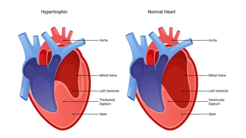
    If you have hypertrophic cardiomyopathy, the muscular wall of your heart becomes thickened, making it difficult for your heart to pump blood effectively throughout your body. This condition is genetic, so if one of your parents has HCM, there is a 50 percent chance that you may have inherited HCM can develop at any age, but it tends to be more severe when it starts during childhood.

    2. Dilated Cardiomyopathy

    In dilated cardiomyopathy, the heart muscle becomes weakened and dilated, decreasing the heart’s ability to pump blood It is the most common type of cardiomyopathy.

    3. Restrictive Cardiomyopathy

    Restrictive cardiomyopathy is a type of myopathy characterized by abnormal stiffness of the heart muscle, which impairs its ability to fill properly with blood during the relaxation phase of the heartbeat. This stiffness is often caused by infiltrating abnormal substances, such as scar tissue or proteins, into the heart muscle, which results in reduced ventricular filling and decreased cardiac output, leading to heart failure.

    Other types of Cardiomyopathy you might want to know:

    • Peripartum Cardiomyopathy: It is a rare variant of cardiomyopathy that begins sometime during the final month of pregnancy and continues about five months after delivery. Risk factors for peripartum cardiomyopathy include multiple pregnancies, advanced maternal age, hypertension, preeclampsia, and the use of certain medications.
    • Takotsubo Cardiomyopathy: It is named after the shape the heart takes on during contraction, resembling a Japanese octopus trap called a “takotsubo.”Some refer to it as “broken heart syndrome” or stress-induced cardiomyopathy. Takotsubo cardiomyopathy is characterized by a sudden and temporary weakening of the heart muscle, typically triggered by severe emotional or physical stress.

    Risk Factors and Causes of Cardiomyopathy

    While the exact cause of cardiomyopathy is often complex and multifactorial, certain risk factors can increase the likelihood of developing this condition. Some of the risk factors are as follows:

    risk-factors-of-cardiomyopathy

    1. Family History

    A family history of cardiomyopathy or sudden cardiac death can significantly increase the risk of developing the condition.

    2. High Blood Pressure

    If you have uncontrolled high blood pressure, it can strain the heart muscle over time, leading to hypertensive cardiomyopathy. Hypertensive cardiomyopathy is a form of secondary cardiomyopathy characterized by thickening and stiffening of the heart muscle.

    3. Alcohol Abuse

    Excessive and prolonged drinking of alcohol can weaken the muscles in your heart and lead to the development of dilated cardiomyopathy.

    4. Heart valve disorder

    Malfunctioning heart valves, such as those affected by conditions like rheumatic fever or infective endocarditis, can disrupt normal blood flow through the heart and contribute to the development of

    5. Viral and protozoal infections

    Certain organisms like the Coxsackie B virus, Human Immunodeficiency virus (HIV), and Chagas disease are known to be associated with the development of cardiomyopathy.

    6. Autoimmune Diseases

    Autoimmune diseases, such as rheumatoid arthritis or lupus, can cause inflammation and damage the heart muscle, leading to secondary

    7. Drug abuse

    Illicit drug use, particularly cocaine and amphetamines, can cause damage to the heart muscle and increase the risk of cardiomyopathy.

    Symptoms of Cardiomyopathy

    The symptoms of cardiomyopathy can vary depending on the type and severity of the condition. Common symptoms include:

    symptoms-of-cardiomyopathy

    1. Shortness of breath during physical activity or when lying down.
    2. Fatigue or weakness, even with minimal exertion.
    3. Swelling in the legs, ankles, or abdomen.
    4. Irregular heartbeat or discomfort.
    5. Chest pain or fainting spells.
    6. Dizziness or fainting spells

    Diagnosis of Cardiomyopathy

    Various diagnostic tests can detect the presence of cardiomyopathy. Common diagnostic tests are discussed below:

    diagnosis-of-cardiomyopathy

    1. Electrocardiogram (EKG)

    EKG is a common diagnostic test used to assess the electrical activity of the heart and can provide valuable information for diagnosing cardiomyopathy. While an EKG alone cannot definitively diagnose cardiomyopathy, it can help detect abnormalities in heart rhythm and conduction, which may indicate underlying cardiac issues.

    2. Echocardiogram

    An echocardiogram is a noninvasive test that uses sound waves to create detailed images of the heart’s structure and function. It can help identify abnormalities in heart size, shape, and function and assess the heart’s pumping ability.

    3. Blood Tests

    Blood tests help assess levels of certain enzymes and proteins that can indicate heart muscle damage. These may include cardiac troponin and brain natriuretic peptide (BNP).

    4. Cardiac MRI (Magnetic Resonance Imaging)

    Cardiac MRI provides detailed images of the heart’s structure and can detect abnormalities in the heart muscle, valves, and blood It is particularly useful for evaluating myocardial tissue characteristics and detecting scar tissue.

    5. Cardiac CT (Computed Tomography)

    CT scans can provide detailed images of the heart and blood vessels, helping to identify structural abnormalities, assess coronary artery disease, and evaluate heart function.

    6. Genetic Testing

    Genetic testing may be recommended in cases where a genetic component to cardiomyopathy is It helps to identify specific genetic mutations associated with the condition.

    7. Cardiac Catheterization

    This procedure involves inserting a tube called a catheter into a blood vessel in the arm, groin, thigh, or neck, which is then guided to the heart. It helps to measure heart chamber pressures to assess for heart failure, obtain blood samples to evaluate oxygen levels and heart function and take an endomyocardial biopsy to diagnose specific types of cardiomyopathy or myocarditis.

    8. Coronary Angiography

    Coronary angiography is a specialized X-ray imaging technique used to visualize the coronary arteries. It helps to rule out CAD as a cause of cardiomyopathy by detecting blockages or narrowing in the coronary It can also guide treatment decisions, such as angioplasty or bypass surgery if significant CAD is present.

    9. Heart Biopsy

    It is a minimally invasive procedure and is sometimes utilized in diagnosing cardiomyopathy. It involves extracting a small tissue sample from the heart muscle. This biopsy aids in identifying specific types of cardiomyopathies or myocarditis, providing crucial insights for accurate diagnosis and treatment planning.

    Treatment of Cardiomyopathy

    The treatment of cardiomyopathy aims to manage symptoms, improve heart function, prevent complications, and enhance overall quality of life.

    Beyond these lifestyle steps, medications, surgery, and implantable devices for cardiomyopathy are key parts of care. Here is a list of five things you can do to help you with your cardiomyopathy.

    treatment-of-cardiomyopathy

    • Heart-Healthy Diet: you can begin by adopting a diet low in sodium, saturated fats, and cholesterol while high in fruits, vegetables, whole grains, and lean proteins. it can help manage blood pressure, cholesterol levels, and overall heart health.
    • Regular Exercise: Regular physical activity, as tolerated, can improve cardiovascular fitness, strengthen the heart muscle, and enhance overall well-being. However, you should be aware that not everybody can do all types of You need to talk to your doctor before you begin any kind of exercise.
    • Smoking Cessation: It is important to note that a reduction in the consumption of cigarettes can reduce your risk of cardiovascular complications.
    • Limiting Alcohol: For individuals with alcoholic cardiomyopathy, abstaining from or reducing alcohol consumption is critical to prevent further damage to the heart muscle.
    • Weight Management: You should aim to achieve a healthy weight. Achieving and maintaining a healthy weight can help decrease the strain on your heart and improve overall cardiovascular health.

    Medications for cardiomyopathy

    Heart Failure Medications include angiotensin-converting enzyme inhibitors (Enalapril, Lisinopril), Angiotensin II Receptor Blockers (Losartan, valsartan), beta-blockers (carvedilol, metoprolol), and diuretics(spironolactone, Eplerenone). These medications are a core part of heart failure treatment, commonly prescribed to alleviate heart failure symptoms, enhance heart function, and decrease fluid retention.

    • Anticoagulants/Antiplatelets: These drugs may be administered to prevent blood clot formation in individuals with specific types of cardiomyopathy at a higher risk of stroke or thromboembolism.
    • Antiarrhythmics: Antiarrhythmic medications are vital in managing abnormal heart rhythms associated with cardiomyopathy. They work by modifying cardiac cell electrical properties to restore and maintain normal heart rhythm. Examples include sodium channel blockers (e.g., flecainide), beta-blockers (e.g., metoprolol), potassium channel blockers (e.g., amiodarone), and calcium channel blockers (e.g., verapamil). The selection of these drugs depends on the type of arrhythmia and patient factors.
    • Blood Pressure Management: Medications like ACE inhibitors, ARBs, calcium channel blockers, or diuretics are prescribed to control elevated blood pressure, which is particularly crucial in managing hypertensive cardiomyopathy.
    • Statins: Statins are prescribed to regulate cholesterol levels and help mitigate the risk of complications associated with coronary artery disease in individuals with cardiomyopathy.

    Implantable Devices for Cardiomyopathy

    1. Pacemakers

    Pacemakers are among the most common implantable devices for cardiomyopathy, essential in managing certain types of cardiomyopathy, particularly those associated with rhythm disorders. Indicated primarily for patients with bradyarrhythmia like sinus node dysfunction or heart block, pacemakers work by monitoring the heart’s electrical activity and delivering electrical impulses to regulate heart rate and rhythm. Pacemaker implantation, a minimally invasive procedure performed under local anesthesia, involves inserting leads into the heart through veins and placing the pulse generator under the skin.

    2. Implantable Cardioverter-Defibrillators (ICDs) 

    IICDs are among the most important implantable devices for cardiomyopathy, playing a crucial role in managing cardiomyopathy, especially in cases with a risk of life-threatening ventricular arrhythmias. These devices continuously monitor the heart rhythm and are designed to deliver a shock when they detect a dangerous arrhythmia such as ventricular tachycardia or ventricular fibrillation. By restoring normal heart rhythm through electrical therapy, ICDs can prevent sudden cardiac arrest and potentially save lives. The implantation procedure involves placing leads into the heart and positioning the device under the skin, typically in the chest area.

    3. LifeVest

    The LifeVest is a wearable cardioverter defibrillator (WCD) designed for patients at risk of sudden cardiac arrest who are not immediate candidates for an ICD or who require temporary protection after an ICD removal. It is a garment worn directly against the skin, typically over the torso, with electrodes integrated into the fabric. The LifeVest continuously monitors the patient’s heart rhythm, and if it detects a life-threatening arrhythmia, it delivers a shock to restore normal rhythm. Unlike permanent ICDs, the LifeVest is non-invasive and can be worn during daily activities, providing continuous protection without surgical implantation. This device serves as a bridge therapy, offering temporary protection while patients await further evaluation or as a transitional measure after ICD implantation or explantation.

    4. Cardiac Resynchronization Therapy (CRT)

    CRT is a specialized treatment for certain types of cardiomyopathy, particularly those associated with heart failure and electrical It involves implanting a device similar to a pacemaker, called a CRT device, which delivers precisely timed electrical impulses to the heart’s ventricles. These impulses help synchronize the contractions of the heart’s chambers, improving its overall pumping efficiency. CRT is typically recommended for individuals with heart failure and a reduced ejection fraction who have a widened QRS complex on their EKG, indicating electrical desynchrony.

    Surgical Interventions for Cardiomyopathy

    Surgical interventions for cardiomyopathy aim to address specific underlying issues contributing to heart dysfunction and improve overall cardiac function.

    • Septal Myectomy: This surgical procedure involves removing a portion of the thickened heart muscle (septum) in individuals with hypertrophic cardiomyopathy to relieve symptoms and improve blood flow.
    • Heart Transplantation: In severe cases of cardiomyopathy refractory to medical therapy, heart transplantation may be considered. It is reserved for people with end-stage heart failure.

    From heart-healthy lifestyle changes and medications to implantable devices for cardiomyopathy and, in severe cases, heart transplantation, today’s treatments can help most people manage cardiomyopathy and protect their quality of life.

    Questions To Ask Your Doctor

    1. If the cause of my cardiomyopathy is genetic, what is the chance that my children will have it?
    2. Are there any diet plans that I can follow to help me with my symptoms?
    3. Since cardiomyopathy affects my heart, can I go to the gym?
    4. Is there anything that I need to watch out for that might need immediate medical attention?
    5. Can I go on a vacation with this cardiomyopathy?

    References


  • Rheumatic Heart Disease – Risks, Signs, Diagnosis, and Empowered Solutions

    Rheumatic Heart Disease – Risks, Signs, Diagnosis, and Empowered Solutions

    Rheumatic Heart Disease (RHD)

    Rheumatic heart disease (RHD) is a chronic condition that develops as a complication of rheumatic fever. Rheumatic fever is an inflammatory disease that can occur after an untreated or inadequately treated streptococcal throat infection, caused explicitly by group A Streptococcus bacterium.
    Rheumatic fever triggers an immune response in the body, where the immune system attacks various tissues, including the heart, joints, skin, and nervous system. If the heart is affected, it can lead to the development of rheumatic heart disease. The most significant impact of rheumatic heart disease is the damage it causes to heart valves, particularly the mitral valve. The inflammation caused by the immune response causes them to become thickened and less flexible. This leads to valvular dysfunction, including regurgitation and narrowing of the valves.

    Key points for Rheumatic Heart Disease

    • Rheumatic heart disease occurs when the heart valves are permanently damaged because of rheumatic fever.
    • Rheumatic fever is a kind of sickness that can affect many parts of the body, especially the heart.
    • If infections caused by strep bacteria aren’t treated well, they can raise the chances of getting rheumatic fever. Kids who often get strep throat are at the most risk for this heart problem.
    • Getting treated for streptococcal throat infection can prevent getting RHD.
      Doctors look for recent cases of strep infections or rheumatic fever to find out if someone has rheumatic heart disease.

    Risk Factors for Rheumatic Heart Disease

    Here are some of the key risk factors for rheumatic heart disease:

    1. Untreated Strep Infections: The primary risk factor for RHD is inadequate treatment or lack of treatment for streptococcal infections. When these infections are not promptly and properly treated with antibiotics, the bacteria can trigger an abnormal immune response in some individuals, leading to inflammation and damage to the heart valves.
    2. Repeated Infections: Recurrent episodes of streptococcal infections, especially in susceptible individuals, increase the risk of developing RHD. Multiple episodes of untreated or poorly managed strep infections can heighten the chances of heart complications.
    3. Age and Gender: RHD is more common in children and young adults, particularly between the ages of 5 and 15. Females also tend to be at a slightly higher risk than males. particularly in areas where access to healthcare and antibiotics is limited.
    4. Socioeconomic Factors: Socioeconomic factors, such as poor living conditions, overcrowding, limited access to healthcare, and inadequate nutrition, can increase the likelihood of exposure to streptococcal infections. It hinders the prompt and effective treatment of these infections, contributing to the risk of RHD.
    5. Environmental Factors: Environmental factors, such as living in densely populated areas with poor sanitation, might contribute to the spread of streptococcal infections, increasing the risk of RHD.

    Symptoms of Rheumatic Heart Disease

    A streptococcal throat infection usually causes the clinical signs of acute rheumatic fever (ARF) to appear two to three weeks later. RHD symptoms may not show up for several years. The kind and extent of the damage to the heart valves and which heart valves are impacted determine the symptoms when they appear. Common symptoms of rheumatic heart disease include:

    1. Shortness of Breath: As heart valve damage progresses and the heart’s pumping efficiency decreases, individuals with RHD may experience difficulty breathing, especially during physical activities or when lying flat.
    2. Fatigue: Reduced heart function can lead to decreased blood flow to the body’s organs and tissues, resulting in persistent fatigue and weakness.
    3. PalpitationsIrregular heartbeats or palpitations can occur due to valve abnormalities and changes in the heart’s electrical conduction system.
    4. Chest Pain: Some individuals with RHD may experience chest discomfort or pain, particularly when the heart’s blood supply is inadequate or when the heart is strained due to valve issues.
    5. Swelling: Swelling of the feet, ankles, or legs (edema) can develop as the heart’s pumping efficiency decreases, causing fluid buildup in the body’s lower extremities.
    6. Coughing: A persistent cough may develop, often accompanied by frothy or pink-tinged sputum, which can indicate fluid accumulation in the lungs (pulmonary congestion).
    7. Lightheadedness or Fainting: Insufficient blood flow to the brain can lead to dizziness, lightheadedness, or even fainting episodes.
    8. Weight Gain: Fluid retention due to heart failure can lead to unexplained weight gain over a short period.
    9. Joint Pain: In some cases, joint pain and inflammation (similar to symptoms of rheumatic fever) might occur, particularly if there is ongoing immune system activity.
    10. Skin Changes: Skin changes such as a bluish tint to the skin (cyanosis) or the development of small, painless nodules (called subcutaneous nodules) under the skin can occur in more severe cases of RHD.

    Diagnosis of Rheumatic Heart Disease

    Diagnosing RHD involves a combination of medical history assessment, physical examination, imaging tests, and sometimes laboratory investigations. The diagnosis of rheumatic heart disease is discussed below:

    1. Medical History and Physical Examination: Individuals with suspected RHD often have a history of rheumatic fever or recurrent streptococcal infections. During a physical examination, a murmur may be detected. The murmur results from blood leaking around damaged heart valves affected by RHD.
    2. Streptococcal Infection Testing: People with RHD may have had a recent or current streptococcal infection. Thus, streptococcal testing is of utmost importance. A swab of the throat can be cultured to identify the presence of streptococcus bacteria.
    3. Blood Test: Blood tests may be performed to measure certain markers of inflammation (such as C-reactive protein or erythrocyte sedimentation rate) and to check for streptococcal antibodies. Elevated levels of these markers can indicate ongoing inflammation and might suggest a recent streptococcal infection.
    4. Chest X-ray: A chest X-ray can provide information about the size and shape of the heart. In cases of advanced RHD, the heart might be enlarged, and there could be signs of fluid accumulation in the lungs.
    5. Electrocardiogram (ECG): An ECG records the electrical activity of the heart. It can help identify any irregularities in the heart’s rhythm or conduction system that might be related to RHD.
    6. Echocardiogram: An echocardiogram is a key diagnostic tool for RHD. It uses sound waves to create detailed images of the heart’s structure and function. It can help identify any damage to the heart valves, such as thickening, narrowing, or leakage, which are common characteristics of RHD.
    7. Cardiac MRI: In some cases, your doctor may recommend obtaining a cardiac MRI for more detailed images of the heart’s structure and function. This can provide additional information about the extent of valve damage.

    Treatment and Management of Rheumatic Heart Disease

    Treating RHD involves addressing the underlying cause, managing symptoms, and preventing further complications. The goals of treatment include:

    1. Antibiotics: Acute rheumatic fever, which is the precursor to RHD, is caused by a streptococcal infection. Antibiotics like penicillin or other appropriate antibiotics treat the active infection and prevent further streptococcal infections, which could exacerbate the condition.
    2. Anti-inflammatory medications: In the acute phase of rheumatic fever, anti-inflammatory medications such as nonsteroidal anti-inflammatory drugs (NSAIDs) are often prescribed to reduce inflammation and manage symptoms like fever, pain, and joint swelling.
    3. Heart Failure Management: For individuals with severe valve damage or other heart complications resulting from RHD, medications such as diuretics to reduce fluid retention, ACE inhibitors to lower blood pressure and improve heart function, and beta-blockers to slow down heart rate and reduce strain on the heart might be prescribed to manageheart failure symptoms.
    4. Surgery: In cases where the heart valves are significantly damaged and not functioning properly, surgical intervention might be necessary. Valve repair or replacement surgeries are common procedures to address this issue and improve heart function.
    5. Preventive Antibiotics: Individuals with rheumatic fever or existing heart valve damage due to RHD are typically advised to take prophylactic antibiotics before certain medical or dental procedures. This is to prevent the risk of bacterial endocarditis, an infection of the heart lining or valves.

    Complications of Rheumatic Heart Disease

    RHD can lead to several complications due to the damage it causes to the heart valves and other heart structures. Some of the complications of rheumatic heart disease include:

    1. Valvular Heart Disease: RHD primarily affects the heart valves, leading to valvular damage. The valves can become thickened, scarred, or distorted, impairing their ability to open and close properly. This can result in conditions such as:

    • Mitral Stenosis: The mitral valve becomes narrowed, making it difficult for blood to flow from the left atrium to the left ventricle.
    • Mitral Regurgitation: The mitral valve doesn’t close properly, causing blood to leak back into the left atrium.
    • Aortic Stenosis: The aortic valve becomes narrowed, obstructing the flow of blood from the left ventricle to the aorta.
    • Aortic Regurgitation: The aortic valve doesn’t close properly, causing blood to flow back from the aorta into the left ventricle.

    2. Heart Failure: As the valve damage worsens, the heart’s ability to pump blood efficiently can be compromised, leading to heart failure. This can result in symptoms like shortness of breath, fatigue, fluid retention, and swelling.

    3. Infective Endocarditis: The damaged heart valves can create an environment where bacteria are more likely to infect the heart lining or valves, leading to infective endocarditis. This is a serious and potentially life-threatening infection.

    4. Arrhythmias: Irregular heart rhythms, or arrhythmias, can occur due to the changes in the heart’s structure and function caused by RHD. These can range from mild palpitations to more severe rhythm disturbances.

    5. Pulmonary Hypertension: Over time, the increased strain on the heart due to valve damage can lead to high blood pressure in the pulmonary arteries which can affect the lungs and heart function.

    6. Stroke: In some cases, blood clots can form on the damaged heart valves. If a clot dislodges and travels to the brain, it can cause a stroke.

    7. Embolism: Blood clots can also travel to other parts of the body, causing blockages in blood vessels (embolism) and potentially affecting various organs.

    Prevention of Rheumatic Heart Disease

    Preventing RHD primarily involves the prevention and prompt treatment of streptococcal infections. Here are key strategies for preventing RHD:

    1. Prompt Treatment of Strep Infections: It’s crucial to promptly and effectively treat streptococcal infections, such as strep throat, with appropriate antibiotics. Treating these infections within ten days of symptom onset reduces the risk of developing rheumatic fever.
    2. Complete Antibiotic Course: Ensure that antibiotic treatments for streptococcal infections are completed as prescribed by a healthcare professional to fully eradicate the bacteria.
    3. Early Detection and Treatment of Rheumatic Fever: Recognizing and promptly treating rheumatic fever is essential. Treating rheumatic fever with anti-inflammatory medications (like aspirin or corticosteroids) and antibiotics can help prevent further heart damage that could lead to RHD.
    4. Regular Antibiotic Prophylaxis: Individuals who have had rheumatic fever are often prescribed ongoing antibiotics as prophylaxis to prevent recurrent streptococcal infections and minimize the risk of developing RHD.
    5. Improving Access to Healthcare: Ensuring access to healthcare, especially in regions with high incidence rates of streptococcal infections and rheumatic fever, is crucial. This includes access to healthcare facilities for diagnosis, treatment, and follow-up care.
    6. Health Education and Awareness: Educating communities, healthcare professionals, and individuals about the importance of seeking medical care for streptococcal infections, completing antibiotic courses, and recognizing symptoms of rheumatic fever can aid in early intervention and prevention.
    7. Promoting Hygiene and Sanitation: Implementing measures to improve hygiene, sanitation, and living conditions can help reduce the spread of streptococcal infections in communities.

    Questions for your doctor

    1. Is RHD curable, or can it be reversed through treatment?
    2. How often should I have follow-up appointments and monitoring for my condition?
    3. Are there any lifestyle changes or self-care measures that can help manage RHD symptoms and prevent complications?
    4. Can you guide me on preventing recurrent episodes of rheumatic fever and RHD?
    5. What should I do if I experience a flare-up or new RHD-related symptoms?
    6. Are there any specific dietary recommendations or restrictions for individuals with Rheumatic Heart Disease?
    7. How does RHD impact physical activity and exercise, and are there any limitations or precautions I should follow?

    References

    1. https://www.ncbi.nlm.nih.gov/books/NBK538286/
    2. https://world-heart-federation.org/what-we-do/rheumatic-heart-disease/

  • Aortic Stenosis – Symptoms, Treatment, Diagnosis and Prevention

    Aortic Stenosis – Symptoms, Treatment, Diagnosis and Prevention

    What is Aortic Stenosis?

    Aortic stenosis is a heart condition that affects the aortic valve, one of the four valves in your heart. Aortic stenosis occurs when the aortic valve narrows, reducing blood flow from the heart into the aorta, the main blood vessel that carries oxygenated blood to the rest of the body.

    Symptoms Of Aortic Stenosis

    Symptoms of aortic valve stenosis range from mild to severe. Most people with aortic valve stenosis would not experience any major symptoms for many years. Symptoms generally occur when the narrowing of the valve is severe:

    • Chest pain

    This is often felt during physical activity or when the heart has to work harder. This pain is often termed as angina.

    • Shortness of breath

    You can have shortness of breath, especially during exercise or when lying flat.

    • Dizziness

    You might feel lightheaded or dizzy due to reduced blood flow to the brain through the narrowed lumen of the valve. You might even have a syncope if it is too narrow.

    • Fatigue

    As the condition progresses, you can tire easily, usually much faster. You might not be able to conduct the regular activities you used to do.

    • Poor growth

    Children with congenital aortic stenosis can have poor growth. They can be low in weight and height about their age.

    • Heart failure

    When the disease progresses to very advanced stages, patients can present with heart failure and have worsening symptoms.

    Risk Factors of Aortic Stenosis

    Several factors can increase the risk of developing aortic stenosis, which often develops gradually over time. Some of them are discussed below:

    • Age

    As we age, the risk of aortic stenosis increases. The valve may naturally thicken and become less flexible over time.

    • Congenital heart conditions

    Some people are born with abnormal aortic valves that may eventually lead to stenosis. The bicuspid aortic valve is the most common congenital heart defect affecting 1% of the general population and is one of the leading causes of stenosis in the young population.

    • Aortic valve calcification

    Calcium is a mineral present in the blood. As blood flows through the valves, calcium can deposit on the heart valves, and the process gets hastened if the valve is highly pressured, like the aortic, and has microtrauma.

    • High cholesterol

    High cholesterol levels can lead to the buildup of fatty deposits on the aortic valve, narrowing it.

    Uncontrolled hypertension can strain the aortic valve and contribute to its deterioration.

    • Rheumatic fever

    Rheumatic fever is one of the most common conditions seen among children. A previous bout of rheumatic fever can damage the aortic valve, increasing the risk of stenosis.

    Treatment Of Aortic Stenosis

    Treatment options for aortic stenosis depend on the symptoms and the severity of the condition. While you might need a regular follow-up with your doctor if there is mild aortic valve disease or no symptoms, you may need surgery to repair or replace the valve for severe disease. There are less invasive options available, too, but that depends on your overall health and your choice.

    The treatment options for aortic valve stenosis are:

    • Balloon valvuloplasty

    This minimally invasive procedure is used in infants and children. The procedure is limited in adults, as the valve gets narrower afterward. So, it is usually only done if you are young or waiting for a valve replacement. In this procedure, a tube with a balloon tip is inserted into an artery through the arm or the groin. It is then taken to the aortic valve. Once it reaches the valve, the balloon is inflated, which widens the valve opening. The balloon is then deflated, and the catheter is withdrawn.

    • Aortic valve replacement

    It is the ultimate treatment for aortic stenosis. The damaged valve is replaced with a mechanical valve made from cows, pigs, or human tissue. While biological valves break down over time and need to be replaced again, mechanical valves remain longer than the biological valve, but the patient will have to take a blood thinning agent.

    The valve can be replaced by either of the following two procedures:

    • Surgical aortic valve replacement (SAVR):

    Aortic valve replacement is done via an open-heart surgical procedure. During an SAVR, you would be put under general anesthesia and placed on a ventilator while on a heart-lung bypass machine. A surgeon would then cut open your breastbone to get to the heart. In some patients, less invasive approaches can be used. The aortic stenosis valve is cut out and replaced by the new valve. Recovery in the hospital usually takes about five days. This procedure can use both bioprosthetic as well as mechanical valves.

    • Transcatheter aortic valve replacement (TAVR):

    TAVR is an alternative to SAVR and is done for those not candidates for open-heart surgery due to other comorbid conditions. This minimally invasive procedure requires no sternotomy or a heart-lung bypass machine. The replacement valve is usually inserted using a catheter into an artery in the leg, then threaded through arteries to the heart. Once within the diseased aortic valve, the folded TAVR valve is opened with a balloon and placed in the right place. Some valves can, however, self-expand and do not need re-positioning. The surgeon then removes the catheter once the new valve is securely placed. It is to be noted that this procedure uses only bioprosthetic valves only.

    Diagnostic Evaluation Of Aortic Stenosis

    Several diagnostic tests are performed to confirm and determine the cause and its severity. Tests for aortic valve stenosis may include:

    • Electrocardiogram (EKG)

    It is one of the routine tests performed that measures the electrical activity of the heart. It can show how fast or slow your heart is beating. Your doctor will look for the strain pattern in the EKG if you have aortic stenosis.

    • Echocardiogram (ECHO)

    In simple terms, it is an ultrasound of your heart, and it creates images of the heart. It is a fast and painless investigation that helps to evaluate the structure and function of the heart. It can recognize a weakened heart muscle, changes in the valves of the heart, and look at how the blood flows through the valves. With an ECHO, your doctor can determine the severity of aortic valve stenosis.

    • Chest X-ray

    Chest X-ray gives you a view of the heart and lungs together. It helps to determine if there is an enlarged heart, a common finding of aortic stenosis. It can also show an enlarged aorta and calcium build-up.

    • Cardiac computed tomography(CT)

    This test utilizes multiple X-rays to obtain detailed heart pictures. It is also utilized to evaluate the size of the aorta and calcium deposits on the aortic valves and to guide surgical management of the condition.

    • Cardiac catheterization

    This is a test where a catheter is guided from a vessel in the wrist or groin leading to the heart, and a contrast dye is injected through it. The dye goes to the arteries of the heart and helps to assess their patency. The test is not used to diagnose aortic valve disease but can be used if other tests are not able to diagnose the condition. A coronary angiogram can be used to identify patients who also might benefit from surgery on the blood vessels around the heart along with an aortic valve replacement procedure. Therefore, every patient with aortic stenosis should undergo a coronary angiogram to detect and treat any coexisting coronary vessel disease.

    Complications Of Aortic Stenosis

    While aortic stenosis can initially be asymptomatic, it can lead to several complications as the condition progresses. Watch out for these complications:

    • Reduced exercise tolerance

    As the condition progresses, reduced blood flow can lead to decreased exercise tolerance, fatigue, and a decline in overall physical capacity.

    • Heart failure

    As the aortic valve narrows further, the heart must work harder to pump blood through the narrowed valve. Over time, this extra strain on the heart can lead to left ventricular hypertrophy and, eventually, heart failure.

    • Arrhythmias

    Aortic stenosis can increase the risk of abnormal heart rhythms or arrhythmias. The irregular heartbeats can further compromise the heart’s ability to pump blood effectively.

    • Infection (Infective Endocarditis)

    Aortic stenosis may make the heart valve more susceptible to infection. Bacterial or microbial infections of the heart valve can lead to a condition called infective endocarditis, which is a serious and potentially life-threatening complication.

    Prevention of Aortic Stenosis

    You can take steps to strengthen your heart and guard against developing aortic stenosis. These include:

    • Make heart-healthy choices 

    Healthy choices such as daily physical exercise and quitting smoking can prevent calcium buildup in arteries and aortic valve. If you have high blood pressure, high cholesterol, diabetes, or kidney problems, you should consult your doctor regarding adequate management of these chronic health conditions.

    • Keep up with dental checkups

    Gum infections can lead to infection and development of aortic valve stenosis. A regular dental checkup can avoid aortic valve disease secondary to gum infections.

    • Treat infections

    The bacteria that cause strep throat can sometimes cause rheumatic fever if left untreated. Rheumatic fever often happens in children and young adults. This is a condition that can be easily treated with antibiotics. So, getting treated for strep throat is important to prevent valvular heart disease.

    Question For Your Doctor

    1. What symptoms of Aortic Stenosis should prompt me to seek immediate medical attention?
    2. Should I limit my physical activity because of Aortic Stenosis?
    3. How often should I have my heart checked yearly?
    4. What are the treatment options?
    5. Are there any medications that I can take for this condition?
    6. How do I know if surgical or Transcatheter Aortic Valve Replacement is a better treatment option for me?
    7. What changes happen to the heart after having aortic stenosis over time?

    References

Sign Up to Our Newsletter

Be the first to know the latest updates