Category: Heart Health

  • Heart Health – Understanding and Managing Coronary Artery Disease

    Heart Health – Understanding and Managing Coronary Artery Disease

    What is coronary artery disease (CAD)?

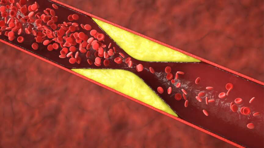
    Coronary Artery Disease (CAD) is a prevalent and potentially life-threatening cardiovascular condition affecting millions worldwide. It is the leading cause of heart-related morbidity and mortality in the US, imposing a significant burden on public health systems and individuals alike. CAD develops when the coronary arteries, the blood vessels responsible for supplying oxygen and nutrients to the heart muscle, become narrowed or blocked due to atherosclerosis, characterized by the accumulation of fatty deposits and plaque on the arterial walls.

    As the disease progresses, it hinders blood flow to the heart, depriving the cardiac muscle of the necessary oxygen and nutrients required for proper functioning. This can lead to various symptoms, such as chest pain (angina), shortness of breath, and fatigue. In severe cases, CAD can result in a heart attack, where a sudden and complete coronary artery blockage leads to the death of a portion of the heart muscle.

    Forms of coronary artery disease

    Coronary artery disease can present in various forms, each with its distinct characteristics and implications for heart health. It is essential to recognize and diagnose the specific form of CAD in each individual, as treatment strategies and management approaches may vary.

    The different forms of coronary artery disease include:

    • Stable Angina: Stable angina is the most common form of CAD. It occurs when there is a partial blockage or narrowing of the coronary arteries, leading to chest pain or discomfort during physical exertion or emotional stress. The pain is predictable and tends to subside with rest or medication.
    • Unstable Angina: Unstable angina is a more severe and unpredictable form of CAD. It happens when a plaque in the coronary artery ruptures, causing a sudden and severe reduction in blood flow. Unstable angina can occur at rest or with minimal exertion and requires immediate medical attention, as it may lead to a heart attack.
    • Acute Myocardial Infarction (Heart Attack): A heart attack occurs when a coronary artery becomes completely blocked by a blood clot, cutting off blood flow to a portion of the heart muscle. This leads to the death of heart tissue. Heart attacks are medical emergencies that require urgent treatment to restore blood flow and prevent further damage.
    • Chronic Total Occlusion (CTO): CTO refers to a complete blockage of a coronary artery that persists for an extended period, often over three months. People with CTO may experience chronic chest pain, reduced exercise tolerance, and, in severe cases, heart failure.
    • Silent Ischemia: Some individuals with coronary artery disease may not experience typical symptoms like chest pain or discomfort. Instead, they may have “silent ischemia,” where the heart muscle does not receive enough blood and oxygen without noticeable symptoms. This type of CAD is concerning as it can lead to heart damage without the person knowing it.

    Risk factors of coronary artery disease

    Coronary artery disease are often associated with modifiable risk factors, such as an unhealthy diet, sedentary lifestyle, smoking, high blood pressure, high cholesterol levels, and diabetes. However, genetic predisposition and older age are the 2 major non-modifiable risk factors that significantly affect the development of coronary artery disease.

    Symptoms of coronary artery disease

    The symptoms of CAD may not be immediately apparent, as it is a chronic condition that progresses slowly over many years, possibly even decades. However, as the arteries narrow due to plaque accumulation, mild symptoms may emerge, indicating that the heart is working harder to supply oxygen-rich blood to the body.

    Common symptoms of chronic CAD include:

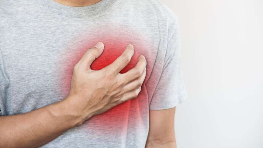
    1. Chest Discomfort: One of the most common and characteristic symptoms of coronary artery disease is chest discomfort, often described as a feeling of pressure, tightness, squeezing, or heaviness in the chest. This discomfort may be mistaken for indigestion or heartburn, but it typically occurs during physical exertion or emotional stress and eases with rest. Such kind of pain is seen with stable angina. It is essential not to ignore persistent or recurrent chest discomfort that does not go away with rest or nitroglycerine then, it might be more severe conditions like unstable angina or myocardial infarction.
    2. Shortness of Breath: People with CAD may experience shortness of breath, especially during activities that once seemed effortless. As the coronary arteries become narrowed, the heart may struggle to pump blood efficiently, leading to reduced oxygen supply to the body. Shortness of breath can occur even during mild physical exertion or at rest in more advanced stages of the disease.
    3. Fatigue: Persistent fatigue is another common symptom of CAD. As the heart’s blood supply diminishes, the heart muscle may not receive enough oxygen, leading to a feeling of constant tiredness and reduced stamina. Fatigue may be particularly pronounced after physical activity or during stressful situations.
    4. Unexplained Pain in Other Areas: In some cases, the pain or discomfort associated with coronary artery disease may radiate to other body parts. Pain may be felt in the arms (usually the left arm), shoulders, neck, jaw, or back. This referred pain can vary in intensity and may come and go, making it important to recognize these unusual symptoms.
    5. Dizziness and Lightheadedness: Insufficient blood flow to the brain due to narrowed coronary arteries can cause dizziness or lightheadedness. People with CAD may experience fainting spells or suddenly feel dizzy when standing up.
    6. Palpitations: CAD can disrupt the heart’s rhythm, leading to palpitations or a sensation of irregular heartbeats.

    Tests to detect coronary artery disease

    Tests play a crucial role in diagnosing CAD and evaluating heart function. Besides a thorough medical assessment, your healthcare provider may recommend the following tests to aid the diagnosis:

    1. Blood Tests: These tests analyze specific substances in your blood that may indicate artery damage or an increased risk of developing CAD. Elevated levels of certain biomarkers like Troponin, CK-MB, and BNP can provide valuable insights into your heart health.
    2. Electrocardiogram (EKG/ECG): By recording your heart’s electrical activity, an EKG/ECG can detect previous or current heart attacks, ischemia (reduced blood flow to the heart), and irregular heart rhythms that might be associated with CAD.
    3. Echocardiogram: Utilizing sound waves, an echocardiogram evaluates the structure and function of your heart. It provides valuable information about heart chamber sizes, pumping capacity, and any abnormalities in heart valves, contributing to diagnosing CAD.
    4. Stress Test: This test assesses how your heart responds to physical exertion. It helps detect angina (chest pain) and can identify potential blockages in the coronary arteries by monitoring the heart’s performance during exercise.
    5. Cardiac Catheterization: Considered the gold standard for diagnosing coronary artery disease, this procedure involves inserting thin tubes (catheters) into the coronary arteries to evaluate their condition. It can confirm the presence and severity of blockages, guiding further treatment decisions.
    6. Computed Tomography Coronary Angiogram: Using advanced imaging technology and contrast dye, a CT coronary angiogram generates detailed 3D images of your heart’s movement. This non-invasive test allows the detection of blockages in the coronary arteries, aiding in CAD diagnosis.
    7. Coronary Calcium Scan: This test measures the amount of calcium in the walls of your coronary arteries, which indicates atherosclerosis. Though it does not directly determine significant blockages, it helps assess your risk for developing coronary artery disease.

    What is Cardiac catheterization?

    Coronary catheterization, also known as cardiac catheterization or coronary angiography, is a widely used and minimally invasive medical procedure that plays a crucial role in assessing the condition of the coronary arteries—the blood vessels responsible for supplying oxygen-rich blood to the heart muscle.

    During this diagnostic test, a skilled medical team performs the procedure in a specialized area called the catheterization laboratory or “cath lab.” The patient is usually awake during the procedure, as it involves only local anesthesia to numb the area where the catheter will be inserted.

    To begin the process, a small incision is made in the wrist or groin area, where a thin, flexible tube called a catheter is carefully inserted into the blood vessels. The choice of insertion site depends on various factors, such as the patient’s medical history and the preference of the medical team.

    Once the catheter is in place, a special dye known as contrast is injected through the catheter and into the coronary arteries. This dye is visible on X-ray images, creating what is called an “angiogram.” As the contrast moves through the arteries, the medical team can visualize the blood flow and identify any potential blockages, narrowings, or abnormalities in the coronary arteries. The X-ray images obtained during the procedure provide valuable information about the condition of the heart’s blood vessels, helping doctors diagnose conditions such as CAD or other heart-related issues.

    What is coronary artery calcium scan?

    A coronary artery calcium scan, also known as a coronary calcium scan or cardiac CT for calcium scoring, is a specialized imaging test that helps assess the amount of calcium present in the walls of the coronary arteries. This test estimates a person’s risk of developing CAD.

    During the scan, the CT machine takes detailed images of the heart, focusing on the coronary arteries. The presence and quantity of calcium deposits in the arterial walls are measured and scored using a numerical scale called the Agatston score.

    A higher Agatston score indicates a greater amount of calcified plaque and suggests an increased risk of CAD. The results of the coronary artery calcium scan can help healthcare professionals identify individuals who may benefit from early intervention or lifestyle changes to reduce their risk of heart-related problems.

    What is the treatment of coronary artery disease?

    CAD is a chronic condition that requires comprehensive management to prevent progression and reduce the risk of complications. The treatment approach for CAD typically involves a combination of medications and, in some cases, surgical procedures.

    Medications

    • Antiplatelet Drugs: Medications like aspirin and clopidogrel help prevent blood clots from forming by inhibiting platelet aggregation. They are often prescribed to individuals with CAD or those who have undergone coronary stent placement.
    • Cholesterol-Lowering Drugs: Statins are the most commonly prescribed medications to lower LDL cholesterol levels, reducing the risk of plaque buildup in the arteries. Other lipid-lowering drugs, such as ezetimibe or PCSK9 inhibitors, may also be used with statins.
    • Beta-Blockers: These drugs help reduce the heart’s workload by slowing the heart rate and reducing blood pressure. Beta-blockers can alleviate angina symptoms and improve heart function too. These drugs help prevent the changes in the heart muscle called remodeling.
    • Angiotensin-Converting Enzyme (ACE) Inhibitors and Angiotensin II Receptor Blockers (ARBs): These medications are used to lower blood pressure and reduce strain on the heart. They are often prescribed to individuals with coronary artery disease and heart failure.
    • Nitroglycerin: Nitroglycerin is used to relieve angina symptoms by relaxing and dilating the coronary arteries, increasing blood flow to the heart.
    • Calcium Channel Blockers: These medications help widen blood vessels, improve blood flow, and reduce blood pressure. They are sometimes prescribed as an alternative to beta-blockers for angina management.
    • Antianginal Medications: Ranolazine is a newer antianginal drug that can manage chronic angina when other medications are insufficient.

    Surgical Procedures

    • Angioplasty and Stent Placement: Percutaneous Coronary Intervention (PCI) involves inserting a catheter with a balloon at the tip into the narrowed coronary artery. The balloon is inflated to widen the artery, and a stent (a mesh-like tube) is placed to keep the artery open and improve blood flow.
    • Coronary Artery Bypass Grafting (CABG): In this surgical procedure, a healthy blood vessel from another part of the body is grafted to the blocked coronary artery, bypassing the blocked segment and restoring blood flow to the heart muscle.
    • Enhanced External Counterpulsation (EECP): EECP is a non-invasive treatment option for individuals with refractory angina. It involves applying external pressure cuffs to the legs to improve blood flow to the heart.
    • Transmyocardial Laser Revascularization (TMR): This procedure involves using a laser to create tiny channels in the heart muscle to improve blood flow to areas with reduced blood supply.

    Importance of blood thinners after angioplasty

    The importance of blood thinners after angioplasty cannot be overstated, as these medications play a vital role in preventing complications and ensuring the success of the procedure.

    Angioplasty involves the insertion of a stent to open narrowed or blocked coronary arteries, improving blood flow to the heart. However, this procedure can cause the inner layer of the blood vessel to be damaged, leading to the formation of blood clots.

    Blood thinners, also known as antiplatelet medications, such as aspirin, Plavix or brilinta, are prescribed after angioplasty to prevent these clots from forming. Your cardiologist will recommend dual antiplatelet therapy like as aspirin and plavix or aspirin or brilinta after angioplasty for at least one year. By inhibiting platelet aggregation and reducing the risk of clot formation, blood thinners help maintain the newly opened artery’s patency and reduce the chance of restenosis (re-narrowing) at the stent site.

    Additionally, these medications lower the risk of heart attacks and other cardiovascular events, providing essential protection to individuals who have undergone angioplasty, and contributing to improved heart health and overall well-being. It is crucial for you to adhere to their prescribed blood thinner regimen even while you are on vacation with your friends.

    What is cardiac rehabilitation?

    Cardiac rehabilitation is a medically supervised program aimed at improving cardiovascular health for individuals who have undergone heart attack, heart failure, angioplasty, or heart surgery.

    The program encompasses three essential components: exercise counseling and training to promote heart health through physical activity, education on heart-healthy living to manage risk factors and make nutritious choices, and counseling to address and reduce stress, which impacts heart health. Cardiac rehab involves a collaborative effort, with doctors, nurses, pharmacists, family, and friends working together to manage lifestyle choices and habits affecting the heart.

    To get started, individuals need to consult their doctor for eligibility, register for a cardiac rehab program, set heart health goals with the medical team, and actively participate in their care to achieve these objectives. Consistent adherence to medication and promptly seeking medical attention for any new or worsening symptoms are also emphasized for a successful cardiac rehabilitation journey.

    What are the ways to prevent coronary artery disease?

    Preventing coronary artery disease is not always entirely within our control, as some risk factors are beyond our influence. However, we can take numerous proactive steps to lower our risk and manage the condition effectively if diagnosed. To reduce the risk of CAD and slow its progression, consider the following strategies:

    1. Quit Smoking and Tobacco Use: Connect with resources and support groups to aid in the challenging process of quitting. Seek recommendations from healthcare providers for assistance in your community.
    2. Adopt a Heart-Healthy Diet: Avoid foods high in saturated and trans fats, sodium, and sugar. Opt for whole grains over refined carbohydrates and learn about nutrition’s impact on cholesterol levels.
    3. Prioritize Quality Sleep: Strive for seven to nine hours of restful sleep per night. Discuss strategies with healthcare providers to ensure adequate sleep to support heart health.
    4. Maintain a Healthy Weight: Work with healthcare providers to determine your ideal weight and set achievable goals. Avoid extreme diets and focus on lifestyle changes for long-term success.
    5. Assess Heart Disease Risk: Undergo regular risk screenings with healthcare providers starting around age 20. These screenings include blood pressure and BMI measurements and may use risk calculators to predict future heart disease risk.
    6. Limit Alcohol Consumption: Be mindful of alcohol intake, aiming for no more than two drinks per day for men and people assigned male at birth (AMAB) and one drink per day for women and people assigned female at birth (AFAB).
    7. Engage in Regular Exercise: Aim for 150 minutes of planned and intentional weekly exercise, such as 30-minute walks on five days. Incorporate additional movement throughout the day, like parking farther away or walking around the house during daily activities.
    8. Adhere to Prescribed Medications: Take medications as healthcare providers recommend to reduce CAD risk and prevent heart attacks if diagnosed. Understand their benefits and how they contribute to prolonging life.

    Questions for your doctor

    1. What are my treatment options?
    2. How often should I have follow-up appointments?
    3. How can I make lifestyle changes to improve my condition and overall health?
    4. How does this condition affect my daily life or work?
    5. Are there any clinical trials or new treatment options I should know?

    References

  • Atrial Fibrillation -Effective Treatment Strategies for AFib

    Atrial Fibrillation -Effective Treatment Strategies for AFib

    What is Atrial Fibrillation (Afib)?

    Atrial fibrillation, also known as AFib, is the most common type of treated arrhythmia, arising from an issue with the heart’s electrical system. In AFib, electrical signals originate from a location other than the sinoatrial (SA) node. These signals are rapid and compete for the conduction pathway, leading to a state of chaos. Subsequently, the normal coordination of the electrical signal that traverses the heart becomes disrupted; instead of a synchronized progression, the electrical impulses become disorganized. This causes the heartbeat to become quick, irregular, and less efficient. Compared to a healthy heart’s rate of 60-100 beats per minute, the atria start contracting over 300 times per minute. Consequently, blood is not effectively pumped through the heart during episodes of atrial fibrillation.

    What Are the Symptoms of Atrial Fibrillation or Afib?

    Many people with atrial fibrillation do not have any symptoms and may not be aware of them. However, you may experience palpitations (fluttering sensation in your chest), shortness of breath, chest pain, dizziness, nausea, fatigue, faintness or confusion, and sweating. You may also have trouble with everyday exercises or activities and experience pain, pressure, tightness, or discomfort over the chest.

    What Are The Risk factors of Atrial Fibrillation?

    Atrial fibrillation is frequently caused by heart disease. These include heart failure, heart valve disease, hypertension, and heart attacks(5 warning signs of heart attack). Obesity, sleep apnea, chronic obstructive pulmonary disease, abnormal heart structure, hyperthyroidism, and alcohol intoxication are other risk factors associated with atrial fibrillation.

    Why is There a Risk of Stroke in Atrial Fibrillation?

    The rapid, chaotic heartbeat means less time for the atria to relax and contract, resulting in slowed blood flow. When blood does not flow smoothly, there is an increased chance of clot formation in the heart. The blood then can carry these clots to other body areas, called as thromboembolism. These moving clots can be dangerous as these clots get wedged when reaching the smaller blood vessels and decrease blood supply downstream. A blood clot lodged in small blood vessels in the brain reduces blood flow to the brain and causes injury to the brain leading to ischemic stroke. Without treatment, AFib can also cause the ventricles to beat too fast. Over time, this weakens the heart muscle and leads to heart failure.

    Your stroke risk depends on age and other risk factors such as heart disease, high blood pressure (hypertension), diabetes, or vascular disease. The CHA2 DS2 -VASc stroke risk tool helps doctors quickly measure your risk of stroke. Each major stroke risk factor carries points. By adding these points, your doctor can determine your stroke risk. Higher total points mean a higher risk of stroke. Your doctor will ask questions about heart and vascular problems and other risk factors for stroke and calculate the risk to see if you need a blood thinner to prevent a stroke.

    How is a Diagnosis of Atrial Fibrillation Made?

    Your doctor can detect atrial fibrillation in many ways:

    • Using a stethoscope to listen to the heart
    • Checking your pulse
    • Electrocardiogram (ECG)- a test that detects electrical activity in the heart using sensors on the skin over the chest,
    • Wearing a Holter monitor, a portable ECG with sensors on the chest, worn around the waist for 24 hours to several days,
    • Several other mobile cardiac monitors, like Holter, are worn for extended periods. It monitors the heartbeat when it is normal and will trigger a recording when it senses an abnormal rhythm.
    • An intermittent event monitor is always carried by the patient and placed on the chest when the symptoms occur. The rhythm recorded during symptoms is transmitted over the phone for review.
    • Echocardiogram: Ultrasound of the heart to view heart function and heart valves and see if there are any clots.

    How is Atrial Fibrillation Treated?

    Your doctor individualizes the treatment strategy, considering your medical history and symptoms. Let’s look at some of the possible strategies:

    • Rate control: Your doctor may prescribe drugs that lower heart rate and alleviate symptoms and stabilize heart function, such as β-blockers (e.g., metoprolol or carvedilol) and calcium channel blockers (e.g., diltiazem) or digoxin. If medications are not working, you might benefit from ablation of the conduction system, or if your heart rate goes too low while on drugs, you might need a pacemaker.
    • Rhythm control: Rhythm control approaches include antiarrhythmic medications, electrical cardioversion, and catheter ablation. These interventions aim to reset the heart’s rhythm and prevent AFib episodes. Rhythm control may be preferred in certain situations, such as when AFib symptoms are severe, recurrent, or significantly impacting the patient’s quality of life. Your doctor may cardiovert your hearts rhythm to a regular sinus rhythm. Electrical cardioversion is a procedure that involves delivering a shock to the heart to restore normal electrical activity. If AFib returns and causes symptoms, your doctor may prescribe antiarrhythmic drugs for long-term use. Antiarrhythmic drugs help maintain a normal rhythm.

    What Are the Surgical Procedures for Atrial Fibrillation?

    1. Catheter Ablation

    Catheter ablation involves inserting catheters into the heart to eliminate AFib sources and reduce the number of AFib episodes. Medications can control and maintain a regular heart rate by slowing the electrical signal traveling through the AV node. For patients who don’t have an optimal response to medications, ablation (a procedure that uses heat or cold energy to create tiny scars in the heart to block faulty electrical signals and restore a typical heartbeat) might be the better option.

    This procedure destroys the tissue responsible for the arrhythmia. Ablation is not always successful and can result in serious complications such as infection, bleeding, or stroke in rare cases. The risk of recurrent atrial fibrillation is most significant in the first few weeks following the procedure. Sometimes, a pacemaker may be implanted in the system to ensure that your heart generally beats adequately after removing the problematic tissue. If this occurs, your provider may decide to repeat the process.

    2. Plugging or Closing Off The Left Atrial Appendage(Watchman Device)

    The left atrial appendage is a small sac in the muscle wall of your left atrium, known as the left atrial appendage. If you cannot take blood thinners, this procedure prevents clots from forming in the area and causing a stroke. This procedure may be performed concurrently with surgical ablation or cardiac surgery by your provider.

    3. Watchman Procedure For Afib

    By closing off the left atrial appendage, blood clots that form within the left atrial appendages are prevented from entering the bloodstream. The device has been shown in clinical trials to provide comparable effectiveness in stroke prevention to warfarin.

    What Are the Measures to Prevent Atrial Fibrillation?

    • Manage high blood pressure.
    • Avoid excessive amounts of alcohol and caffeine- Overusing alcohol and stimulants can lead to abnormal heart rhythms, such as AFib and stroke.
    • Eat a heart-healthy diet low in salt, saturated fats, trans fats, and cholesterol.
    • Don’t smoke
    • Control blood cholesterol
    • Maintain a healthy weight
    • Manage sleep apnea- If you have sleep apnea, your doctor may prescribe a mask or a treatment called a continuous positive airway pressure (CPAP) machine to ensure you get average amounts of oxygen while you sleep.

    What is an Arrhythmia?

    The heart normally beats 60 to 100 times per minute, with the beats occurring at regular intervals, creating a steady rhythm. However, when there is any disruption or abnormality in the usual sequence, rate, or coordination of the electrical impulses that regulate the contraction and relaxation of the heart chambers, the beats become irregularly spaced. This irregularity in the heartbeat is known as an abnormal heart rhythm or an arrhythmia.

    What Are a Pulse, Heart Rate, and Heart Rhythm?

    The blood circulation that occurs with each contraction of the heart is what we feel as the pulse in our body. The number of times the contraction of the heart occurs in a minute is what we call heart rate. When the SA node generates regular electrical impulses, the heart’s chamber contracts consistently and in a steady pattern, referred to as a regular heart rhythm. In simple terms, the regularity of your heartbeat is commonly called heart rhythm.

    How Does the Electrical System in a Healthy Heart Work?

    The heart consists of four chambers, i.e., two atria and two ventricles. Electric signal regularly originates in a small bundle of tissue known as the sinoatrial (SA) node, located in the right atrium- the upper chamber of the heart. It travels along a particular route in the heart called the conduction pathway.

    Following the generation of the signal at the SA node, the atria contract, causing blood to flow from the atria into the ventricles. The signal is then routed through the atrioventricular (AV) node and into the muscle surrounding the ventricles leading to the contraction of the ventricles. When the ventricles contract, blood is pumped to the lungs and the rest of the body. This completes one cycle of heart contraction, and the next occurs with the generation of electrical impulses from the SA node.

    Questions For Your Doctor

    • If you have been diagnosed with AFib or suspect that you may have the condition, here are some questions that you may want to ask your physician:
    • What is the cause of my AFib?
    • How can I be sure I have AFib, not another severe heart rhythm problem?
    • Will my condition go away on its own?
    • What risks will it become worse (impact my life more)?
    • Am I at increased risk of having a stroke?
    • What are my treatment options?
    • What are the risks and side effects of medications to control my condition or reduce the stroke risk?
    • What are the risks and benefits of other treatment options?
    • Should I see an electrophysiologist (a specialist in heart rhythm disorders)

    References

  • Heart block- Do you need a pacemaker?

    Heart block- Do you need a pacemaker?

    Mr. Anderson, a 58-year-old schoolteacher, complains of dizziness. He has been experiencing fatigue and chest pain since. He has a history of hypertension and has been taking his medication regularly. His general health is otherwise good. He is worried that he has a heart block and needs a pacemaker.

     What is Heart Block?

    It is a problem in the electrical system of the heart. The heart has four chambers and four heart valves, which regulate the blood supply in the body normally. Typically, the electrical signal starts from the sinoatrial node (SA node) at the right atrium to the atrioventricular node (AV node). From the AV node, it goes to the Bundle of His, and ultimately, it extends into the right and left ventricles. Heart blocks occur when these electrical signals, which move from top to bottom, are disrupted or delayed.

    There are three types of heart block, from first-degree heart block, which is the mildest, to third-degree, which is the most severe type.

    What Are The Symptoms of Heart Block?

    • Tiredness and fatigue
    • Dizziness or lightheaded, feeling of fainting
    • Unsteady gait, shortness of breath, tiredness, and fatigue.
    • You may need help in doing day-to-day activities.
    • You may have shortness of breath.
    • Chest discomfort

    What Are The Causes of heart Block?

    • Age: Heart block is more common in older adults (>65 years).
    • Sex: Heart block is more common in males.

    Medical Conditions and Medicines

    • Certain medical conditions: If you have ischemic heart disease, hypertrophic obstructive cardiomyopathy, amyloidosis and sarcoidosis, congenital heart disease, family history of heart block, hypothyroidism, hyperkalemia, Lyme disease.
    • Use of certain medications: You are at higher risk if you are on beta blockers, digoxin, adenosine, and antiarrhythmic drug, most commonly amiodarone.

    What Are the Types of Heart Block?

    There are mainly three types of heart block, first-degree, the mildest type, and third-degree, the most severe type.

    1. First-Degree Heart Block

    It usually doesn’t have any symptoms. All electrical impulses reach the ventricles despite being slowed through the conduction system. Well-trained athletes may have first-degree heart block. Some drugs can also bring on this syndrome. First-degree heart block typically does not require any treatment.

    2. Second-Degree Heart Block

    • In this type, the electrical impulse doesn’t reach the heart’s lower chamber, and the heart may escape beats and become irregular. You may feel dizzy, faint, or have other symptoms.
    • This may be serious in some cases.

    3. Third-Degree Heart Block

    • In this type, the upper and lower chamber of the heart doesn’t beat in a synchronized manner.
    • The upper and lower chambers do not beat sequentially (one after the other) as they ordinarily do, and the lower chambers beat at a significantly slower rate.
    • The heart is unable to supply the body with enough blood. Breathlessness and dizziness may result from this.
    • This is an emergency that needs medical help right away.

    How Will Your Doctor Diagnose Heart Block? 

    • Your doctor will talk to you about the symptoms that a heart block could cause.
    • Your doctor will do ECG to look for any irregularities in the heart.
    • It’s crucial to record your activities and symptoms accurately while using a Holter monitor; therefore, keep a journal. Write down the time of day they started and what you did in your diary if you experience symptoms like chest discomfort, shortness of breath, irregular heartbeats, or dizziness. The variations in your ECG captured by the Holter monitor will be compared to the entries in your diary. Sometimes your doctor also may ask you to wear a Holter machine for 24-48 hours to monitor any irregularities.

    How Will Your Doctor Treat Heart Block?

    Treatment depends upon the type of heart block. Firstly, your doctor finds out about the kind of heart block.

    • If you don’t have any severe symptoms, you need regular follow-ups with your doctor.
    • Your doctor may prescribe a wearable device to monitor your heart’s electrical activity, even at home.
    • If you have second or third-degree heart block, you may need a pacemaker. It is a small battery-operated device that senses when your heart is beating too slowly or irregularly. It sends a signal to your heart which will correct the heart’s pace.

    Questions For Your Doctor

    1. What is the cause of my heart block?
    2. What is the type of heart block?
    3. What therapy or medication will help me?
    4. Do I need to stop any medications?
    5. Do I need to use a wearable device to monitor my heart at home?
    6. When should I visit the emergency room or call 911?

    References

    1. Heart block: MedlinePlus Medical Encyclopedia
    2. Arrhythmia | American Heart Association
    3. Heart Block | UpBeat.org – powered by the Heart Rhythm Society

  • What are Varicose Vein and Venous Leg Ulcers?

    What are Varicose Vein and Venous Leg Ulcers?

    What is Varicose Veins?

    Varicose veins are swollen and enlarged veins visible just below the skin’s surface. They are dark blue to purple and may appear lumpy or bulging out from the skin, making patients physically conscious of them.

     Healthy leg veins carry blood from the legs to the heart. Leg veins have a small one-way valve that helps move the blood in the right direction (towards the heart). However, if these valves become faulty or if the vein walls weaken or become twisted, the leg veins become swollen as blood pools in them. This is how varicose veins are formed and are quite painful. Essentially, varicose veins are caused when the veins in our legs do not work correctly. 

    When more blood pools in the legs due to faulty or weak veins, there is an increase in venous pressure. Venous pressure causes blood proteins and fluid to leak outside of the vein. This eventually causes inflammation and tissue breakdown, including the blood vessels in your skin which then cause venous ulcers. 

    What is Venous Leg Ulcers?

    Venous leg ulcers are unhealed sores or wounds on the leg’s skin, especially around the ankle. It usually starts with a slight sore that gradually increases in size.

    You may develop stasis dermatitis or stasis eczema in the lower legs because of tissue fluid pooling within the legs. The skin in the lower legs may become swollen, itchy, and discolored.

    What Are The Signs and Symptoms of Varicose Veins and Venous Ulcers?

    In the beginning, you may have swollen, heavy, tired, and aching legs. Over time, however, you may develop blue to purple bulky and twisted veins in your legs which can be painful. You may have wounds on your mid-calf area to the ankles which are hard to heal. People who develop stasis dermatitis experience dry and itchy skin with a scaly and dry rash.

    Risk Factors for Varicose Veins & Venous Ulcers

    • Prolonged sitting or standing
    • If your biological sex is female
    • Multiple pregnancies
    • Obesity
    • Previous blood clots in legs
    • Previous traumatic damage to valves has weakened their ability to move blood back to the heart.

    How Will Your Doctor Diagnose Varicose Veins and Venous Leg Ulcers?

    If you have blue or prominent veins in your legs or minor wounds in your leg, start by making an appointment with your primary care physician.  Your doctor will then conduct a thorough physical exam of the skin and veins of the legs.  You will need to talk about your symptoms, family history, activity levels, and lifestyle to determine your risk factors for developing varicose veins.

    An ultrasound machine can be used to take pictures of your veins to determine whether your veins are functioning properly or not. Sometimes your doctor may order another test like a Computed Tomography Scan (CT) of the legs to further assess detailed pictures of your veins. If you have a non-healing wound, your doctor can take a small skin sample from your wound (called a skin biopsy) if they suspect an infection.

    Depending on your veins’ condition, your primary care physician may also refer you to a vascular surgeon, a dermatologist, or a radiologist.

    Treatment of Varicose Veins and Venous Leg Ulcers

    Treating varicose veins and venous ulcers involves identifying the cause of the venous disease and taking active steps to reduce high pressure in these leg veins. Here are some steps that help reduce swelling and discomfort in your legs as much as possible;

    • Elevate your leg often. One way to do this is to lie down with your feet propped up on pillows.
    • Raise and rest your legs 3 to 4 times daily for 30 minutes.
    • Point and flex your feet 10 to 15 times.
    • Take a walk or exercise every day. Being active helps improve blood flow to and from the heart. Walk around and try not to sit or stand in one place for an extended period of time.
    • Wear compression stockings or bandages as instructed. They help prevent blood from pooling, reduce swelling, help with healing, and reduce pain.

    Many patients prefer surgical procedures for varicose treatment for cosmetic reasons. If you develop venous ulcers that do not heal well, your provider may recommend specific surgical procedures to improve blood flow through your veins. The surgical procedures might involve;

    • Heat or laser application to close off a varicose vein
    • Surgical removal
    • Stripping of  prominent varicose veins

    How to Take Care of a Venous Ulcer or Wound Care?

    • Always keep the wound clean.
    • You will need to bandage the wound to prevent infection.
    • Work with your doctor to determine how often you need to change your dressing.
    • Keep the dressing and the surrounding skin dry. Try not to get healthy tissue around the wound too wet. This can soften the healthy tissue, causing the wound to get bigger.
    • Wear compression stockings or a bandage over the dressing. This helps to drain blood away from the legs, reduce swelling, and heal the ulcer.
    • Protect the skin around the wound by keeping it clean and moisturized.
    • Use gentle, fragrance-free cleansers for the skin and moisturize with creams or ointments such as petrolatum.
    • Ask your doctor about suitable creams or ointments to apply to avoid a rash or irritation.
    • Your doctor may prescribe medicated steroid creams or ointments to help reduce skin inflammation or itchiness.
    • Avoid using topical antibiotics containing neomycin or bacitracin, as you may develop a skin allergy that may worsen your rash. Avoid using topical ointments and creams without consulting your doctor first.
    • You might need a wound care nurse to take care of your wound if your wounds are large and non-healing.
    • Seek immediate medical care if it is too painful, your legs have reddened, and you have symptoms like fever and chills. This could be a sign of an infection of the skin’s deeper layers caused by bacteria, which could require treatment with antibiotics.
    • Your doctor may also prescribe painkillers for your pain, blood thinning medications for those with a blood clot disorder, and some oral pills and ointment to heal your wound faster.

    How Do We Prevent Varicose Veins and Venous Leg Ulcers?

    • Minimize your standing or sitting time.
    • Elevate your legs while lying down.
    • Weight loss: If you are overweight, aiming for a healthy weight could improve blood flow and pressure in your veins.
    • Eat a healthy diet and exercise regularly. Physical activity helps to move blood through your veins. Strenuous or vigorous exercise might make varicose veins worse, however. Ask your doctor about what level of physical activity is right for you.
    • Keep control of health conditions such as heart disease, diabetes, and peripheral arterial disease.
    • Check the skin on your legs frequently. Moisturize any dry or scaly areas. Everyone with venous insufficiency will not develop stasis dermatitis, but poor blood flow will increase the risk of getting it.

    Questions For Your Doctor

    • What is the cause of my varicose veins and ulcers?
    • Why is it not healing on its own?
    • What lifestyle modifications do I need to make?
    • What should I do if my varicose ulcers keep coming back? Do I need surgery?
    • How do I care for my leg ulcer?
    • Do I need to wear compression stockings all day and night?
    • Which cream should I apply and which ones should I avoid?

    References

    1. Venous Ulcers | Dermatology | JAMA Dermatology | JAMA Network
    2. Venous Leg Ulcers – Annals In the Clinic Patient Information (acpjournals.org)
    3. Stasis Dermatitis | Dermatology | JAMA Dermatology | JAMA Networ
    4. Varicose Veins – Varicose Veins | NHLBI, NIH
    5. Venous ulcers – self-care: MedlinePlus Medical Encyclopedia
    6. Eczema types: Stasis dermatitis overview (aad.org)

  • Heart Failure – Symptoms, Diagnosis and Treatment

    Heart Failure – Symptoms, Diagnosis and Treatment

    John is a 65-year-old man. He has recently noticed swelling in his legs and gets tired more often than he used to. He gets shortness of breath more often when he goes for a short walk. His doctor thinks he could have heart failure.

    What is Heart failure?

    Our heart acts as a pump. It fills blood while resting and empties when squeezing. The pump doesn’t work properly when it’s too weak or stiff. This failure of the heart’s pumping mechanism is called heart failure. It doesn’t mean the heart has completely “failed” or stopped working.

    Heart failure can affect one or both sides of the heart. Our heart has two working parts- the left and the right. In left heart failure, the left side of the heart must work harder to pump the same amount of blood. Similarly, in right heart failure, the right side of the heart has to work harder and is under stress from pumping blood in your body. You will hear your doctor talk about two types of heart failure: systolic heart failure and diastolic heart failure.

    1. Systolic heart failure or Heart failure with reduced ejection fraction (HFrEF): Here, the left ventricle loses its ability to contract normally. The heart can’t pump with force to push enough blood into the bloodstream.
    2. Diastolic heart failure or  Heart failure with preserved ejection fraction (HFpEF)Here, The left ventricle loses its ability to relax, and as a result, the heart can’t appropriately fill with blood during the resting period between each heartbeat.

    What Are The Signs of Heart Failure?

    The symptoms of heart failure depend on how much your heart fails to pump blood in the body. The more your heart grows weaker, the more you may become short of breath, sometimes even when lying down. You may experience fatigue and generalized weakness when you have heart failure. For instance, you will feel short of breath after routine activities like climbing stairs or working in an office or kitchen.

    Some people struggle with weight gain due to water retention and abdominal and leg swelling. The symptoms of heart failure can be very slow to show. However, you will know your heart is weakened if you have a heart attack.

    How Will Your Doctor diagnose Heart Failure?

    Your doctor might suspect you have heart failure based on the symptoms such as swelling of the body or shortness of breath during activities that require low stamina. Most people who develop heart failure might have other underlying heart conditions too. Your doctor will ask you if you have coronary artery disease, high blood pressure, irregular heart rhythm, or a previous heart attack to ascertain if you have heart failure or are at risk.

    Your doctor can order a chest x-ray, electrocardiogram (electrical tracing of the heart), and echocardiogram (ultrasound of the heart) to evaluate your heart failure. Blood tests are also available to check if there is an increased strain on the heart.

    How Does an Echocardiogram Help?

    An echocardiogram is simply an ultrasound of your heart. It uses sound waves to take detailed pictures of your heart which help your doctor evaluate how much your heart is pumping blood and if it is functioning correctly.

    What is Ejection Fraction?

    An ejection fraction compares the amount of blood in the heart to the amount of blood pumped out. Your doctor might tell you that you have an ejection fraction (EF) in percentage. This percentage helps describe how well the heart pumps blood to the body.

    What’s a Normal Ejection Fraction?

    • A normal heart’s ejection fraction can be between 50 and 70 percent.
    • You can have a normal ejection fraction measurement and still have heart failure. This is known as heart failure with preserved ejection fraction (HFpEF).
    • You usually feel fine during regular activities if you have a normal ejection fraction.

    How Will Your Doctor Treat Heart Failure?

    Your primary care provider or cardiologist will need to tailor your treatment plan for heart failure depending on its cause, symptoms, and ejection fraction. Your symptoms can improve quickly after starting a treatment plan, but you must not leave your medication plan or stop seeing your doctor if this happens. You will benefit from following up with your doctor every 3 to 6 months, even if you show no symptoms. Many hospital systems and cardiologists have special heart failure clinics that will follow up with you for a long time to ensure your heart is functioning properly.

    Many patients with heart failure take diuretics, also called water pills, to remove excess fluid, which helps alleviate symptoms. Many other medications have proven beneficial for patients and allow them to live longer and avoid hospitalization. The doctor must gradually introduce these medications to you so you can tolerate them. Your doctor will slowly add these medications to your treatment plan. Your doctor must ensure your blood pressure, kidney functions, and electrolytes are well maintained while heart failure medications are introduced.

    Depending on your ejection fraction, your cardiologist may recommend pacemakers and implantable cardioverter-defibrillators. These devices can help strengthen your heart and prevent life-threatening abnormal heart rhythms. In very advanced heart failure, you might need an artificial heart pump (also known as a left ventricular assist device) or a heart transplant.

    When you have a new diagnosis of heart failure, your doctor will evaluate you for coronary artery disease. This disease causes the narrowing of the arteries that supply blood to the heart. Evaluating the coronary arteries might involve taking more images of the heart, called a ‘stress test’ or a percutaneous coronary angiogram. In a coronary angiogram, an interventional cardiologist will introduce a catheter and use dyes to assess flow efficiency in arteries supplying blood to the heart. If the heart’s arteries are blocked or narrowed, the cardiologist can inflate the narrowed arteries with a balloon or put in a permanent stent. This specialized procedure is called Percutaneous Coronary Intervention (PCI). A stent can permanently hold your artery open, resulting in adequate blood flow to the arteries and helping prevent further narrowing of the artery. This specialized procedure is called Percutaneous Coronary Intervention (PCI).

    Don’t forget to talk to your doctor regarding lifestyle changes you can make to limit the progression of your heart failure. Diet and exercise are critical to managing heart failure.

    Your doctor can prescribe:

    • Specific medicines to control your blood pressure.
    • Treat your diabetes to lower blood glucose with diet, exercise, and certain anti-diabetic medications.
    • Prescribe drugs such as statins (cholesterol-lowering medicines) and aspirin.

    What is Cardiac Rehabilitation?

    Cardiac Rehabilitation is a program for the heart that involves various activities to stop the progression of cardiovascular disease. This multidisciplinary program helps change the lifestyle of afflicted individuals and, ultimately, decreases hospital readmission and death. It is usually located in or affiliated with your hospital and can consist of 2 to 3 hours sessions per week and lasts 12 to 18 weeks. Talk to your doctor regarding Cardiac Rehabilitation.

    Cardiac Rehabilitation activities can include:

    • Aerobic exercise training
    • Nutrition counseling
    • Psychological support
    • Stress management
    • Tobacco cessation
    • Cardiac Health education (especially to manage blood pressurediabetes mellitus, and high cholesterol)

    When do I Join Cardiac Rehabilitation?

    Your doctor will recommend you join a Cardiac Rehabilitation Program if you have one or more of the following conditions:

    • Any heart attack or acute coronary syndrome has occurred within the past year.
    • Angina (Chest pain or pressure in the chest)
    • Heart failure
    • Recent coronary artery stent placement, coronary artery bypass graft, cardiac valve surgery, or heart transplant.

    How do I Prevent Heart Failure?

    Heart failure prevention involves living a healthy lifestyle and proper medical treatment for conditions predisposing one to heart failure.

    Some ways to prevent heart failure are:

    • Regular physical activity (exercise ≥ five days/wk)
    • Not smoking cigars or cigarettes
    • Eating fruits and vegetables (4 servings/day)
    • Having a moderate alcohol intake (a maximum of 1 drink a day). Heavy alcohol usage and binge drinking can weaken your heart muscles.
    • Avoiding cocaine, amphetamines, or any form of drugs

    Other ways to manage conditions that make one more likely to have heart failure are:

    • Talk to and work with your doctor to manage high blood pressure if you have it.
    • Managing your diabetes mellitus (high blood glucose) with diet, exercise, and certain anti-diabetic medications with supervision from your physician.
    • Lowering your blood cholesterol which helps prevent coronary artery disease. Blood cholesterol can build up in the arteries that supply blood to the heart and can result in heart attacks that damage and weaken heart muscles.
    • A history of heart failure in the family. Your doctor can suggest screening for heart failure and further genetic studies if heart failure runs in your family.
    • Your heart can be at risk if you have certain cancers and undergo chemotherapy or immunotherapy.

    Talk to your doctor about whether your heart function needs to be monitored if you have any of the above underlying conditions.

    Salt Restriction in Patients with Heart Failure

    Traditionally, patients with heart failure are suggested to restrict their salt intake. Some studies have revealed no reduction of significant adverse effects and hospitalization reduction in patients who restricted their salt intake. However, there was a significant improvement in their quality of life, and progression of the heart disease was slowed down in patients who restricted salt intake. While we suggest people limit their salt intake, it is unnecessary to completely omit salt in their diets.

    How Can I Manage Heart Failure at Home?

    Heart failure treatment plans need regular monitoring at home. You can make a log book of your weight at least a few times weekly to ensure you have not gained excess water. The logbook can help your doctor decide on what dose of diuretics (water pills) to prescribe to you to help improve your symptoms and alert you about when to call your doctor.

    It would be best if you tracked the following in your logbook.

    • Is my shortness of breath worsening?
    • Is my normal level of physical activity manageable for me?
    • Is there any swelling in my body? Do my feet, ankles, and legs look normal or swollen?
    • Is my weight stable?
    • Do I have any chest pain? If so, when do they occur?

    Call Your Primary Care Provider or Cardiologist’s Office if You Have The Following Symptoms

    • Frequent, dry cough.
    • Shortness of breath during normal physical activity.
    • Increased discomfort or swelling in the lower body.
    • Sudden weight gain of 2 to 3 lbs within the last 24 hrs
    • Dizziness or confusion
    • Trouble sleeping or cannot lie flat due to trouble breathing.

    Take Home Message

    Heart failure treatment involves a multidisciplinary team, and so, your participation and home monitoring are vital for you to recover. When heart failure is left untreated, your heart can weaken severely and cause life-threatening complications. You will need regular follow-ups with your doctor to control heart failure.

    Heart failure can happen to anyone. Do not hesitate to contact your doctor if you suddenly have any new or unexplained symptoms that may indicate a heart problem. It is always better to be safe than sorry, after all.

    Questions For your Doctor?

    1. How severe is my heart failure?
    2. What did you learn from my tests and imaging studies? What is my ejection fraction?
    3. What treatment options should I consider for managing my HF?
    4. Are there medications that can help? Do they have any side effects?
    5. How often do I need to see a healthcare professional to monitor my condition?
    6. What symptoms or problems would you want me to notify you of?
    7. Am I a candidate for a heart transplant?
    8. Am I a candidate for a cardiac rehabilitation program? If yes, where can I go for one?
    9. What lifestyle changes will make the most significant difference to my health?
    10. What foods should I avoid?
    11. Do I need to restrict my activities or increase them?
    12. How much water can I take daily, and how do I measure this?

    References

    1. American College of Cardiology. (2022, April 2). Cutting salt intake shows some benefits for people with heart failure. https://www.acc.org/About-ACC/Press-Releases/2022/04/02/12/21/Cutting-Salt-Intake-Shows-Some-Benefits-for-People-with-Heart-Failure
    2. American Heart Association. (n.d.). Discharge packet for patients diagnosed with heart failure. Retrieved October 12, 2022, from https://www.heart.org/-/media/files/health-topics/heart-failure/hf-discharge-packet.pdf?la=en
    3. American Heart Association. (2017). Causes and risks for heart failure. Www.heart.org. https://www.heart.org/en/health-topics/heart-failure/causes-and-risks-for-heart-failure
    4. Baman, J. R., & Ahmad, F. S. (2020). Heart failure. Journal of the American Medical Association, 324(10), 1015. https://doi.org/10.1001/jama.2020.13310
    5. Horwich, T. B., & Fonarow, G. C. (2017). Prevention of Heart Failure. JAMA Cardiology, 2(1), 116. https://doi.org/10.1001/jamacardio.2016.3394
    6. IIonze, O., & Lala, A. (Anu). (2022, July 15). Efficacy of dietary sodium restriction in heart failure: urban legend or scientific fact? American College of Cardiology. https://www.acc.org/Latest-in-Cardiology/Articles/2022/07/15/12/15/Efficacy-of-Dietary-Sodium-Restriction-in-Heart-Failure

Sign Up to Our Newsletter

Be the first to know the latest updates